
Rondonops biscutatus, Colli, Guarino R., Hoogmoed, Marinus S., Cannatella, David C., Cassimiro, José, Gomes, Jerriane Oliveira, Ghellere, José Mário, Sales Nunes, Pedro M., Pellegrino, Kátia C. M., Salerno, Patricia, Souza, Sergio Marques De & Rodrigues, Miguel Trefaut, 2015
|
publication ID |
https://doi.org/ 10.11646/zootaxa.4000.4.1 |
|
publication LSID |
lsid:zoobank.org:pub:9D8F0DD1-B28B-4E43-8817-0E165467D68B |
|
DOI |
https://doi.org/10.5281/zenodo.6120940 |
|
persistent identifier |
https://treatment.plazi.org/id/038E592C-9F6A-FFEE-8FDF-0BF2A066FDF9 |
|
treatment provided by |
Plazi |
|
scientific name |
Rondonops biscutatus |
| status |
sp. nov. |
Rondonops biscutatus , sp. nov.
( Figs. 1–2 View FIGURE 1 View FIGURE 2 )
Gymnophthalmidae sp.— Gainsbury and Colli (2003):509. Colobosaura sp. nov. — Hoogmoed et al. (2007):147, 151, 152. Gymnophthalmidae sp.— Garda et al. (2013):247, 248.
Holotype. CHUNB 18739 (field number GRCOLLI 06106); adult male; from Pimenta Bueno (11°35’S, 61°10’W), RONDÔNIA, BRAZIL; leg. G. R. Colli, F. G. R. França, A. M. Gainsbury, A. A. Garda and H. C. Wiederhecker; 1 August 2000.
Paratypes. BRAZIL: MATO GROSSO: Alta Floresta: Parque Estadual do Cristalino (9°34'38.77"S, 55°55'18.49"W): CHUNB 47042, leg. J. P. Caldwell, G. R. Colli, F. G. R. França, D. L. P. Leite, D. B. Shepard, M. M. Vasconcellos and L. J. Vitt, 11 November 2005. Pará: Tapajós region, Jacareacanga-Itaituba: MPEG 31102, leg. Team UFPA /Herpetologia; Itaituba, APA Tapajós, Mina do Tocantinzinho (6°02'45.41"S, 56°18'13.38"W): MPEG 28555, leg. A. Lima, A. Araújo and S. R. dos Anjos, 23 October 2010; (6°4'58.68"S, 56°15'14.34"W): MPEG 28556, leg. J. O. Gomes and F. Chagas, 13 August 2010; (6°3'48.00"S, 56°16'58.68"W), MPEG 28557, leg. J. O. Gomes and F. Chagas, 9 August 2010; Mina do Palito (6°18'48.9"S, 55°47'02.7"W): MPEG 28558, leg. J. Gomes and A. D'Angiolella, 19 November 2010; (05°13'49.18"S, 56°55'91"W): MPEG 31095, leg. Team UFPA / Herpetologia, 28 October 2013; (5°17'57.01"S, 56°58'52.57"W) MPEG 31096, leg. Team UFPA /Herpetologia, 11 January 2013; (5°22'41.99"S, 56°54'50.65"W): MPEG 31098, leg Team UFPA /Herpetologia, 16 August 2013; Jacareacanga (05°45'59.9”S, 57°1714.6”W): MPEG 31097, 31099, 31100-01, all leg. Team UFPA /Herpetologia, 23 January, 21–22 August 2013; Novo Progresso (7°08.061'S, 55°24.888'W): MPEG 24127–24130, leg. M. S. Hoogmoed, M. A. Ribeiro Jr., C. O. Araujo and D. G. Nascimento, 22–26 November 2005. RONDÔNIA: Alta Floresta d’Oeste, Parque Estadual do Corumbiara (12°54'21.10"S, 62°4'6.90"W): CHUNB 52868, leg. R. J. Bosque and G. R. Colli, 9 March 2008; Cerejeiras, Parque Estadual do Corumbiara (13°6'47.87"S, 61°28'37.65"W): CHUNB 50539–50569, leg. R. J. Bosque and G. R. Colli, August 2007; Guajará-Mirim (10°46’S, 65°20’W): CHUNB 23454–23458, leg. G. R. Colli, G. C. Costa, A. M. Gainsbury, A. A. Garda, F. P. Werneck and H. C. Wiederhecker, December 2000 – January 2001; Pimenta Bueno (11°35’S, 61á10’W): CHUNB 18738, leg. F. G. R. França, A. M. Gainsbury, A. A. Garda and H. C. Wiederhecker, 31 July 2000.
Etymology. The specific epithet is an adjective formed from Bi (from the Latin: two) and scutatus (from the Latin: shield-shaped). The name refers to the arrangement of nuchals in two longitudinal rows, characteristic of the genus, but first noted in this species.
Diagnosis. Body robust; tail up to 3 times longer than body. Limbs pentadactyl, slender; first finger lacking claw. Ear openings and eyelids distinct. Frontonasal single; prefrontals, frontal, frontoparietals, parietals and interparietal present; parietals longer than wide. Collar fold absent. Three pairs of chinshields; anteriormost two enlarged, posteriormost reduced. Three supraoculars, anteriormost smallest. Dorsals (including nuchals) in 26–32 rows; anteriorly smooth, wide, imbricate, with rounded posterior margins, in two longitudinal and 6–10 transverse regular rows; posteriorly to arm level becoming progressively narrower, mucronate, with broad and flat keels, and then lanceolate, strongly keeled, imbricate and mucronate. Occipitals absent. Ventrals very wide, smooth, imbricate, in two regular longitudinal and 14–20 transverse rows, identical in size and shape to nuchals. Scales around midbody 23–30; subdigital lamellae under finger IV and toe IV, respectively 11–15 and 16–20. Fingers and toes short and robust. Males with a continuous series of 17–22 pores, with no gap between preanal and femoral pores; femoral and preanal pores absent in females.
Description of holotype ( Fig. 1 View FIGURE 1 ). Adult male, in good state of preservation. Snout-vent length 56 mm, intact tail length 170 mm. Rostral broad, wider than high, contacting first supralabials, nasals, and frontonasal. Frontonasal pentagonal, wider than long, contacting rostral, nasals, and prefrontals. Prefrontals wider than long, in contact at midline and contacting frontonasal, nasals, loreals, first supraoculars (and second supraocular on the right side) and frontal. Frontal hexagonal, with parallel lateral margins, longer than wide, anteriorly indenting prefrontals and, posteriorly, frontoparietals. Frontoparietals roughly pentagonal, as wide as prefrontals, in broad contact, strongly indented by interparietal, in contact with second and third supraoculars and parietals. Interparietal longer than wide, longer and narrower than frontal, as long as and narrower than parietals. Parietals heptagonal; bordered laterally by three enlarged, longer than wide, temporals; bordered anteriorly by third supraocular and frontoparietal, medially by interparietal, and posteriorly by first dorsals. Posterior margin of parietals and interparietal contacting first row of nuchals. Three supraoculars; first smallest; second largest, with longest suture with frontal, narrowly contacting frontoparietals and right prefrontal; third about the same size as frontoparietals, rounded posteriorly, in broad contact with frontoparietal, parietal, and temporal. Nasal dorsal to first supralabial, large, slightly longer than high; nostril in the middle of lower part of nasal, indenting suture with first labial. Loreal posterior to nasal, higher than long, diagonally oriented; contacting nasal, prefrontal, first supraocular, first superciliary, preocular, frenocular, and first and second supralabials. Frenocular small, below preocular, followed posteriorly by four suboculars; second and third suboculars elongate; fourth longer, followed by slightly smaller postoculars. Seven supralabials; suture between third and fourth below the center of eye; fifth largest, contacting third and fourth subocular; seventh smallest, contacting granules surrounding anterior margin of ear. Three superciliaries; first largest, higher anteriorly, longer than first supraocular, contacting first and second supraoculars, loreal, preocular, second superciliary and upper eyelid; second superciliary smallest, contacting second supraocular. Enlarged quadrangular scale posterior to third superciliary and contacts postocular. Central part of lower eyelid with semitransparent, undivided disc surrounded by small, slightly pigmented, granular, smooth scales and eleven moderately pigmented palpebrals. Lower eyelid with eleven moderately pigmented palpebrals. Temporals smooth, juxtaposed, irregular in size and shape, largest about the same size of sixth supralabial. Ear opening surrounded by series of very small, juxtaposed, rounded granules; external auditory meatus shallow; tympanum distinct, subovoid. Scales on sides of neck in approximately 12 irregularly transverse series between ear and arm level; those next to ear small, smooth, rhomboid, becoming gradually more elongate, mucronate, striated and imbricate near arm. All head scales smooth and juxtaposed, with many scattered sensorial pits.
Mental broad, wider than long. Postmental heptagonal, wider than long, contacting first and second infralabials. Two pairs of enlarged chinshields, in broad contact along midline; first contacting second to fourth infralabials; second largest, contacting fourth and fifth infralabials. Third pair of chinshields reduced, narrow, chevron-like, laterally contacting a scale slightly longer than wide, about the same size as fifth supralabial, separated from sixth infralabial by elongate scale. Six infralabials; third, fifth, and sixth largest, subequal in size. Gulars enlarged, wider than long, smooth, imbricate, most rounded posteriorly and a few with irregular margins, in two longitudinal and seven transverse rows; scales in first row smaller. Interbrachial region distinct, with seven smooth, strongly imbricate scales; central scale subtriangular, contacting larger scales laterally; lateral interbrachials smaller, longer than wide. Collar fold absent.
Nuchals large, wider than long, smooth, imbricate, rounded posteriorly, in two longitudinal and nine transverse, regular rows between parietals/interparietal and arm level. Occipitals absent. Dorsals becoming progressively narrower, imbricate, mucronate, lanceolate, and strongly keeled posteriorly of forelimb level; keels appearing gradually, broad and flat anteriorly, becoming thin, high and sharp posteriorly. Twenty-nine transverse rows of dorsals between parietals/interparietal and posterior level of hind limbs. Scales on flanks smaller and more diagonally arranged than dorsals; smooth, wide, imbricate, and posteriorly rounded in the row bordering ventrals, becoming progressively narrow, elongate, keeled, and mucronate dorsally. Distinctive area with small, smooth, and rounded granules around arm insertion. Twenty-four scales around midbody. Ventrals smooth, imbricate, wider than long, rounded posteriorly, in two longitudinal and 17 transverse rows from interbrachials to preanals. Five preanals; central one rhomboidal (posterior part wider), not reaching preanal border, which is formed by two enlarged scales in broad median contact and two smaller external paramedials. Pores 20 (total), opening in centre of scales, continuous; no gap between femoral and preanal pores. Dorsal scales on tail elongate, lanceolate, keeled, strongly imbricated, smaller than midbody dorsals; ventral scales on tail smooth, rounded and enlarged near preanal area, becoming gradually keeled, elongate, lanceolate; dorsals and ventrals undifferentiated near tail tip.
Forelimbs with large, smooth, imbricate scales; scales on ventral part of upper arm smaller, rounded, rhomboid, juxtaposed; scales on ventral part of forearm almost granular. Anterior and ventral parts of hind limbs with irregularly large, smooth, imbricate scales, identical to those on corresponding parts of forelimbs, except for some on posterior dorsal part that are keeled. Scales on posterior part of hind limbs granular, juxtaposed, becoming larger, imbricate, and keeled on anterior part of tibia and femur. Carpal and tarsal scales large, imbricate, smooth; supradigital lamellae smooth, imbricate. Palmar and plantar surfaces with smooth, small, tuberculate granules. Fingers and toes relatively short and robust. First finger cylindrical, distally only slightly compressed laterally. First two-thirds to three-quarters of the 14 subdigital lamellae of finger IV single, bulbous, rounded, sometimes separated by a longitudinally divided rounded tubercle from the most distal scales, the latter being mostly single and smooth. Toe IV laterally compressed over its entire length, 19 subdigital lamellae, proximal six or seven bulbous, followed by seven or eight pairs of smaller bulbous scales that are arranged in zigzag pairs, distal scales single, smooth, narrow with a median keel. Toes and fingers with claws, except finger I; relative sizes: 1 <2 = 5 <3 = 4 and 1 <2 <5 <3 <4, respectively.
Dorsal surfaces of body and tail and lateral part of tail dark brown with irregularly distributed dark brown dots, generally concentrated on anterior part of some dorsal scales ( Fig. 2 View FIGURE 2 C–D). Flanks dark brown due to more intense pigmentation in anterior part of scales, becoming more conspicuous laterally. This darker pattern strongly mottled with scattered, cream yellow spots concentrated on central part of lateral scales, and extending to hind limb level. Lateral parts of head with similar pattern, with irregular dark brown blotches concentrated in central parts of supra- and infralabials, with yellow dominating in sutures. Ventral parts of body cream-yellow, immaculate. Ventral part of tail dark-brown near its tip. Tail dark brown dorsally, lighter ventrally. Limbs dark brown dorsally, irregularly mottled with cream yellow pattern similar to flanks; ventrally cream yellow, immaculate.
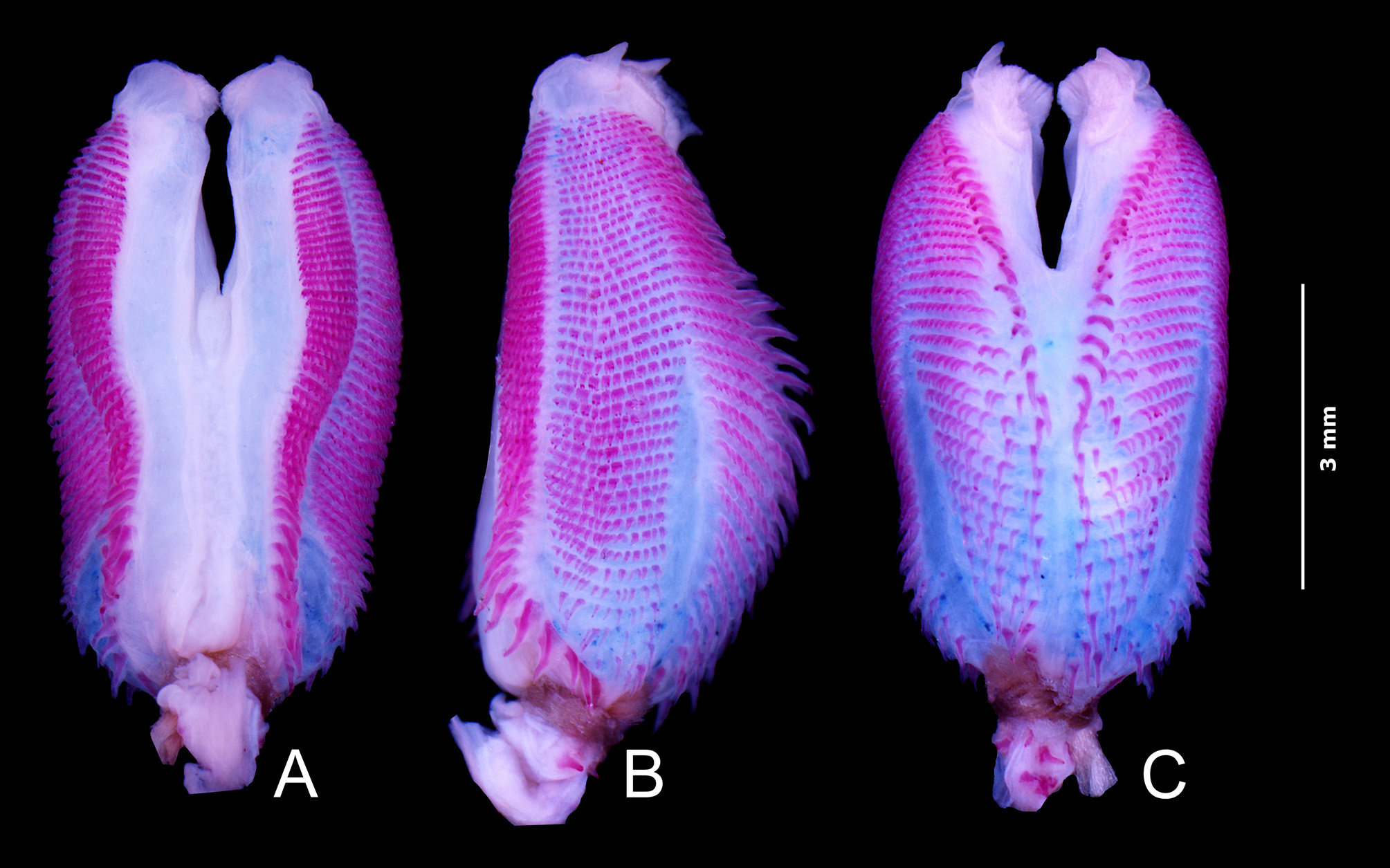
Description of hemipenis ( Fig. 3 View FIGURE 3 ). We prepared the left hemipenis of two paratypes (CHUNB 50548, 18738). In normal position (retracted), the organ is up to 7 mm long, extending for about six subcaudal rows. Hemipenial body roughly globular, with a slight median constriction and clearly bilobate, ending in two pronounced lobes with approximately one-third of total length of the organ. Lobes noncapitated; apex ornamented with a few small folds. Sulcus spermaticus in midline of sulcate face, extending straight from base of organ to lobes. At distal part of hemipenial body, the sulcus is divided by a small fleshy fold at base of a lobular crotch in two branches, each running on medial surface of lobes and ending in their tip among lobular folds. Two large naked areas parallel to sulcus spermaticus in sulcate face of hemipenial body. Each naked area bordered externally by isolated longitudinal ornamented area composed by single spines or series of spicules, arranged in about 45 transverse rows from base of organ to about second third of each lobe. Basal region of longitudinal ornamented area with about 10 single enlarged spines, the latter gradually giving way to more complex transverse rows with up to 10 small and bicuspidate spicules. Lateral face of hemipenis adorned by about 40 transverse rows of spicules; the more apical bi- or tricuspidate, basal ones longer, unicuspidate, enlarged. Lateral series of spicules separated from those on sulcate face by narrow longitudinal nude area extending from base to lobes of organ. Another bare area, curved and wider medially, separates the 20 more basal rows of lateral spicules of ornamented asulcate face of organ. Asulcate face of hemipenis adorned by two longitudinal ornamented areas, separated from each other by bare sagittal area running from base towards lobular crotch, and then bifurcating along medial area of lobes. This sagittal bare area is slightly wider in CHUNB 50548. Ornamented areas of asulcate face composed by about 30 rows of spines and/or spicules; spines of these rows bordering the bare area enlarged, hook-shaped, gradually decreasing in size towards sulcate face. Spicules of superior third of asulcate side bi- or tricuspidate.
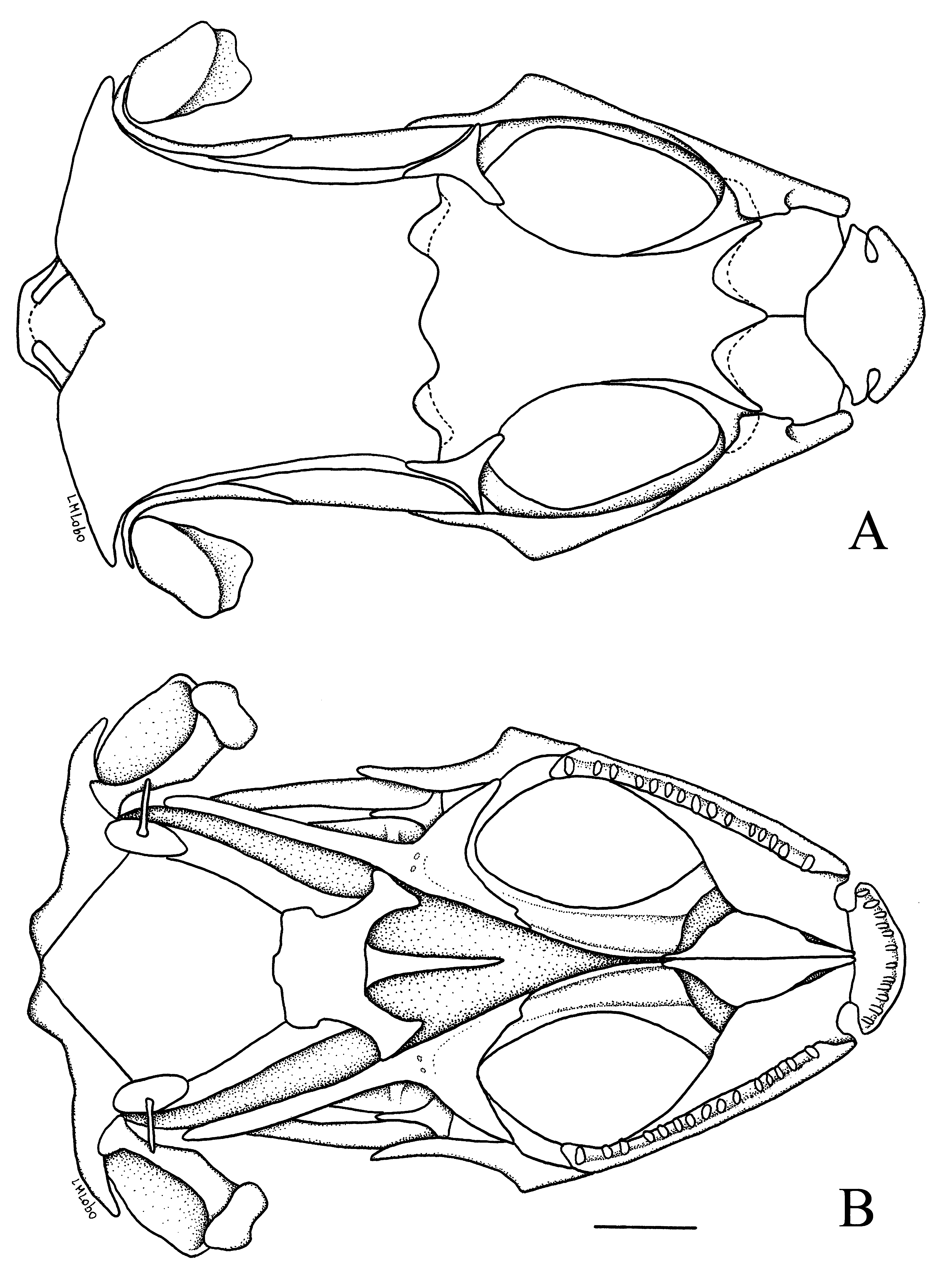
Osteological description ( Figs. 4–5 View FIGURE 4 View FIGURE 5 ). Premaxillary as long as large, touching but not articulating with the maxillary laterally. Its dorsal lamina triangular posteriorly, long, covering the nasals slightly anteriorly the nasals and deeply indenting their suture, preventing their anterior contact. Thirteen conical premaxillary teeth. Nasals large, slightly longer than wide, wider anteriorly, diagonally arranged, widely separated anteriorly, in midline contact in posterior third, covering the frontal anteriorly. Frontal longer than wide, strongly constricted between orbits, wider posteriorly, covering parietal and articulating laterally with it by a pair of frontoparietal tabs. Parietal longer than wide, wider and concave posteriorly, covering occipital region laterally. Lateral expansion of parietal absent, leaving supratemporal fenestra open. Epipterygoid contacting superficially a descending epipterygoid process of parietal. Maxillary contacting nasal dorsally, parts of frontal and lacrimal laterally, but not overlapping, and extensively covering prefrontal and jugal; 24 maxillary teeth. Prefrontal large, its posterior process long but not reaching level of middle of orbit; in broad contact with frontal. Lacrimal small, rod shaped, very conspicuous, contacting prefrontal and maxillary along the inferoanterior part of orbit. Postfrontal and postorbital single. Postfrontal roughly triangular, contacting jugal, frontal, postorbital and parietal, closing the orbit posteriorly. Posterior part of postfrontal wider, longer, almost straight, preventing contact between frontal/parietal and postorbital and concealed marginally by postorbital. Postorbital long and wide, slightly expanded, contacting posteriorly squamosal but leaving supraorbital fenestra widely open. Squamosal long, posteriorly curved and articulating with dorsal end of quadrate. Supratemporal fenestra widely open. Supratemporal present, small, in close contact with posterior part of parietal and squamosal. Fifteen scleral ossicles. Vomer, palatine, pterygoid and ectopterygoid present. Vomer, palatine, premaxillary and maxillary in contact, restricting fenestra exochoanalis. Infraorbital fenestra large, bordered posteriorly by ectopterygoid and pterygoid. Pterygoid teeth present. Stapes rod-like, wider and rounded at the base. Sutures between supraoccipital, exoocipital, basioocipital and otic area of skull not clearly visible in articulated skeleton, as well as those between basioccipital and basisphenoid. Dentary, articular, splenial, angular, and supraangular distinct; 25 dentary teeth, conical anteriorly, bicuspid or tricuspid posteriorly. Glossohyal long, fused to basihyal. First ceratobranchial curved posteriorly; hypohyal and ceratohyal present. A second short pair ceratobranchials present and positioned parallel to anterior part of trachea.
Anterior part of clavicle greatly enlarged, flattened, enclosing a fenestra. Interclavicle long, cruciform, with very long lateral processes reaching sternum but not sternal fenestra. Scapulocoracoid with coracoid, scapular and scapulocoracoid fenestra; suprascapula present. Sternum with large fenestra invaded by long sternal process; three sternal ribs; xiphisternum with two inscriptional ribs. Ilium, ischium and pubis present, the latter with a conspicuous pectinate apophysis. Hypoischium long, larger at the base, almost reaching preanal border; preischium small, elongate; prepubis small, quadrangular and ossified.
Twenty-seven procelous presacral vertebrae, neural spines low, higher anteriorly hypapophyses present in first eight vertebrae; zygantrum-zygosphene present. Last presacral vertebra lacking ribs. Two sacral vertebrae. First four caudal vertebrae lacking autotomic processes, with long and wide transverse processes and wide and high neural spines. From fifth vertebra on, intravertebral autotomic septa present, transverse processes narrow and neural spines decreasing in height. Humerus and femur slightly longer than radius and ulna, and tibia and fibula, respectively. Remaining elements of forelimbs and hind limbs as in Fig. 5 View FIGURE 5 .
Variation and sexual dimorphism. Variation in external morphology is summarized in Table 1. All specimens have three supraoculars and three superciliaries, except CHUNB 52868 where the first and second superciliaries are fused on the right side. Most specimens have 6 or 7 supralabials and infralabials; variation is due to fusions or subdivision of scales and frequently asymmetric. Likewise, most specimens have five suboculars, although the number may vary between three and seven, frequently asymmetrically in the same individual. Gulars also vary between six and eight. Other variations include: the loreal and frenocular are fused on the left side in MPEG 24128; four specimens (CHUNB 50551, 50545, 50553, 50563) have a small azygous scute behind frontal that separates frontoparietals (not in CHUNB 50563); the central scale of the preanal plate is absent in three specimens (CHUNB 50549, 50557, 50565); in CHUNB 50552 the semitransparent disc of the left lower eyelid is divided medially; in CHUNB 23458 prefrontals are not in contact; and CHUNB 50545 has anomalous chinshields. The sexes can be readily separated by the absence of preanal and femoral pores in females. Pores are highly conspicuous, placed in distinctively elevated scales, and aligned on each side without gaps between preanal and femoral. In addition to the presence of pores, there is sexual dimorphism in the number of ventrals, which are significantly more numerous in females ( Table 1, t -test p <0.05), and adult males have bright orange bellies. In one male (MPEG 24129), a distinct white ocellus is present above the insertion of the hind limb. Ventral parts in life are pale reddish orange, in one male (MPEG 24129) with vertical, pale reddish orange bands reaching dorsolateral area. Tongue is dark grey.
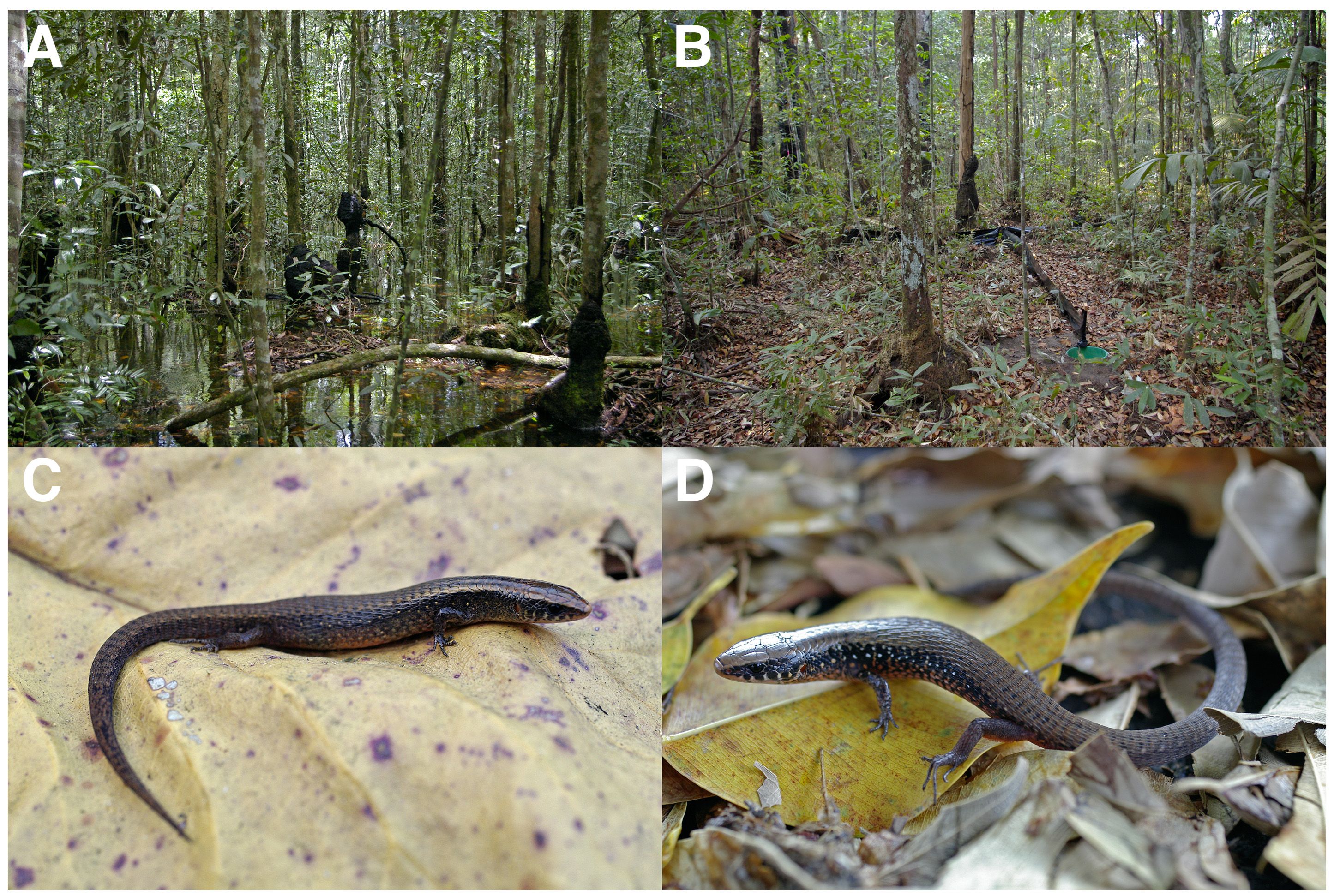
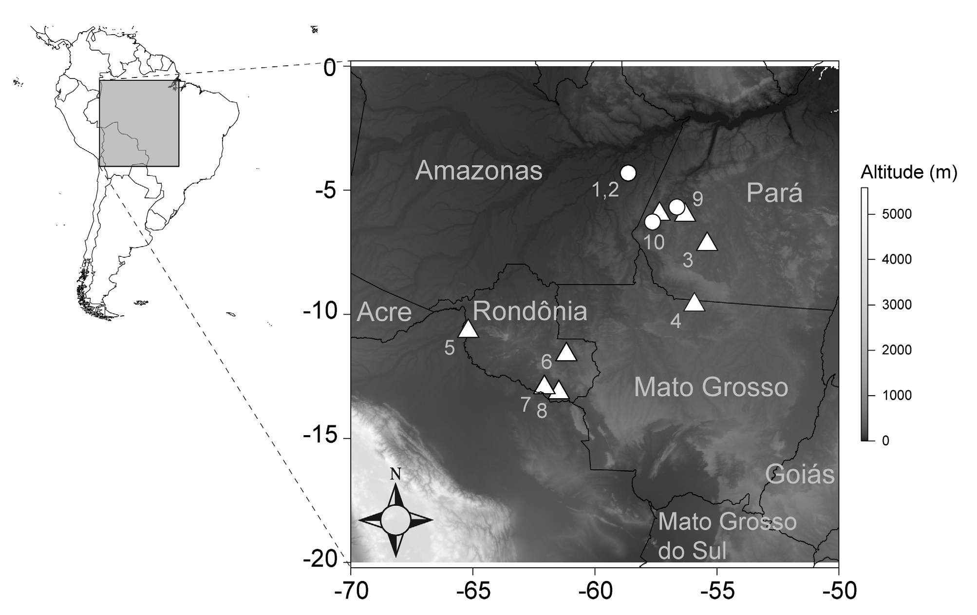
Distribution, habitat and natural history. Rondonops biscutatus is known only from forests in southwestern Amazonia in the states of Rondônia, Mato Grosso and Pará ( Fig. 6 View FIGURE 6 ). This region is part of the "arc of deforestation" ( Aldrich et al. 2012; Fearnside & Graça 2006; Ferreira et al. 2005). Specimens from Parque Estadual do Corumbiara (Cerejeiras and Alta Floresta d'Oeste) were collected in seasonally flooded forest ( Fig. 2 View FIGURE 2 ab); specimens from Guajará-Mirim and Parque Estadual do Cristalino (Alta Floresta) were collected in terra firme forest. Specimens from Pimenta Bueno were collected in Cerrado enclaves within terra firme forest. All these individuals were collected in pitfall traps ( Fig. 2 View FIGURE 2 b), consisting of four 30-liter buckets, arranged in a Y, separated by 6 m drift fences, except CHUNB 52868 that was hand collected at 13:23 h. This individual was in a flooded forest, on top of a termite nest at the side of a large tree, ca. 60 cm above the water ( Fig. 2 View FIGURE 2 a). Specimens from Novo Progresso were collected in logged primary terra firme forest with many Bertholletia excelsa ( Brazil nut) trees. One specimen from Itaituba (Mina do Tocantinzinho) was collected at the margin of a creek in an açaí ( Euterpe oleracea ) forest. The other specimens from Itaituba (Mina do Tocantinzinho and most material collected by the UFPA/Herpetologia Team) were collected in terra firme forest. MPEG 31101 from Jacareacanga and MPEG 31098 from Itaituba were collected in riparian plots. MPEG 24128–9 and 31095–31102 were collected in Y-shaped pitfall traps consisting of four 60-l iter buckets placed 10 m apart and connected by 50 cm high drift fences. MPEG 24127 and 24130 were collected by hand during active searching in leaf litter of terra firme forest at 10:20 and 11:25 h. MPEG 28555–6, 28558 were all collected by hand in leaf litter, between 9:00 and 13:50 h, MPEG 28557 also was collected by hand in leaf litter, but at 22:15 h. Thus, R. biscutatus is an inhabitant of forest floor leaf litter in terra firme forest, transitional areas between Amazon forest and Cerrado, açaí forest, riparian areas and seasonally flooded forest. Specimens seem to be mostly diurnal (9:00–13:50 h), but one specimen was caught while actively moving in leaf litter at night (22:15 h), indicating some nocturnal activity as well. Three females (CHUNB 50544, 50562, 50563) from Cerejeiras, Rondônia, collected in August 2007 (dry season) contained one egg each, whereas none of the adult females collected during the wet season contained eggs. Presumably reproduction takes place during the dry season.
Remarks. The species was first mentioned by Gainsbury and Colli (2003) as Gymnophthalmidae sp., from Cerrado enclaves on latosols and sandy soils in Pimenta Bueno, Rondônia. Hoogmoed et al. (2007) reported the species as Colobosaura sp. nov., but provided no further details except the locality of collection and its general habitat (terra firme forest). Garda et al. (2013) studied the effects of microhabitat variation on lizard distribution in a terra firme forest in Guajará-Mirim, Rondônia. The species, reported as Gymnophthalmidae sp., is associated with sites distant from large trees, with few fallen logs and burrows, less canopy cover, thicker understory, thinner leaf litter, and numerous termite nests.


No known copyright restrictions apply. See Agosti, D., Egloff, W., 2009. Taxonomic information exchange and copyright: the Plazi approach. BMC Research Notes 2009, 2:53 for further explanation.
|
Kingdom |
|
|
Phylum |
|
|
Class |
|
|
Order |
|
|
Family |
|
|
Genus |
Rondonops biscutatus
| Colli, Guarino R., Hoogmoed, Marinus S., Cannatella, David C., Cassimiro, José, Gomes, Jerriane Oliveira, Ghellere, José Mário, Sales Nunes, Pedro M., Pellegrino, Kátia C. M., Salerno, Patricia, Souza, Sergio Marques De & Rodrigues, Miguel Trefaut 2015 |
Gymnophthalmidae
| Gainsbury 2003: 509 |
| Hoogmoed et al. (2007) :147 |
| Garda et al. (2013) :247 |