
Speleogobius llorisi Kovačić, Ordines & Schliewen, 2016
|
publication ID |
https://doi.org/ 10.11646/zootaxa.4066.3.6 |
|
publication LSID |
lsid:zoobank.org:pub:5FFB8910-51C4-4021-B428-B42D94EDCE19 |
|
DOI |
https://doi.org/10.5281/zenodo.5684283 |
|
persistent identifier |
https://treatment.plazi.org/id/03983615-FFD1-FA54-FF34-F897FCD8F79D |
|
treatment provided by |
Plazi |
|
scientific name |
Speleogobius llorisi Kovačić, Ordines & Schliewen |
| status |
sp. nov. |
Speleogobius llorisi Kovačić, Ordines & Schliewen , sp. nov.
Figs. 2–4 View FIGURE 2 View FIGURE 3 View FIGURE 4
Holotype. PMR VP 3510, female, 25.6 + 5.6 mm, Spain, Balearic Islands, Mallorca, 39º14.23'N, 002º58.66’E, red algae beds, 67 m depth, coll. Instituto Español de Oceanografía-Centre Oceanogràfic de Balears (IEO-COB), 13 April 2015. The collecting positions for the type material and for the additional material are presented in Fig. 1 View FIGURE 1 .
Paratypes. PMR VP 3511, female, 18.7 mm SL, caudal fin damaged, Spain, Balearic Islands, Mallorca, 39°30.34’N, 002°25.03’E, red algae beds, 67 m depth, coll. IEO-COB, 12 September 2014; ZSM 43988, female, 27.2 mm SL, caudal fin damaged, Spain, Balearic Islands, Mallorca, 39°22.09’N, 002°41.12’E, red algae beds, 51 m depth, coll. IEO-COB, 12 September 2014; PMR VP 3512, male, 24.4 + 5.5 mm, Spain, Balearic Islands, Mallorca, 39°21.06’N, 002°41.74’E, red algae beds, 56 m depth, coll. IEO-COB, 14 September 2014; PMR VP 3513, female, 24.5 + 5.6 mm, Spain, Balearic Islands, Mallorca, 39°30.34’N, 002°25.03’E, red algae beds, 46 m depth, coll. IEO-COB, 12 September 2014, ZSM 43989, juvenile male, 14.3 + 4.0 mm, Spain, Balearic Islands, Mallorca, 39º14.28'N, 002º58.70’E, red algae beds, 67 m depth, coll. IEO-COB, 13 April 2015.
Additional material. ZSM 43990, undetermined sex, 17.7 + 3.1 mm, Spain, Balearic Islands, Mallorca, 39°22.05’N, 002°39.288’E, red algae beds, 55 m depth, coll. IEO-COB, 5 September 2014; ZSM-PIS-GO-1065/ 1066, undetermined sex, 17.6 + 3.2 mm SL, Spain, Balearic Islands, Mallorca, 39°24.75’N, 002°31.578’E, red algae beds, 69 m depth, coll. IEO-COB, 5 September 2014; ZSM 43991, female, 23.8 + 4.9 mm SL, Spain, Balearic Islands, Mallorca, 39°16.482’N, 002°57.078’E, red algae beds, 56 m depth, coll. IEO-COB, 7 September 2014; ZSM-PIS-GO-1067/1068, undetermined sex, 21.1 + 4.1 mm SL, same data as holotype.
Diagnosis. (1) Preopercular head canal present with pores γ, δ, ε; (2) snout long, equal or longer than eye, 1.0– 1.1 in eye length, dorsal profile of snout gently sloping; (3) lower lip ends anteriorly slightly in front of upper lip; (4) scales in lateral series 28 or 29; (5) scales in transverse series 6; (6) head length as percentage of standard length: 28.7–30.0% adults, 32.2% juvenile; (7) greatest body depth, measured in the new species at the pelvic fin origin as percentage of standard length: 15.0–16.8%; (8) eye diameter (as percentage of standard length): 6.9– 7.7%; adult female coloration: (9) head and body to pectoral and pelvic fins orange reddish, the rest of body whitish transparent with a dark reticulate pattern and small amount of orange red pigment; (10) fins mostly transparent and only partially pigmented; (11) caudal peduncle at caudal fin base with a dark orange vertical band.
Description. General morphology. Body proportions are given in Table 1 View TABLE 1 . Body moderately elongate, laterally compressed with a slender caudal peduncle. Head long, 28.7–30.0% adults, 32.2% juvenile of SL, slightly depressed, with a nearly horizontal predorsal profile. Snout moderately long and pointed, equal or longer than eye, 1.0–1.1% in eye length, dorsal profile of snout gently sloping. Eyes dorsolateral, extending above dorsal profile. Anterior nostril short, tubular, erect, without process from rim; posterior nostril pore-like, near orbit. Mouth oblique, posterior angle of jaws below anterior edge of eye, anterior tip of mouth in the horizontal level of eye. Lips wide, lower lip ends anteriorly slightly in front of upper lip. Rows of pointed teeth in both jaws, outer row with largest teeth. Tongue truncated. Branchiostegal membrane attached to entire lateral margin of isthmus.
Fins. D 1 VI (VI:6); D 2 I /6–7 (6: 1, 7: 5); A I/6–7 (6: 5, 7: 1); C 11–12 branched rays (11:1, 12: 2, C of paratypes PMR VP3511, ZSM 43988 and ZSM 43989 too damaged to count), 14–15 segmented rays (14: 3; 15: 3); P 15 (15: 11, right P of holotype PMR VP3510, too damaged to count), V I/5 + 5/I. Fin-bases and lengths in proportion to standard body length are given in Table I. D 1 II and III the longest, longest spines of D1 backwards nearly reaching origin of D2 when folded down in male adult and juvenile, not reaching in females. Fin membrane of D 1 VI not connected with base of D 2 I. D2 commences over vertical of anus to vertical of urogenital papilla, with last ray over vertical of last A ray. A commences below second to third segmented ray of D2. C posterior edge truncate. P uppermost rays within membrane, P rays all branched except uppermost and lowermost rays, ending back before vertical of D2 spine. V emarginated (shortest branches of V 5 compared to longest branches of V 4: 75%–87%), reaching behind anus, to A spine in juvenile, no anterior transverse membrane.
Squamation. Body covered with ctenoid scales. Predorsal area scaly, scales along dorsal midline 5–7 (5:1, 6:2, 7:1, PMR VP3511, and ZSM 43989 too damaged for count). Opercle and cheek naked. Breast scaly with cycloid scales, scales along ventral midline 3–6 (3:1, 5:3, 6:1, ZSM 43989 too damaged for count). Scales in lateral series 28–29 (left and right side: 28:5, 29:4, PMR VP3511, too damaged to count on one side, ZSM 43989 on both sides), in transverse series 6 (left and right side: 6:8, PMR VP3511, and ZSM 43988 too damaged to count on one side, ZSM 43989 on both sides).
LL system ( Fig 4 View FIGURE 4 ). Head with anterior oculoscapular and preopercular canals, carrying pores σ, λ, κ, α, β, ρ and γ, δ, ε respectively (ZSM 43989 with surface too dried and damaged to see head canals and pores clearly). Posterior oculoscapular canal absent. Rows and number of sensory papillae as follows (all type material checked, but usually only part of LL system visible on specimens due to the skin damage, entire LL system visible in paratype PMR VP3512): (1) preorbital: snout with single row rs as 2 papillae close to pore σ and posterior nostril. Lateral series c1,2 as two or three papillae in longitudinal row starting behind anterior nostril and going backwards above upper lip. (2) suborbital: rows without transverse proliferation; a below rear part of eye starting posteriorly at pore α (3); c below eye (4–5), anteriorly beginning below front border of eye, posteriorly ending before a row; papillae cp’ and cp longitudinally arranged in front of b. Row b single papilla behind rear border of eye and bellow level of it. Longitudinal row d as two papillae above upper lip posterior part. (3) preoperculo-mandibular: anterior part of external row e not visible in all specimens, anterior part of internal row i (5) and row f (2) present. Posterior sections of row e (8–12) and row i (12–13). (4) oculoscapular: anterior longitudinal row x1 (2–3) above pore ρ, row z (2) in the middle between pores β, ρ and γ, row q (2–3) behind pore ρ, posterior longitudinal row x2 and row y not visible. Axillary rows as1 (3), as2 (3), as3 (2–4), la1 (1) and la2 (1) present. (5) opercular: transverse row ot (6–10); superior longitudinal row os (3); inferior longitudinal row oi (2). (6) anterior dorsal: row n as single papila behind eye, rows g and m just as single papilla, row o absent and longitudinal row h (3–4) poorly visible among scales.
Colour of freshly collected material. Freshly collected female ( Fig 2 View FIGURE 2 A)—most scales had already been lost on the freshly collected specimens, so coloration may have been influenced. Head and body to V and P bases orange red, rest of body whitish-transparent with a pattern of melanophores concentrated at posterior edges of scales, resulting in an imbricated pattern. Predorsal area pink whitish, underside of head and cheek mottled orange red and whitish, anterior chin with two partially contingent blotches of melanophore; a transverse orange-red band on chin posterior part of chin below anterior eye margin; opercle and prepelvic area orange red. On body melanophores more concentrated along lateral midline forming four blotches. Reddish to grey silhouette of vertebral column visible behind. Caudal peduncle with dark blotch below midlateral line, expanding vertically backwards into dark orange vertical stripe with a narrow dark and well defined margin at C base. Small amount of orange red to pink pigment also visible scattered on body, mostly along lateral midline. Dorsum with three broad, transverse dark bands of similar intensity and on flank contiguous with lateral side blotches: the first over D1 base, the second over D2 base and the third over caudal peduncle; dark bands alternating with four whitish saddles. Prepelvic area and V base orange red. Ventral midline and belly whitish. Silvery peritoneum visible bellow skin on belly. Fins mostly transparent and partially pigmented. D1 and D2 with melanophores arranged to an ill-defined band in the middle part and at the fin base and with erythrophores distributed mainly in the upper part of the fins. C at base contingent with posterior dark edge of dark orange vertical caudal peduncle stripe; main part of C with orange yellow dots arranged in ill-defined narrow bands over the rest of the fin. A transparent. P with orange mark in anterior part and dark mark in the middle of the fin, the rest of the fin transparent with scattered erythrophores. V with red orange base, dark middle part and white or transparent tips.
Colour preserved specimens in alcohol ( Figs. 2 View FIGURE 2 B and 3 — most scales missing on the preserved specimens, so preserved coloration may have been influenced). Body yellowish white. Melanophores on body concentrated on posterior edges of scales (or scale positions when scales are lost) resulting in an imbricated pattern on lateral and upper sides of body. Melanophores more concentrated along lateral midline forming elongate blotches in adults, in juvenile as a continuous longitudinal stripe along posterior part of lateral midline, and on dorsal side forming dark bands. Caudal peduncle with larger dark blotch in lower part. Predorsal area, opercle and cheek mottled to dotted with pigments. Head underside mostly whitish with various quantities of pigments present. D1 pigmented with melanophores, but no melanophores visible in the juvenile male. D2 and A pigmented in adult males, not in the juvenile male. D2 poorly pigmented and A transparent in females. C transparent, except for dark pigments at the fin base. P transparent, with dark blotch at lower part of the fin center, not visible in the juvenile. Most of the V pigmented in adult male, females with pigmentation only at anterior half of the fin, the juvenile almost without pigment.
Vertebral column and pterygiophore insertion pattern (pty). 12 precaudal and 17 caudal vertebrae (including urostyle); total count 29. One epural; pty 3–2211000; two pre-haemal pterygiophores (based on four non-type specimens listed under “Additional Material“). These counts are identical with those of four specimens of S. trigloides ( ZSM 35457).
Etymology. The species is named llorisi in honor of Dr. Domenec Lloris, an ichthyologist from the Institut de Ciències del Mar, in Barcelona, in recognition for his outstanding contribution to the knowledge of fish species.
Habitat. All individuals of the new species of Speleogobius were collected from sampling stations located on red algae beds. These beds are one of the most widespread types of habitats of the shallow continental shelf of the Balearic Islands in depths between 50 and 80m ( Ballesteros 1994; Ordines & Massutí 2009). The sampling stations where the specimens of the new species occurred presented very similar epibenthic communities in which predominant algae species on the basal stratum belong to the families Corallinaceae (with Lithothamnion corallioides , Lithothamnion valens , Phymatolithon calcareum and Spongites fruticolus among the most abundant) and to Peyssonneliacea (with Peyssonnelia rubra and Peyssonnelia inamoena among the most abundant), and on the erect stratum to Phyllophoraceae (with Phyllophora crispa ). As a result of the high biomass of Peyssonnelia spp., the habitat present at the sampling stations can be classified as Peyssonnelia beds, a frequently occurring habitat in the south west of Mallorca, and the south of Menorca, that presents the highest biomasses of algae in the circalittoral sea bed of the Balearic Islands. The Peyssonnelia beds are frequently associated with open bays characterized by fine sediments, even mud, where calm periods alternate with periods of eddy forming currents ( Pérès 1985; Ordines et al. 2011; Joher et al. 2012). This was the location of the sampling stations where the new Speleogobius species was found: a sheltered area, without significant currents, and at the lee of the north winds, which cause the most important storms in the area ( Ordines et al. 2011).
Comparison with S. trigloides . Speleogobius llorisi sp. nov. is easily separated from S. trigloides ( Figs 2 View FIGURE 2 , 3 View FIGURE 3 and 5 View FIGURE 5 ) by several morphological characters: preopercular head canal present with pores γ, δ, ε vs. preopercular head canal absent; snout long, equal or longer than eye, 1.0– 1.1 in eye length, dorsal profile of snout gently sloping vs. snout short, shorter than eye, 0.6–0.8 in eye length (measured on comparative material), snout dorsal profile steep; lower lip slightly protruding upper lip (prognathous) vs. upper lip slightly protruding lower lip (retrognathous); scales in lateral series 28 or 29 vs. 26; scales in transverse series 6 vs. 7–8 (7 in holotype); head length as percentage of standard length: 28.7–30.0% adults, 32.2% juvenile vs. 25.5%–29.1% in comparative material, holotype recalculated to percentages 27.9% (overlap present only with S. trigloides of standard length 15 mm or smaller due to allometry), i.e. the new species has longer head compared to body length; body depth at the pelvic fin origin as percentage of standard length: 15.0–16.8% vs. 19.3–20.6% in comparative material, holotype recalculated to percentages 25.1% i.e. body of new species is more slender; eye diameter (as percentage of standard length): 6.9– 7.7% vs. 8.1%–9.7% in comparative material, holotype recalculated to percentages 9.3%, i.e. the new species has smaller eyes compared to body length.
S. llorisi sp. nov. is also easily separated from S. trigloides by several coloration characters ( Figs 2 View FIGURE 2 , 3 View FIGURE 3 and 5 View FIGURE 5 ), at least with regard to adult females (coloration of adult S. llorisi males remains unknown): head and body to pectoral and pelvic fins bright orange reddish, pale orange stripes on C, blackish pelvic fin centers with white margins, and posterior part of body whitish-transparent with a dark reticulate pattern and small amount of orange red pigment vs. female dark red, with several white patches on dorsal and ventral sides, a yellow caudal fin and posterior caudal peduncle; unpaired fins predominantly transparent with orange pigments on distal parts and one dark band vs. D1 and D2 of S. trigloides reddish with white bands (D1) or dots (D2); caudal peduncle at caudal fin base with a dark orange vertical band posteriorly fringed by a narrow dark line vs. caudal peduncle at caudal fin base uniformly orange-yellow without orange dark vertical stripe.

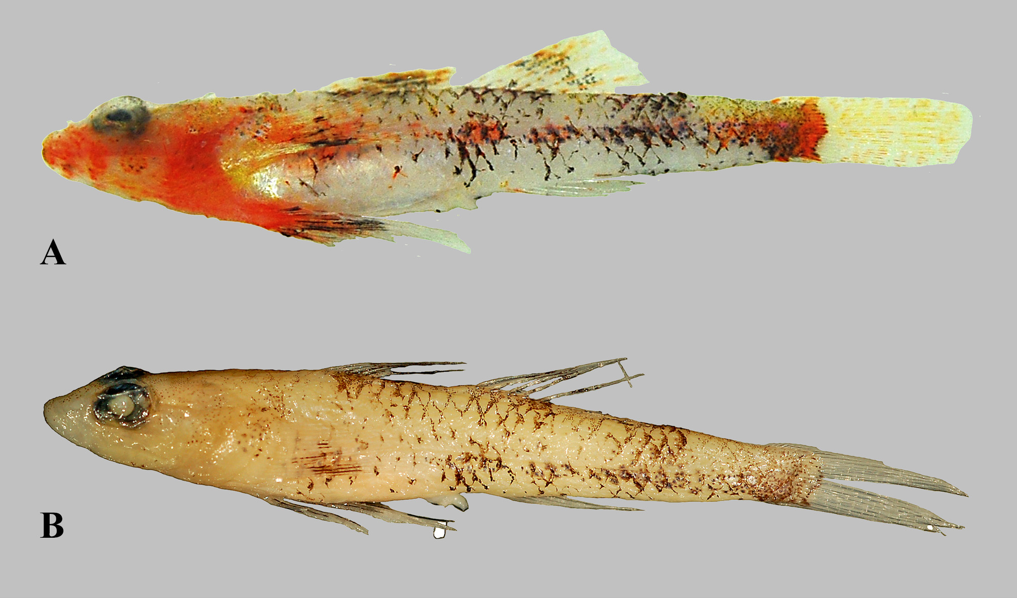
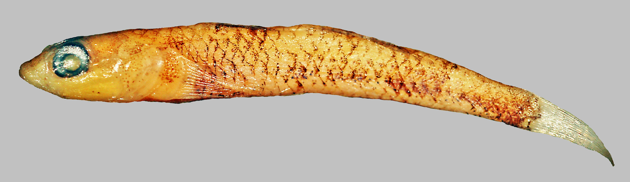
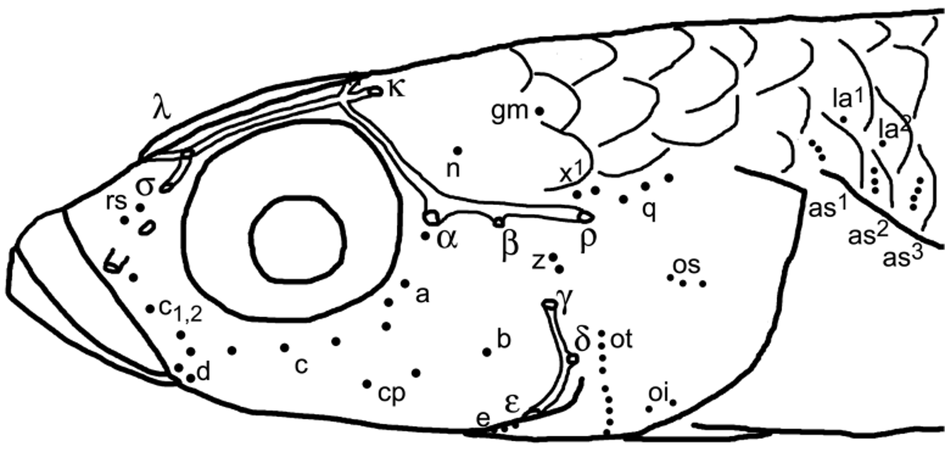
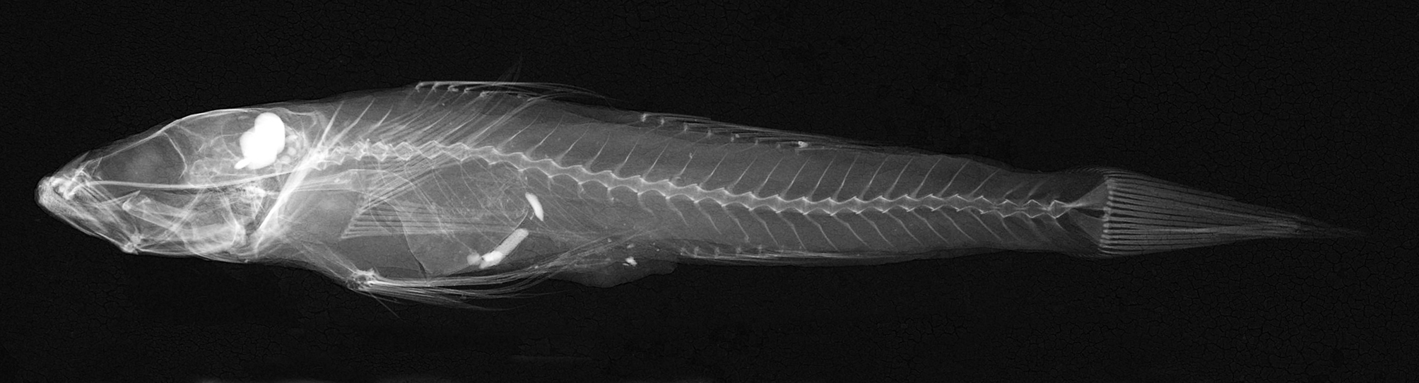
TABLE 1. Morphometric characters (as proportional measurements in %) of Speleogobius llorisi sp. nov. Characters are sorted in alphabetic order.
| Specimen | P3 PMR VP3510 holotype | P47 P57 PMR ZSM 43988 VP3513 paratype paratype | P45 PMR VP3511 paratype | P54 PMR VP3512 paratype | P2 ZSM 43989 paratype |
|---|---|---|---|---|---|
| sex | female | female female | female | male | male juvenile |
| Standard length (Sl) in mm | 25.6 | 27.2 24.5 | 18.7 | 24.4 | 14.3 |
| % of standard length: | |||||
| 1st spine length of first dorsal fin | 13.3 | 12.1 13.9 | 11.8 | 12.7 | 13.3 |
| 2nd spine length of first dorsal fin | 14.8 | 14.3 damaged | damaged | 14.3 | 18.2 |
| 3rd spine length of first dorsal fin | 13.7 | 13.6 15.9 | 16.0 | 13.5 | 16.8 |
| Anal fin base | 11.7 | 9.6 11.8 | 10.7 | 11.9 | 9.1 |
| Anal fin spine length | 9.4 | 9.6 11.0 | 9.1 | 9.4 | 10.5 |
| Body depth at anal fin origin | 14.1 | 15.1 14.3 | 13.9 | 13.5 | 14.0 |
| Body depth at pelvic fin origin | 16.8 | 16.2 15.5 | 15.0 | 16.0 | 16.8 |
| Body width at anal fin origin | 9.8 | 9.2 10.2 | 10.2 | 10.2 | 9.1 |
| Body width at pelvic fin origin | 15.2 | 14.7 15.5 | 13.9 | 13.9 | 16.1 |
| Caudal fin length | 21.9 | damaged 22.9 | damaged | 22.5 | 28.0 |
| Caudal peduncle depth | 8.6 | 8.1 8.2 | 7.5 | 7.8 | 8.4 |
| Caudal peduncle length | 26.6 | 25.4 26.9 | 28.3 | 28.3 | 28.0 |
| First dorsal fin base | 10.5 | 9.9 9.8 | 9.1 | 9.0 | 9.1 |
| Head length | 29.3 | 28.7 29.4 | 29.9 | 28.7 | 32.2 |
| Head width | 17.2 | 15.4 15.9 | 14.4 | 15.6 | 16.1 |
| Pectoral fin length | 23.4 | 21.3 22.4 | 23.5 | 21.3 | 21.7 |
| Pelvic fin length | 27.0 | 25.4 26.9 | 28.3 | 27.0 | 32.9 |
| Pelvic to anus | 23.0 | 23.9 22.0 | 25.7 | 21.7 | 24.5 |
| Second dorsal fin base | 14.5 | 12.9 13.9 | 15.5 | 14.8 | 13.3 |
| Second dorsal fin spine length | 13.3 | 11.8 13.9 | 11.8 | 13.1 | 14.0 |
| Snout to origin of first dorsal fin | 37.5 | 36.8 38.4 | 38.0 | 38.1 | 38.5 |
| Snout to origin of second dorsal fin | 56.6 | 55.1 57.1 | 55.6 | 55.7 | 57.3 |
| Snout to vertical of anal fin origin | 60.5 | 61.8 61.2 | 62.0 | 59.0 | 62.9 |
| Snout to vertical of anus | 53.5 | 54.0 53.1 | 54.5 | 52.0 | 56.6 |
| Snout to vertical of pelvic fin origin | 31.3 | 30.9 34.3 | 31.0 | 33.2 | 34.3 |
| % of caudal peduncle: | |||||
| Caudal peduncle depth | 32.4 | 31.9 30.3 | 26.4 | 27.5 | 30.0 |
| % of head length | |||||
| Cheek depth | 13.3 | 11.5 13.9 | 12.5 | 11.4 | 13.0 |
| Eye diameter | 25.3 | 24.4 23.6 | 25.0 | 24.3 | 23.9 |
| Head depth | 42.7 | 42.3 43.1 | 39.3 | 42.9 | 43.5 |
| Head width | 58.7 | 53.8 54.2 | 48.2 | 54.3 | 50.0 |
| Postorbital length | 52.0 | 48.7 51.4 | 51.8 | 50.0 | 52.2 |
| Snout length | 25.3 | 24.4 26.4 | 26.8 | 25.7 | 26.1 |
| % of eye diameter | |||||
| Interorbital width | 31.6 | 36.8 35.3 | 35.7 | 29.4 | 36.4 |
No known copyright restrictions apply. See Agosti, D., Egloff, W., 2009. Taxonomic information exchange and copyright: the Plazi approach. BMC Research Notes 2009, 2:53 for further explanation.