
Onchimira cavifera
|
publication ID |
https://doi.org/ 10.5281/zenodo.188931 |
|
publication LSID |
lsid:zoobank.org:pub:179F84F7-CB72-4AB7-877A-BAD332E57FEE |
|
DOI |
https://doi.org/10.5281/zenodo.5694796 |
|
persistent identifier |
https://treatment.plazi.org/id/03DD87DF-4011-FFA5-FF51-FD6EFD8BCBEF |
|
treatment provided by |
Plazi |
|
scientific name |
Onchimira cavifera |
| status |
|
Onchimira cavifera View in CoL gen. et sp. nov.
( Figures 2 View FIGURE 2 ; 3A, E; 4A–C, F, J–K; 5A, B; 6A, B; 7A. Table 1 View TABLE 1 )
Type material. Holotype, ZMMU Lc-37446, NW Pacific near Kamchatka peninsula, Starichkov Id., 20–26 m, large boulders, collected by T.A. Korshunova and A.V. Martynov. 14.08.2008. Paratypes, ZMMU Lc- 37447, 11 specimens (three dissected), same locality and collectors as holotype, 14.08.2008. Paratypes, ZMMU Lc-37448, three specimens (one dissected), same locality and collectors as holotype, 18–24 m, 19.08.2008. Paratypes, ZMMU Lc-37449, three specimens (one dissected), same locality and collectors as holotype, 19.08.2008. Paratypes, ZMMU Lc-37450, three specimens (one dissected), same locality and collectors as holotype, 19.08.2008.
Type locality. NW Pacific, SE Kamchatka, Starichkov Id., 18–26 m depth.
Etymology. The species epithet from the Latin cavi (= cavity) and fero (= to bear) refers to the presence of a well-defined gill cavity.
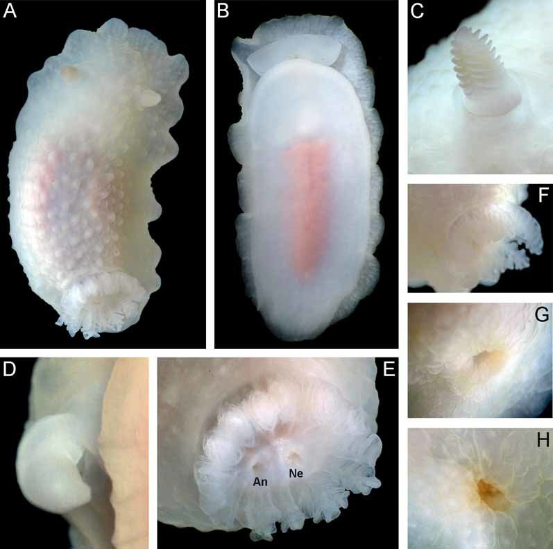
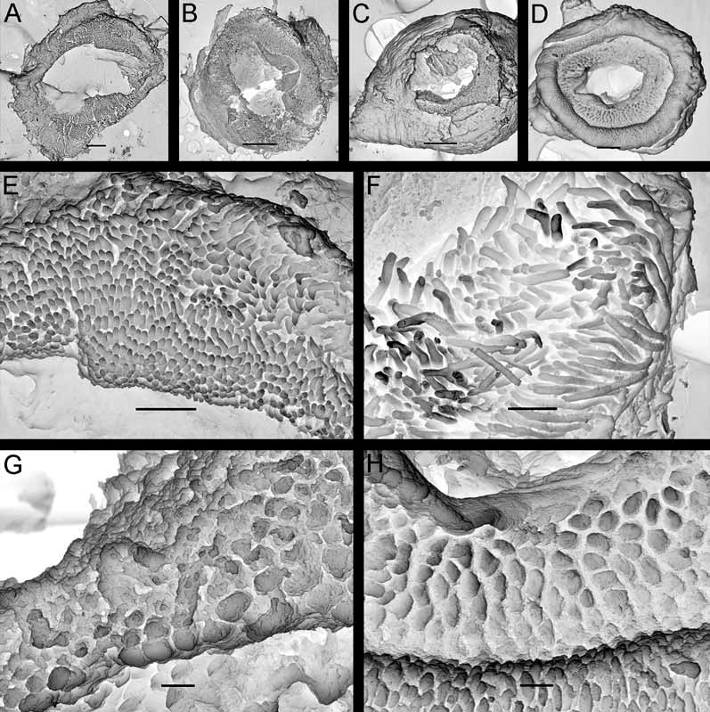
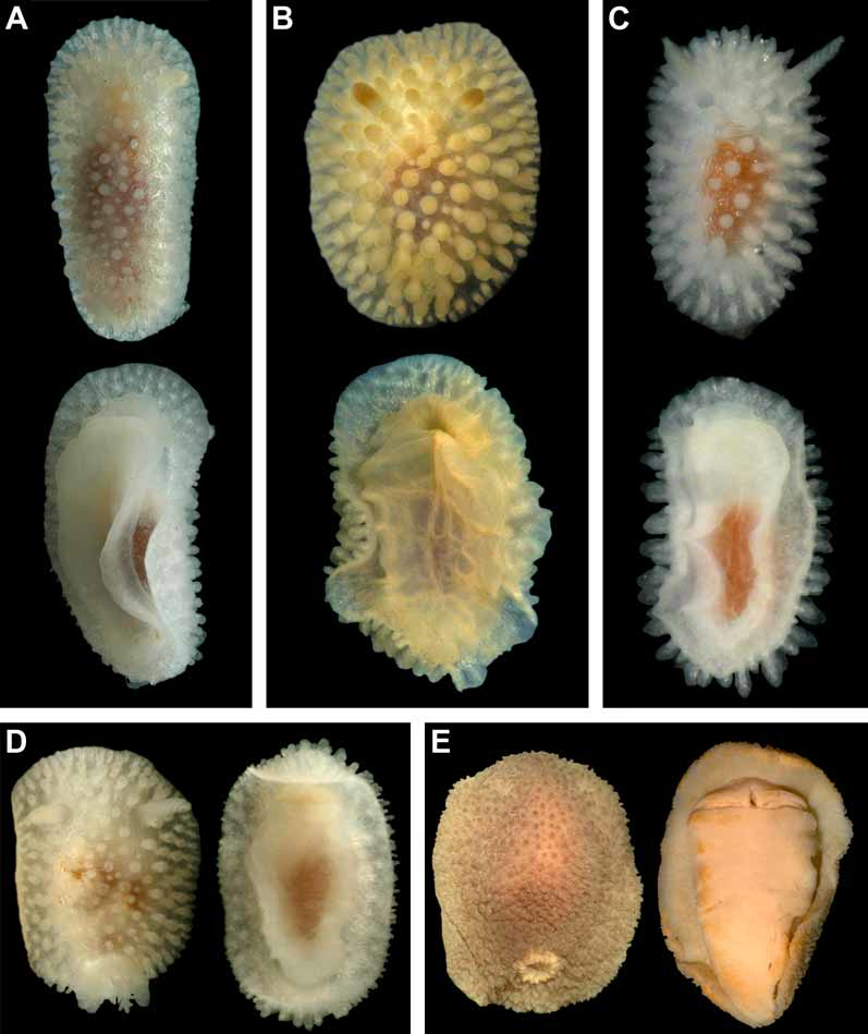
Description. External morphology. The dimensions of the holotype are 22 mm x 12 mm ( Fig. 2 View FIGURE 2 ). The length of fifteen measured living specimens ranged from 8.5 to 25 mm, the width ranged from 4.5 to 14 mm. The consistency of the living animals is soft. The notum is rather broad, rounded in front and posteriorly. The rhinophores are long and retracted into raised sheaths with smooth, soft, sometimes slightly crenulate edges, not bearing tubercles ( Fig. 2 View FIGURE 2 C). The rhinophoral sheath edges are capable of considerable contraction in living specimens. There are 5–9 rhinophoral lamellae. The rhinophore clavus lacks a posterior ridge. The notum is almost smooth, sparsely covered with wrinkled low elevations, sometimes raised to very low tubercles ( Fig. 2 View FIGURE 2 A). Rays of spicules radiate from the bases of such elevations and form a sparse network in the notum ( Fig. 6 View FIGURE 6 A), but spicules are not conspicuous externally ( Fig. 2 View FIGURE 2 A). Each elevation contains sparsely placed spicules, which do not protrude from the tubercles. The strongly calcified spicules are of various size, most with a narrow channel inside ( Fig. 6 View FIGURE 6 B). 10–15 (usually 12) unipinnate gills are united by a common membrane into a circle around the anus. Gills are retractable into a common true gill cavity, which is capable of closing over the gills completely ( Figs. 2 View FIGURE 2 E–H). The border of the gill cavity is moderately raised and has a smooth edge ( Figs. 2 View FIGURE 2 E, F). The oral veil is well defined, trapezoid, with oblique lateral sides and convex anterior edge ( Fig. 2 View FIGURE 2 B). The foot is broad, anteriorly rounded and not thickened; posteriorly it projects slightly from the notum in crawling animals, forming a rounded tail.
Colour. The living specimens are grayish with creamy tinge ( Fig. 2 View FIGURE 2 ). The rhinophores (including lamellae) are similar to the ground colour but more intensively cream. The gills are semitransparent white, similar to the ground colour. The pinkish digestive gland is slightly visible through the notum dorsally and shines more clearly through the foot ventrally. A purple-blackish female gland mass is visible through the anterior part of the right side in some specimens.
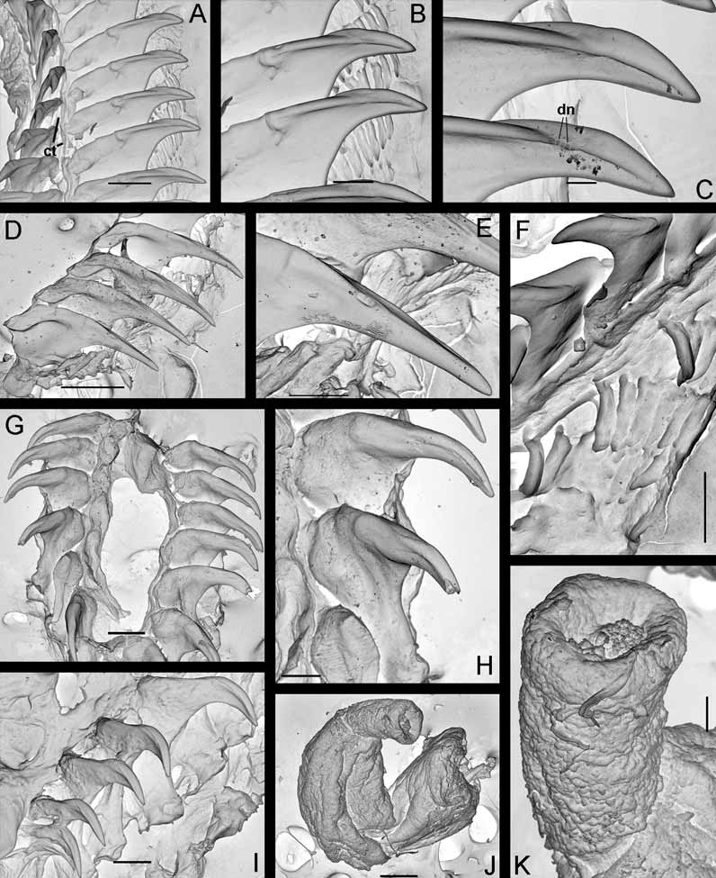
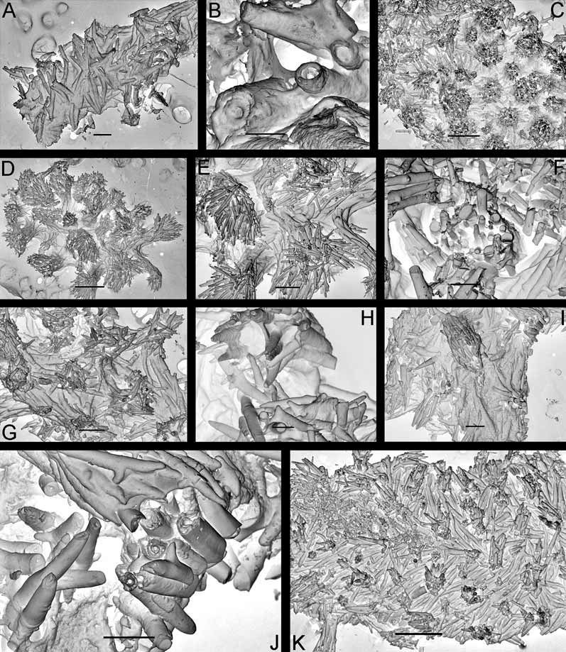
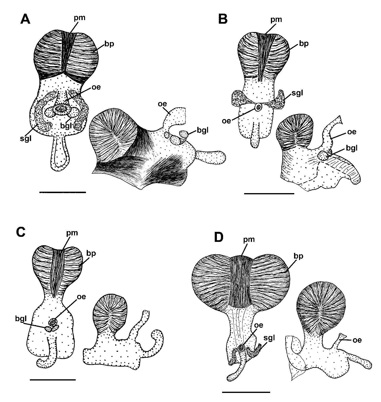
Anatomy. Digestive system. The anterior part of the buccal bulb is modified into the sessile, large, buccal pump which is medially fully banded by a broad peripheral muscle ( Fig. 7 View FIGURE 7 A). The lateral sides of the buccal pump are provided with thin muscular fibres. The rounded labial disk is covered by a brown cuticle bearing distinct, rod-shaped labial elements with bent tips ( Figs. 3 View FIGURE 3 A, E). The radular formula in four specimens (17–23 mm length) is 25- 28 x 7–9.1.1.1.9–7. The radular teeth are slightly yellowish. The central tooth is small, elongate, rectangular and folded on the lateral edges ( Fig. 4 View FIGURE 4 A). The first lateral tooth is large and provided with a long, wide base and a strong, slightly curved beak-shaped cusp ( Figs. 4 View FIGURE 4 A–C, F). The cusp sometimes bears small faint denticles ( Fig. 4 View FIGURE 4 C). Outer lateral teeth are elongated small plates without cusps, all similar in size and shape ( Figs. 4 View FIGURE 4 A, B, F). The salivary glands are relatively long and narrow ( Fig. 7 View FIGURE 7 A). The stomach is relatively large and broad, then rapidly narrowing to the intestine. The stomach caecum is absent.
Circulatory system. In the pericardial sac the broadly triangular posterior auricle and the smaller sized oval ventricle are present. The rather massive, irregularly rectangular blood gland is located above the central nervous system.
Central nervous system. The cerebral and pleural ganglia are well separated, the latter being somewhat smaller in size. The optic nerve is very short. The eyes are relative large, with black pigment in all studied specimens. The pedal ganglia are smaller than the cerebrals, lay below them and are connected to them by very short connectives. The rhinophoral ganglia are rather irregular, rounded or elongate. The buccal ganglia are rounded-oval ( Fig. 7 View FIGURE 7 A). Gastro-esophageal ganglia are present. Five pairs of cerebral nerves, two pleural and three pedal ones are detected.
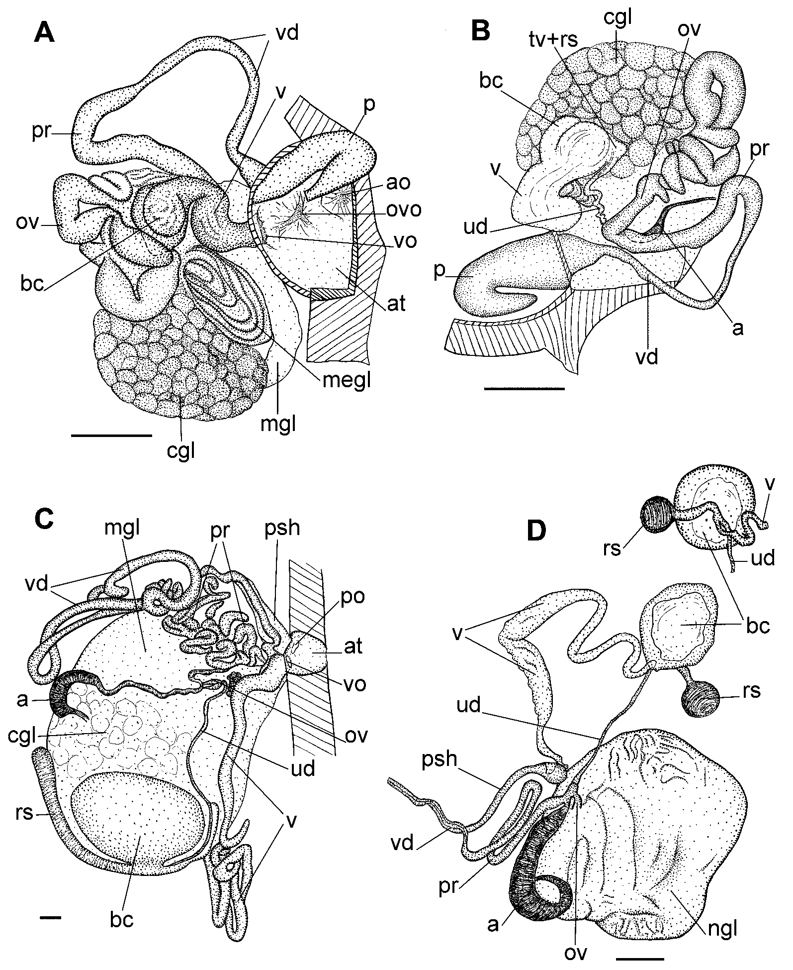
Reproductive system. ( Figs. 5 View FIGURE 5 A, B). The ampulla is relatively short and narrow, not filled by sperm in all studied specimens ( Fig. 5 View FIGURE 5 B, a). The ampulla trifurcates into the moderately long vas deferens, uterine duct and oviduct ( Fig. 5 View FIGURE 5 B, pr, ud and ov). The prostatic part of the vas deferens is a very short, slightly swollen and bending duct, which does not encircle the bursa copulatrix ( Figs. 5 View FIGURE 5 A, B, pr). The prostate transits to a moderately long and narrow single-looped vas deferens ( Figs. 5 View FIGURE 5 A, B, vd), which rapidly widens and enters a common genital atrium ( Fig. 5 View FIGURE 5 A, at), terminating into the large, wide, and prominent penis, which contains a smaller evertable part ( Figs. 4 View FIGURE 4 J, K; 5A, B, p). The vagina is relatively wide, moderately convoluted ( Figs. 5 View FIGURE 5 A, B, v), and enters a rather small, flattened bursa copulatrix ( Figs. 5 View FIGURE 5 A, B, bc). The uterine duct is long and narrow ( Fig. 5 View FIGURE 5 B, ud); it begins at the ampulla bifurcation and then enters the terminal part of the vagina forming a small pointed elevation. A separate seminal receptacle is absent, but the terminal part of the vagina forms a large swollen area ( Fig. 5 View FIGURE 5 B, tv+rs), which may serve as a receptacle. In freshly dissected living specimens, a tiny, almost inconspicuous knob-shaped structure was found on the vagina at the bursa base.
This structure possibly is a vestige of a seminal receptacle, but in the ethanol-fixed specimens it was no longer detectable. The oviduct is well defined, wide ( Figs. 5 View FIGURE 5 A, B, ov). It starts at the ampulla, and is an irregularly convoluted duct. The capsular gland is unusually purplish-blackish in colour, having an alveolar surface ( Figs. 5 View FIGURE 5 A, B, cgl).
Biology. Specimens were found predominantly on large boulders covered with several species of encrusting bryozoa, at 18–26 m depth, considerably less commonly than Adalaria slavi sp. nov.
Distribution. Presently known only from the type locality.
Remarks. Onchimira cavifera gen. et sp. nov. possesses all the usual onchidoridid characters, e.g. a welldefined buccal pump which is fully banded by the peripheral muscle, a rectangular rachidian tooth (when present), a distinct, hooked, first lateral teeth, and a number of elongate, reduced, outer laterals. The new species differs from all known onchidorids by possessing a true gill cavity of a cryptobranch dorid type. The gills are capable of complete retraction into the gill cavity, the edges of which may fully contract over the gills ( Figs. 2 View FIGURE 2 E-H). These features clearly delineate Onchimira gen. nov. from other onchidoridid genera ( Table 1 View TABLE 1 ).
The only genus of the family Onchidorididae , which also demonstrates the presence of a well defined gill cavity, is Calycidoris Abraham, 1876 . In the present study, numerous specimens of the single known species of this genus, Calycidoris guentheri Abraham, 1876 , were examined for comparison ( Fig. 8 View FIGURE 8 E). It is confirmed that C. guentheri possesses a well defined gill cavity, which even can contract to some degree. However, no specimens were found with a completely closed gill pocket, i.e. with edges of the cavity fully contracted over the gills. The general external appearance of Onchimira cavifera is similar to that of cryptobranch dorids, e.g. Cadlina , in having an elevated body, and markedly differs from Calycidoris guentheri , which has a flattened notum ( Fig. 8 View FIGURE 8 E). The notum spicule pattern of the genera Onchimira and Calycidoris is also different ― the former has a soft notum that is sparsely filled with spicules ( Figs. 2 View FIGURE 2 A, 6A) whereas in the latter genus the notum is hard and contains a dense spicule network ( Fig. 6 View FIGURE 6 C).
Internally, Onchimira gen. nov. differs from Calycidoris by the presence of a large distinct penis ( Figs. 2 View FIGURE 2 D, 5A, B, p) instead of a long, narrow ejaculatory duct ( Fig. 5 View FIGURE 5 C, psh). An especially distinctive feature is the seminal receptacle that is apparently fused with the terminal part of the vagina forming a large swollen area ( Fig. 5 View FIGURE 5 B, tv+rs). This part of the vagina is similar to the pattern found in the genera Adalaria and Onchidoris , where possibly all known species have a wide, swollen seminal receptacle that directly transits into the vagina (e.g. Figs. 12 View FIGURE 12 B, rs and 15A, C, rs). In contrast, members of the genera Calycidoris and Acanthodoris possess a well-defined, long-stalked seminal receptacle ( Figs. 5 View FIGURE 5 C, D, rs), which is distinct from the vagina, as is also found in many cryptobranch dorids. The radula of Onchimira gen. nov. has central teeth ( Fig. 4 View FIGURE 4 A), while in Calycidoris central teeth are absent ( Figs. 4 View FIGURE 4 H, I). The lateral teeth of Calycidoris are very massive ( Figs. 4 View FIGURE 4 G–I), whereas those of Onchimira gen. nov. are considerably thinner ( Figs. 4 View FIGURE 4 A–C). The general radular appearance of Onchimira gen. nov. is rather similar to the genus Acanthodoris ( Figs. 4 View FIGURE 4 D, E), except for the presence of the central tooth.
The poorly described Lamellidoris beringi Volodchenko, 1941 was indicated as having a common gill sheath ( Volodchenko 1941). However, a single specimen personally identified by N.I. Volodchenko as Onchidoris beringi stored in the Zoological Institute, St. Petersburg, showed an external morphology that is typical for the genus Onchidoris (including the presence of numerous mushroom-shaped notal tubercles) and lacking a common gill sheath. Other features of Lamellidoris beringi indicated by Volodchenko (1941), i.e. long cylindrical notal tubercles, smooth rhinophores, narrow conical oral tentacles, short thick cusp of the first lateral tooth and 5 outer laterals, significantly differ from Onchimira cavifera .
Another genus traditionally placed within family Onchidorididae , Diaphorodoris Iredale & O’Donoghue, 1923 , also possesses a small gill cavity ( Millen 1985; present study), but other external and internal characters are very different from the genera Onchimira , Calycidoris , and from any other onchidoridid taxa ( Table 1 View TABLE 1 ). Earlier it was suggested ( Martynov 1999a, b) that Diaphorodoris is closely related to the phanerobranch family Anculidae.
Onchimira cavifera thus is a member of Onchidorididae but cannot be incorporated into any existing genus ( Table 1 View TABLE 1 ). There are a number of important differences regarding external features as well as digestive and reproductive organ systems. Rather than widening and confusing the current generic diagnoses we establish the new genus Onchimira . Phylogenetic analyses including characters of the newly described taxa will support or reject this hypothesis.

| ZMMU |
Zoological Museum, Moscow Lomonosov State University |
No known copyright restrictions apply. See Agosti, D., Egloff, W., 2009. Taxonomic information exchange and copyright: the Plazi approach. BMC Research Notes 2009, 2:53 for further explanation.