
Munneurycope persephone, Mursch, Andre, Brenke, Nils & Wägele, Johann Wolfgang, 2008
|
publication ID |
https://doi.org/ 10.5281/zenodo.183789 |
|
DOI |
https://doi.org/10.5281/zenodo.5676892 |
|
persistent identifier |
https://treatment.plazi.org/id/09748790-A226-7C66-FF12-BB27A234F8B8 |
|
treatment provided by |
Plazi |
|
scientific name |
Munneurycope persephone |
| status |
sp. nov. |
Munneurycope persephone View in CoL sp. nov.
Material examined
Holotype ( ZMH K 41803), female (5.83 mm), Angola Basin, 17 06.2’ S, 4 41.7’ E to 17 07.5’ S, 4 42.3’ E, 5415 m, Station No. 344, expedition M48:1 RV “ Meteor ”. Paratype ( ZMH K 41804), female (5.50 mm), Angola Basin, 22 20.0’ S 3 18.3’ E to 22 20.2’ S 3 18.4’ E, 5125–5144 m, Station No. 318, expedition M48:1 RV “ Meteor ”.
Diagnosis
Frons dorsally strongly arched, forming a quarter circle in lateral view. First article of antenna 1 with small medial lobe, lateral lobe absent. Article 3 of antenna 1 longer than article 2. Epipodite of maxilliped slender, 3.5 times as long as wide. Ambulatory pereonites closely packed. Pereonite 1 1.5 times as long as pereonite 2, length of pereonite 1> 2> 3> 4. Basis of pereopod 1 narrower and slightly longer than on the following limbs, 6 times longer than wide. Exopod of pleopod 3 without plumose setae.
Description of female holotype
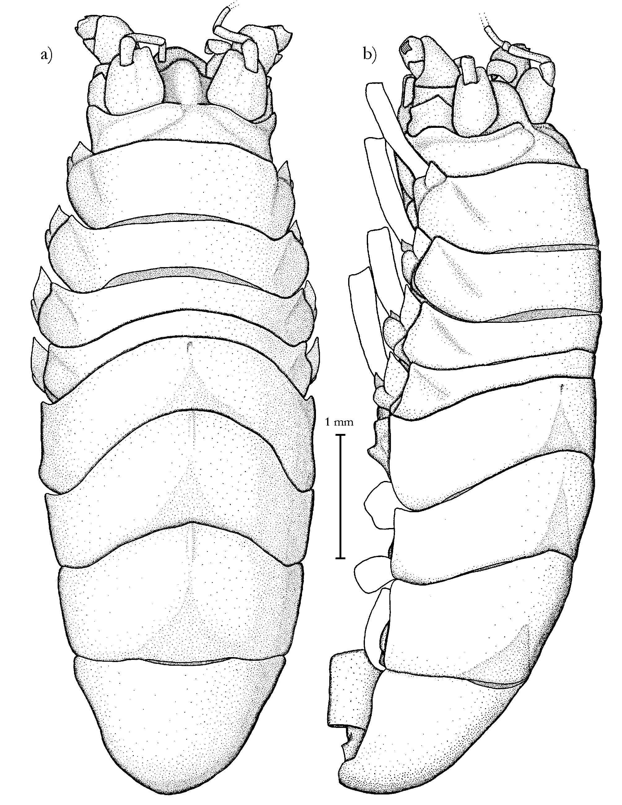
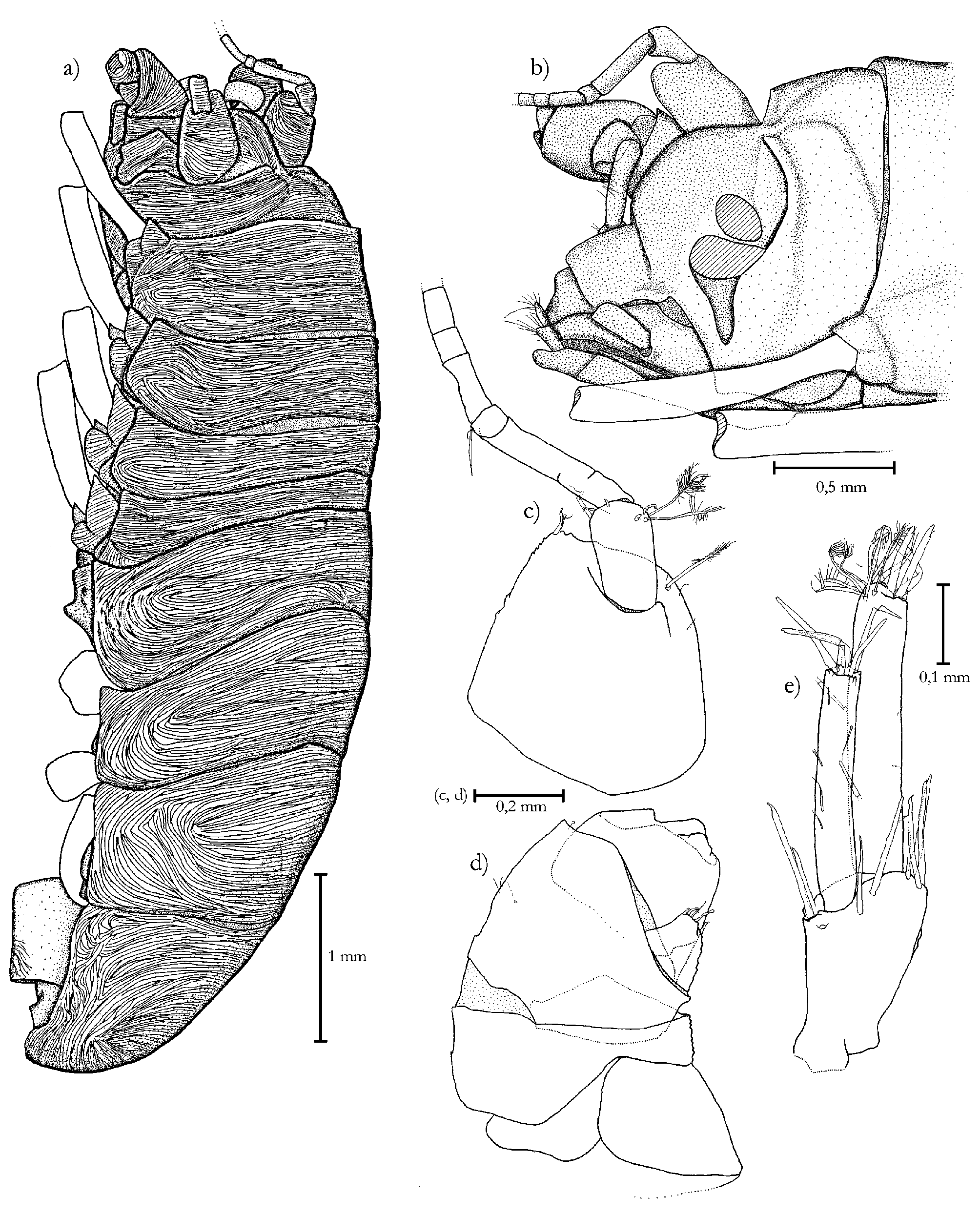
Body ( Fig. 21 View FIGURE 21. M ; Fig. 22 View FIGURE 22. M a): Length 5.83 mm, length: width ratio = 2.66. With weakly sclerotized cuticle, entire surface sculptured by pattern of cuticular grooves ( Fig. 22 View FIGURE 22. M a). Body nearly circular in crosssection, only ventral side of posterior part (pereonite 6 to pleotelson) flattened, in dorsal view evenly broadening from head to pereonite 3, the latter as wide as pereonites 4 and 5, caudally narrowing to tip of pleotelson. Pereonites 3 and 4 seemingly wider than 5 due to extension of coxal lobes. Natasome slightly less than two thirds of total length. Pleotelson 1.08 times wider than long, less than a fifth of total length, caudal end rounded, bent ventrally. Body height evenly increasing from pereonite 1 to 4 (0.26 to 0.30 times body length), maximum height at pereonite 5 (0.31 times body length), caudally evenly flattening.
Head ( Fig. 21 View FIGURE 21. M ; Fig. 22 View FIGURE 22. M b): 0.11 times body length, length: width = 0.43, widest at level of insertion of antenna 1. Frons vaulted, strongly arched to a quarter circle in lateral view, projecting dorsally beyond basal articles of antenna 1, width of this elevation 0.5 times width of antenna 1 article 1, almost entirely covering area between bases of antenna 1. Clypeus 1.31 times as long as labrum, separated from frons, less distinct on lateral side on a level with fossa, distal margin convex in dorsal view ( Fig. 22 View FIGURE 22. M b). Labrum separated from clypeus, frontal margin with medial incision (not figured).
Ambulosome: All pereonites with convex anterior and concave posterior margins in dorsal view. Pereonite 1 0.1 times body length, 0.32 times as long as wide, tergite laterally rounded, without anterolateral angles, fused with coxa, trace of suture remaining. Pereonite 2 0.18 times, pereonite 3 0.12 times, pereonite 4 0.1 times as long as wide. Length ratio of prm 1 to 4 = 2.8: 2.2: 1.3: 1. Anterolateral angles of tergites 2 to 4 pointed anteriorly. Coxal lobes prominent, directed anteriorly, separated from rest of coxa by incision also visible in dorsal view.
Natasome: All tergites with convex anterior margin. Posterior margin of prm 5 concave, on prm 6 less distinctly concave, on prm 7 nearly straight. Pereonites with anterolateral projecting angles, most distinct on prm 5. Medial length evenly increasing from prm 5 to prm 7, width evenly decreasing: length: width prm 5 = 0.27, prm 6 = 0.42, prm 7 = 0.571. Length ratio of pereonites 5 to 7 = 1: 1.5: 1.9.
Pleotelson ( Fig. 21 View FIGURE 21. M ; Fig. 26 View FIGURE 26. M a): Length: width = 0.93, medially 1.44 times as long as pereonite 7, lateral margin 1.35 times as long as lateral margin of pereonite 7, ventrally angled in posterior quarter, dorsal view seemingly triangular with rounded caudal end. Insertion of uropods caudomedial to sharpedged ventral elevation and laterally of anal valves. Left uropod of paratype preserved ( Fig. 22 View FIGURE 22. M e), exposed and visible in dorsal view, caudally projecting beyond tip of pleotelson.
Antenna 1 ( Fig. 22 View FIGURE 22. M c): On both sides incomplete. Peduncular article 1 0.37 times as wide as head, length: width = 0.95. Medial lobe inconspicuous, its place indicated by two spinelike setae, lateral lobe absent, a long lateral broom seta and 3 short, simple setae. Article 2 inserting dorsally in distal third of first, 0.45 times as long as article 1, 4 distodorsal spinelike setae, one distomedial and two distolateral long broom setae. Article 3 elongated, 0.63 times as long as article 1, length: width = 5. 4 flagellar articles preserved, nearly as wide as third basal article. Article 1 stout, with one short and one longer seta, length: width = 0.6, second 2.25, third 1.17 and fourth 1.67 times as long as flagellar article 1, all without setae.
Antenna 2 ( Fig. 22 View FIGURE 22. M d): Only 4 peduncular articles preserved, distal articles lost. Greatest width: head width = 0.33. Article 1 trapezoidal, laterally shifted sclerite, length: width = 0.74. Article 2 widest, length: width = 0.48. Article 3 nearly as wide as 2, medial side 5 times longer than lateral side. Lateral scale on article 3 broad, roundly triangular, anteriorly projecting to middle of fourth article, one distal seta, 4 distal spinelike setae, two medial spinelike setae. Article 4 length: width = 0.95, distal margin with 3 flattened lobes.
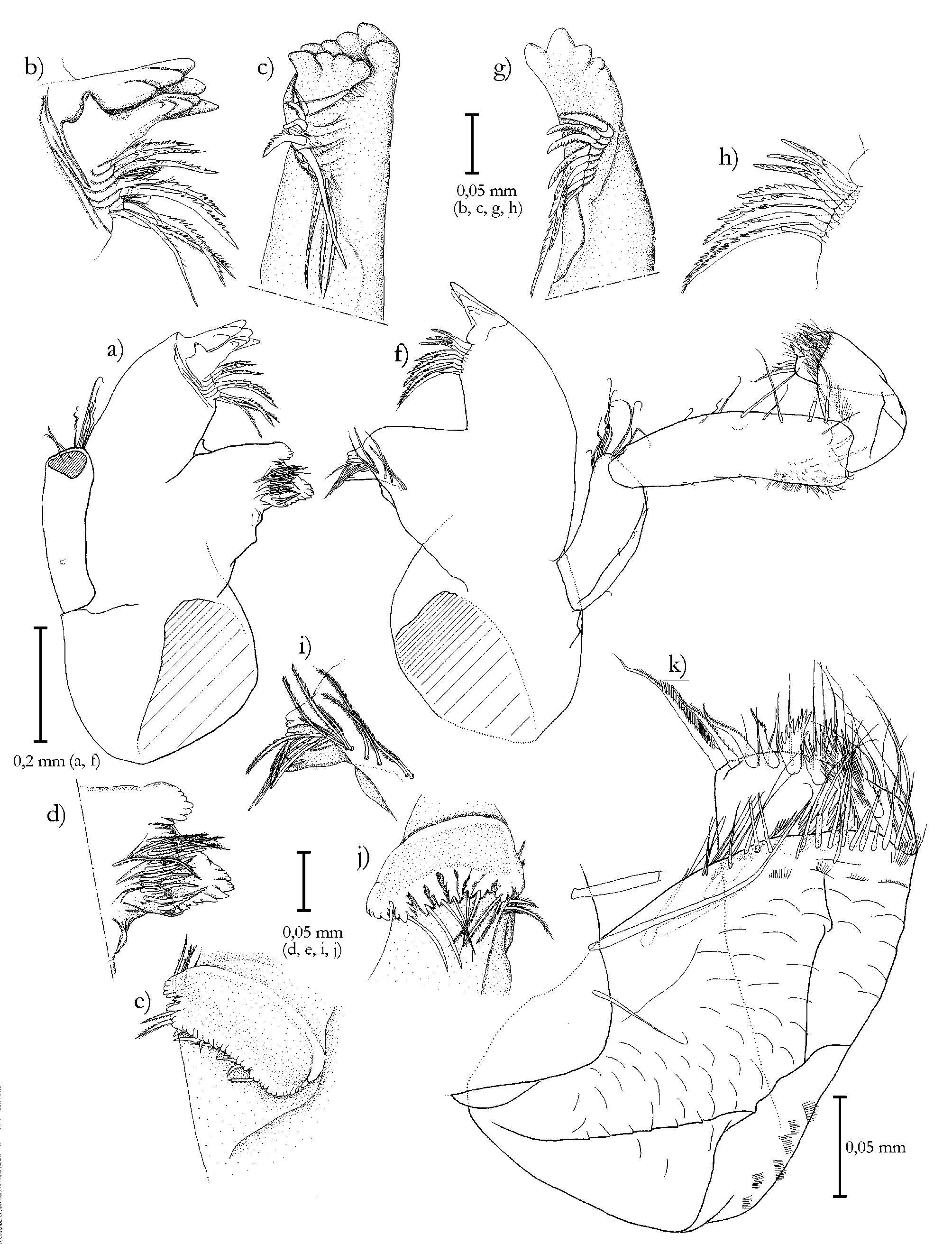
Mandibles ( Fig. 23 View FIGURE 23. M a–k): Dorsoventrally flattened between incisor and molar process. Entire cuticle heavily sclerotized. Palp only on left mandible preserved entirely, 1.04 times as long as body of mandible. First article cylindrical, slightly curved medially, left palp with 9 long distoventral setae, 11 dorsal and 3 lateral short setae. Right palp 8 long distoventral setae, 5 dorsal, one proximolateral and one ventral short setae. Second article about 1.5 times as long as first, anteromedial side with 9 setae, distal ones increasingly longer and stronger. Two strong distoventral setae of similar size, further proximally numerous scale setae. Posterolateral side with 3 shorter setae. Third article as long as and slightly wider than first, laterally flattened with torsion around longitudinal axis; left mandible anticlockwise (supposedly clockwise on right mandible). Dorsomedial surface with numerous scales, distally and laterally gradually replaced by scale setae. Ventromedial margin with 12 cleaning setae in a row. 4 strong subapical, onesidedly finely serrated stout setae, outermost 3 times as long as innermost. Entire ventromedial margin additionally with numerous long setae. Incisor process of both sides with 5 sclerotized cusps, the second (on ventral side) largest, lacinia mobilis of left mandible as long as sclerotized cusps of incisor process. 4 distal cusps, gradually increasing in size from dorsal to ventral side, more proximally an additional cusp pointing laterally. Spine row slightly shifted ventrally and curved caudally, on left mandible consisting of 8 serrated stout setae, further ventrally numerous fine setae, on right mandible 9 serrated stout setae. Molar process oval chewing area 0.45 times as long as wide, posterior margin concave. Chewing area dorsal, posterior and ventral margin of left mandible and posterior margin of right mandible strongly elevated, with numerous cusps and a row of 16 setae: left mandible with 3 simple, 6 serrated and 7 setulate setae, right mandible with 3 simple and 13 setulate setae. Condylus indefinable due to brittle, cracked cuticle.
Maxilla 1 ( Fig. 24 View FIGURE 24. M a–c): Both endites slightly curved medially. Outer endite (=exopod) 1.25 times as long and twice as wide as inner endite with 13 strong apical stout setae curved medially, 7 of which dentate, one very small. Ventral side of apex with 5 slender setae. Dorsal surface with 23 shorter setae, further proximally 3 scale setae. Proximal half of lateral margin with 37 setae. Medial margin with 20 subdistal setae. Inner endite with one short and two long strong stout setae, the latter ones curved medially, finely serrated in distal half. Distal half of lateral margin with 13 long setae, dorsal and ventral side with numerous further setae subapically and along medial margin, here the longest.
Maxilla 2 ( Fig. 24 View FIGURE 24. M d, e): Inner endite dorsoventrally flattened and apically rounded, slightly shorter and about 1.5 times as wide as both rami of exopod. Apex with 12 simple and 5 onesidedly pectinate, strong stout setae, from lateral to medial side gradually decreasing in length. Further proximally medial margin with conspicuously long plumose stout seta. Dorsal and ventral side subapically and in medial half with numerous long stout setae. Medial and lateral margin with numerous long setae. Middle endite weakly curved medially, distally evenly narrowing. Apex truncate with 4 pectinate stout setae. Most dorsomedial stout seta shifted slightly proximally, with longer setulae than on the others. Distal third with numerous ventrolateral simple setae. Dorsomedially in distal half 14 stronger, flattened setae in two irregular longitudinal rows, the 4 largest ones pectinate. Further proximally 17 simple setae. Outer endite slightly curved medially, distally evenly narrowing. Apex truncate with 4 pectinate stout setae. Most dorsomedial stout seta from a subapical lobe, longer setulae than on others. Dorsolaterally and along lateral margin numerous simple setae. Dorsomedially in distal half 23 stronger, flattened setae.
Paragnath ( Fig. 24 View FIGURE 24. M f): With unsclerotized cuticle, subdivided into narrower pair of inner lobes and wider, dorsoventrally flattened pair of outer lobes with rounded lateral margins. Inner lobes half as long as outer lobes, with rounded apex, dorsally densely setose except for bald proximal third. Outer lobes ventrally overlapping inner lobes, with slightly acute apices curved medially. Dorsal surface in distal third with an inconspicuous row of 4 setae on left and 6 stronger setae on right lobe. Distolateral margin with numerous long setae. Medial margin in distal half with dense row of longer setae.
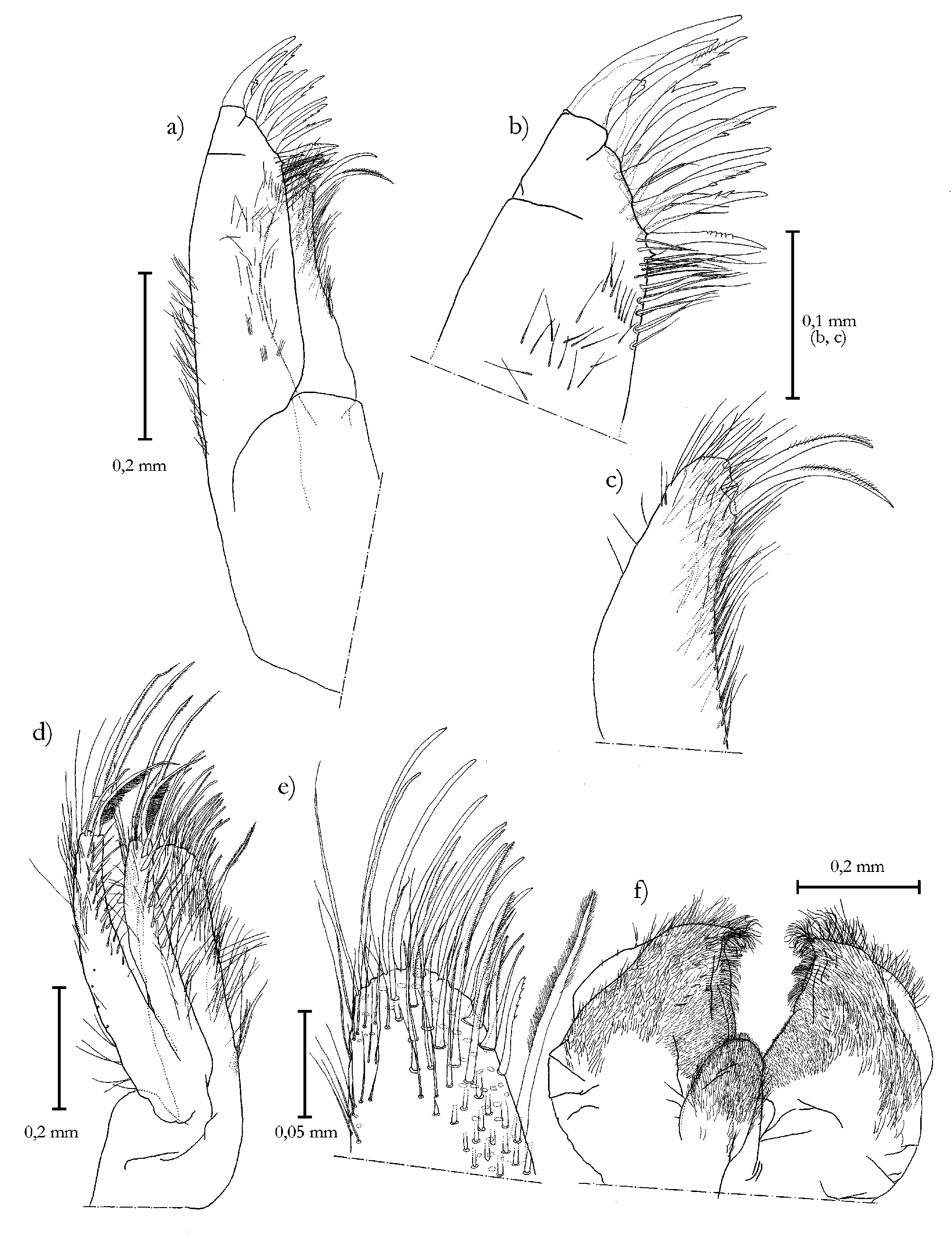
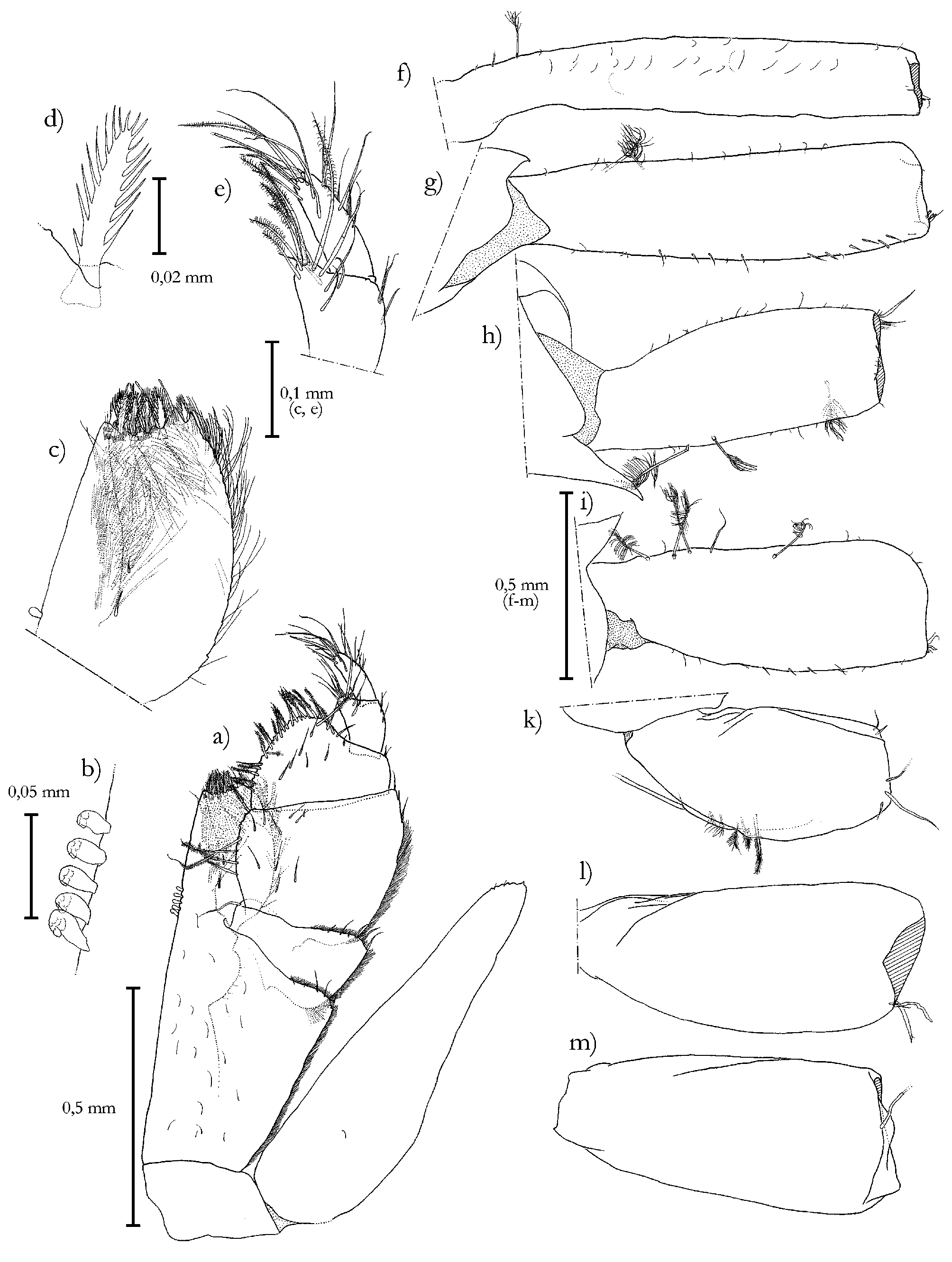
Maxillipeds ( Fig. 25 View FIGURE 25. M a–e): Coxa more than 1.5 times wider than long, 0.22 times as long as basis. Basis 2.33 times as long as wide, widest at insertion of palp, ventral surface with 20 setae, lateral row of fine setae, continued on lateral third of distal margin, here 3 long and further medially two short setae. Medial margin bent dorsally, distal third with laterally pointing lobe bearing 5 short setae. Row of 5 retinacula on a level with first article of palp. Retinacula clubshaped, each with two distodorsal pairs of elongated tubercles, proximalmost retinaculum with 3 pairs. Endite spatulate, about half as long as rest part of basis, medial margin bent dorsally. Two short proximoventral setae. Truncate apical margin at an angle of about 120 to medial margin, with a row of 6 fan setae and dorsal row of 5 stout setae. Ventral surface with numerous scale setae. Lateral margin convexly arched, with numerous long setae. Dorsal surface with triangular area of several hundred setae and 4 long stout setae. Palp 0.89 times as long as maxilliped basis. Article 1 shortest, 0.81 times as wide as basis, lateral margin with row of setae as on basis, continued on lateral third of distal margin, here 5 longer and two short setae. Article 2 largest, 3 times as long as article 1, distally increasing in width, distal margin diagonally truncate, lateral margin 1.54 times longer than medial margin, lateral setose row continued in first two thirds of its length, distally replaced by 3 larger setae. Medial margin with 5 distomedially pointing pappose setae with bald proximal third, demarcated by an annulus. 10 short ventral setae. Article 3 slightly shorter than article 2, lateral margin 0.36 times as long as medial margin, two distolateral seta. Medial margin up to distally rounded tip with 16 shorter pappose setae. Ventral surface with 6 short setae. Article 4 proximally tapering, one third as wide as article 3 and inserting distolaterally, 3 lateral, at distomedial lobe 7 long setae and 7 pappose setae. Article 5 cylindrical, twice as long as wide, seemingly shifted laterally due to distolateral lobe of article 4, setose apices of both lying parallel to each other. Distally and subdistally 10 setae, the 4 distalmost longest. Epipodite 1.04 times as long as basis, nearly 3.5 times as long as wide, without conspicuous proximolateral projection. Distomedial margin with 6 cusps, the distalmost acute, 2 short setae, single seta ventrally in proximal third.
Pereopods 1–4 ( Fig. 25 View FIGURE 25. M ): All broken off distally of basis, basis described here due to its diagnostic importance. Length ratio of bases: pereopod 1> 2> 3 <4. Basis of pereopod 1 6 times longer than wide, narrowest, one basal broom seta, further proximal insertion of a second one. Basis 2 4 times longer than wide, two dorsal broom setae. Basis 3 2.7 times longer than wide, 3 dorsal broom setae. Basis 4 2.7 times as long as wide, 5 dorsal broom setae.
Pereopods 5–7 ( Fig. 25 View FIGURE 25. M k–m): Length ratio of bases: pereopod 5 <6 <7. Pereopod 5 2.1 times longer than wide, with 5 swimming setae, further proximally 1 long seta. Pereopod 6 2.3 times longer than wide, with short distal seta and two spinelike setae. Pereopod 7 2.4 times longer than wide, one long distal seta.
Pleopods ( Fig. 26 View FIGURE 26. M a–e): Pleopod 2 (=operculum) inserting on a level with coxae of pereopods 7, slightly wider than long when spread out, in situ bent by ventral medial keel and seemingly longer than wide. Cuticular grooves of ventral surface only in distal third. Medial keel distinct, not sharp. Its distal end with a singular insertion of a (lost) seta of 5 µm diameter. Lateral margins with short setae, in distal half 21 hemiplumose setae on each side. Pleopod 3 endopod length: width = 2.03, with 3 plumose setae. Exopod curved medially, width: endopod width = 0.25, slightly shorter than endopod, tip lancetshaped. Margins with rows of fine setae, apex with stronger simple seta. Pleopod 4 bald, endopod length: width = 1.51, medial margin nearly straight. Lateral margin convexly arched, dorsally partially overlapping exopod. Exopod twojointed, basal article a third as wide as endopod, lateral margin with seam of fine, short setae. Article 1 3 times as long as wide, proximolateral and distomedial margins slightly rounded. Article 2 half as long and a third as wide as article 1, evenly narrowing, with apical plumose seta. Pleopod 5 uniramous, delicate and lobiform, nearly oval, length: width = 1.17, 0.95 times as long and 1.23 times wider than endopod of pleopod 4. Lateral margin slightly more rounded than medial margin.
Uropods ( Fig. 22 View FIGURE 22. M e): Lost on holotype, left uropod of paratype ventrolaterally inserting in shallow cavity in posterior third of pleotelson. Protopodite trapezoidal, broadened distally, 1.5 times as long as wide. 9 strong setae in transversal distal row, 5 around exopod, the others around endopod. Both rami slender and cylindrical, with truncate apex. Exopod 1.24 times as long as protopodite, 6 times as long as wide. Ventrolaterally and ventrally 6 setae. Apex with 5 strong setae. Endopod length: width = 5.38, about 1.2 times longer than exopod, 3 very short dorsomedial setae, two lateral broom setae. Apex with one simple seta, 9 broom setae and 3 spinelike setae.
Remarks
The paratype’s head seems to be shorter than that of the holotype as it is further retracted under pereonite 1. Whether the head is actively retractile and if there is a relation to the pereonite 1 being elongated cannot be answered. It is also possible that the retraction is an artifact due to dehydration of specimens stored in ethanol.
There is an articulation of a lost and probably long seta at the apex of the operculum ( Fig. 26 View FIGURE 26. M a, b) which has to be considered when determining the species.
A taxonomic feature most previously described species of Munneurycope have in common is the absence of the lateral and medial lobes on the first peduncular article of antenna 1. The new species still has a remaining – yet small – medial lobe although a distinct reduction compared to the lobes within the genus Eurycope is obvious. Therefore, the possible presence of a medial lobe was included in the generic diagnosis of the genus Munneurycope .
Comparing the length ratios of the ambulatory pereonites, Aydogan et al. (2000) postulate the elongation of pereonite 1 to be a possible autapomorphy of the genus. This is the case for Munneurycope persephone as well as for M. hadalis Aydogan et al., 2000 , M. nodifrons ( Hansen, 1916) , M. menziesi Wolff, 1962 and less significantly for M. curticephala Birstein, 1963 , but not for the other species of the genus. As this feature is not applicable to all species currently included in this genus this may lead to the supposition of Munneurycope being a paraphylum until some species are transferred into separate genera ( Aydogan et al. 2000).
Several species differ highly from the new species in their general appearance: M. murrayi (Walker, 1903) , M. harrietae Wolff, 1962 , M. incisa (Gurjanova, 1946) and M. glacialis Malyutina and Kussakin, 1996 are obviously not conspecific with M. persephone ; none of them has the tapering pleotelson seen in the latter. The habitus of the present species is similar to that of M. hadalis , M. nodifrons , M. menziesi and M. curticephala . A detailed comparison is summarized in table 2.
The new species lacks an anteromedian concavity of the frons typical for M. hadalis , described as “prominent rostrum, distally with deep concavity”, the “rostrum” being the medially swollen frons ( Aydogan et al. 2000). Besides, M. hadalis has a relatively longer first pereonite and a pereopod 1 with a wider basis. M. curticephala has equally long pereonites 1 and 2 and a different insertion of uropods which are therefore invisible in dorsal view. Both species lack – as M. menziesi – the medial lobe of antenna 1. It is described for M. nodifrons ( Wolff 1962) which has a very similar frons and general appearance compared to the new species. Whereas the new species has an elongated article 3 of antenna 1 which is longer than article 2, in M. nodifrons it is shorter than article 2. The truncate tip of the maxilliped endite of M. nodifrons forms an angle of ca. 90° with the medial margin and is narrower ( Wolff 1962: Fig. 104 a) than in M. persephone and M. menziesi where the tip forms an angle of ca. 120° with the medial margin. The endite's lateral margin is much more convexly rounded than in the latter two species.
The presence of a plumose seta on the exopod of pleopod 3 has been considered a character that separates Munneurycope and other genera from the Eurycopinae that lack this seta ( Wägele 1989), however the seta is also reduced in some Munneurycope ( M. murrayi , M. nodifrons , M. hadalis and M. persephone ), while it is present in some Eurycopinae : Eurycope septentrionalis Malyutina and Kussakin, 1996 , E. vasinae Malyutina and Kussakin, 1996 , E. baea Wilson, 1983 , E. cryoabyssalis Just, 1980 , E. dahli Svavarsson, 1987 , E. producta Sars, 1868 and Tytthocope megalura (Sars, 1872) (the latter with two plumose setae, in Wilson and Hessler 1981). Presence or absence of plumose setae on the exopod of pleopod 3 seems to be a useless character for diagnoses of subfamilies within the Munnopsididae . The remaining reason for exclusion of Munneurycope from Eurycopinae – the reduction of the rostrum as synapomorphy of the genus – is uncertain, as it is a weak homology. Furthermore, not all species without rostrum were consequently excluded from the genus Eurycope (compare Munneurycope antarctica Schultz, 1977 and Eurycope acutiperaeons Schultz, 1978 which could be a Munneurycope due to the absence of a rostrum and the elongation of pereonite 1). So the actual systematic position of Munneurycope remains uncertain until revision.

| ZMH |
Zoologisches Museum Hamburg |
No known copyright restrictions apply. See Agosti, D., Egloff, W., 2009. Taxonomic information exchange and copyright: the Plazi approach. BMC Research Notes 2009, 2:53 for further explanation.