
Anoplodelphys laubieri, Boxshall, Geoff A. & Marchenkov, Andrey, 2007
|
publication ID |
https://doi.org/ 10.5281/zenodo.176361 |
|
DOI |
https://doi.org/10.5281/zenodo.5661719 |
|
persistent identifier |
https://treatment.plazi.org/id/C03D8785-0450-FFB9-FF2D-FB29FC26FBEC |
|
treatment provided by |
Plazi |
|
scientific name |
Anoplodelphys laubieri |
| status |
sp. nov. |
Anoplodelphys laubieri n. sp.
Type material: Holotype female in alcohol, reg. no. MNHN-Cp2315 with antenna removed from one side and mounted on slide (MNHN-Cp2316).
Type locality: Sodwana Bay, Kwazulu-Natal, Republic of South Africa. Depth 0– 22 m.
Host: Didemnum rodriguesi Rocha and Monniot, 1993 [ MNHN reg. no. A2 DID C 361]
Locality in host: within the common tunic between the zooids (F. Monniot, pers. comm.)
Etymology: this species is named for Dr Lucien Laubier in honour of his contributions to knowledge of parasitic copepods.
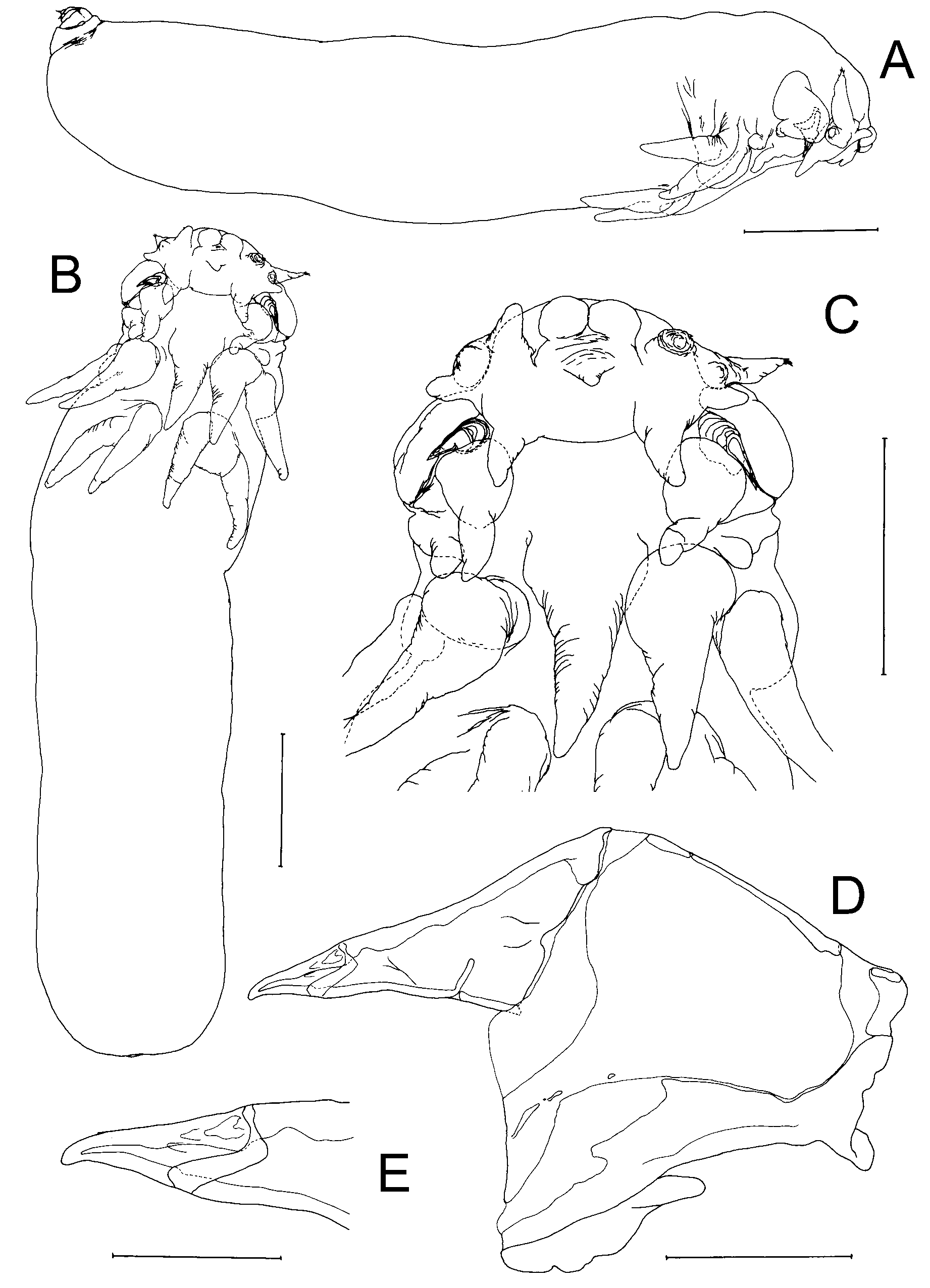
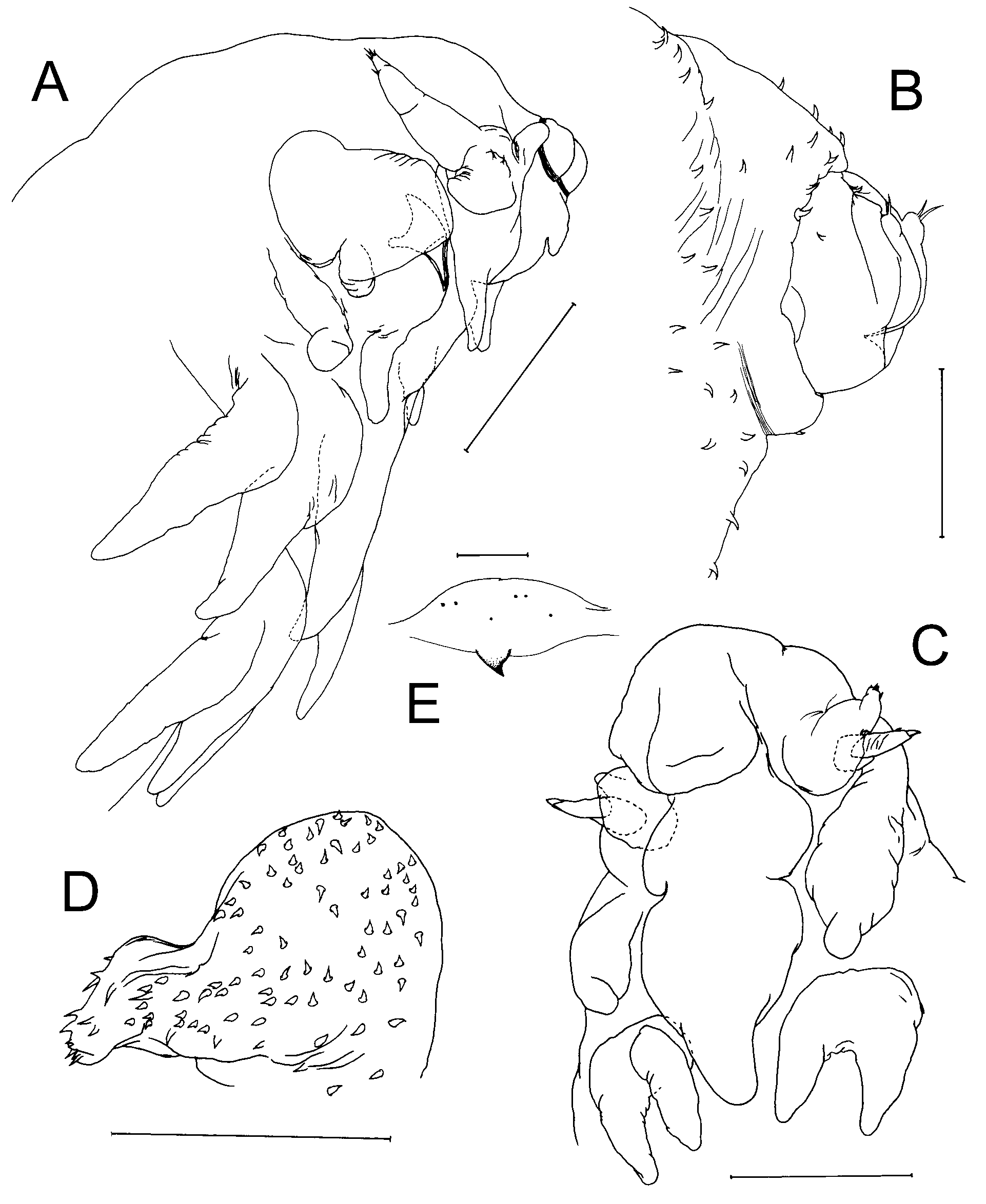
Description: Adult female body ( Fig. 3 View FIGURE 3 A–B) highly transformed, vermiform and lacking external segmentation: indistinctly divided into 3 regions, cephalosome, metasome and urosome, by shallow superficial furrows. Entire body surface densely ornamented with relatively short setules. Body length of holotype female 3.10 mm. Cephalosome slightly dorsoventrally flattened, tapering anteriorly; bearing paired antennules, antennae, and 1 pair of lobes which probably represent reduced and modified oral appendages. Lateral margins of head expanded ventrally to form ridge-like swelling produced into thumb-like process at posterior extremity ( Figs 3 View FIGURE 3 C, 4A). Rostrum well developed on frontal margin of cephalosome ( Fig. 3 View FIGURE 3 C), forming 2 broad, hemispherical lobes and with small, median ventral lobe. Metasome more or less cylindrical, comprising 4 somites but lacking surface expression; first 2 somites only pedigerous, each bearing pair of modified, biramous thoracic legs. Urosome ( Figs 3 View FIGURE 3 A, 4B) indistinctly 2-segmented, inserted on posterior margin of metasome and directed somewhat dorsally. Caudal rami ( Fig. 4 View FIGURE 4 B) largely incorporated into urosome but each armed with 2 caudal setae terminally. Surface of urosome sparsely ornamented with setules.
Antennules ( Figs 2 View FIGURE 2 F, 4A) inserted just lateral to rostrum; divided into swollen basal part bearing 4 rounded processes, orientated anteriorly, anterolaterally, laterally and posteroventrally, and conical, tapering distal part armed with 5 setal elements, 4 apical and 1 subapical ( Fig. 4 View FIGURE 4 A). Surface of basal part of antennule ornamented with isolated spinules ( Fig. 2 View FIGURE 2 F) on anterior and anterolateral processes.
Antenna ( Fig. 3 View FIGURE 3 D–E) 2-segmented: proximal segment, robust, triangular in shape and largely concealed between lateral cephalosomic process and antennomedial lobe located just posterior to base of antenna on ventral surface of cephalosome ( Figs 3 View FIGURE 3 C, 4A): distal segment forming strongly sclerotised subchela. Subchela with defined apical section (terminal claw) retaining traces of 3 weakly sclerotised spines ( Fig. 3 View FIGURE 3 E). Adjacent antennomedial lobe comprising inflated basal part and long, posteromedial process extending posteriorly as far as anterior border of leg 1.
Labrum ( Fig. 3 View FIGURE 3 C) elongate, conical lobe, reaching to mid-level of leg 2; surface ornamented with setules.
One pair of oral appendages (?mandibles) present, reduced to unarmed lobes located about at mid level of labrum ( Fig. 3 View FIGURE 3 C).
Legs 1 and 2 only present ( Figs 3 View FIGURE 3 B, 4A); legs 3 and 4 absent. Leg 1 originating posterior and lateral to base of labrum; leg 2 originating close to leg 1, but slightly nearer midline. Each leg biramous, with unsegmented, lobate rami forming elongate conical processes, lacking setal armature but densely ornamented with surface setules. Exopodal lobe slightly longer than endopodal lobe, arising from common protopodal part, largely incorporated into somite.
Leg 5 absent.
Male unknown.
Remarks: As for the preceding species, Anoplodelphys africana n. sp., this new species differs from A. corneci , A. galli and A. incerta by the presence of only two leg pairs rather than four, and in details of shape of the rostrum and structure of the antennule. This species is closely related to A. africana n. sp., sharing the bipartite antennule and the bilobed rostrum. It can be distinguished from A. africana by the presence of two caudal setae, a small median post-rostral process, a fourth process (located anterolaterally) on the inflated basal part of the antennule, and a well developed posterior process on the large ventral lobe located immediately medial to the antenna.
The single pair of lobes in the oral region is here interpreted as probably representing vestigial mandibular palps, from their position just anterolateral to the base of the labrum.

| MNHN |
Museum National d'Histoire Naturelle |
No known copyright restrictions apply. See Agosti, D., Egloff, W., 2009. Taxonomic information exchange and copyright: the Plazi approach. BMC Research Notes 2009, 2:53 for further explanation.