
Ephesiella australiensis Hartmann-Schröder, 1982
|
publication ID |
https://doi.org/ 10.11646/zootaxa.4019.1.9 |
|
publication LSID |
lsid:zoobank.org:pub:D4ECFEE2-BF9D-4B5F-8128-EFAAC2B728C4 |
|
DOI |
https://doi.org/10.5281/zenodo.6095999 |
|
persistent identifier |
https://treatment.plazi.org/id/C22DFF62-FFF3-FF8E-FF21-FA9EFE8C9BE9 |
|
treatment provided by |
Plazi |
|
scientific name |
Ephesiella australiensis Hartmann-Schröder, 1982 |
| status |
|
Ephesiella australiensis Hartmann-Schröder, 1982 View in CoL
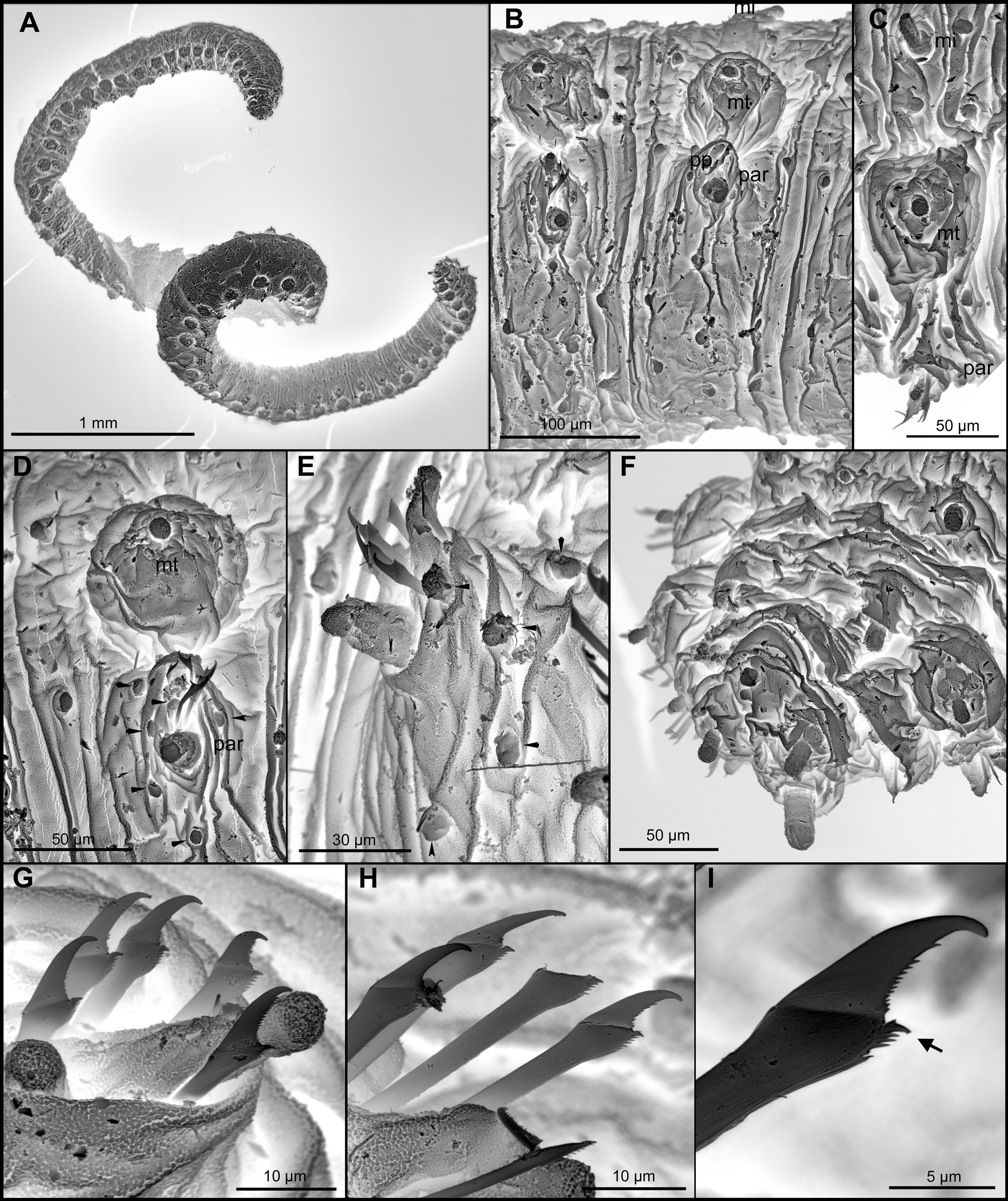
( Figs 1 View FIGURE 1 , 2 View FIGURE 2 A, B)
Ephesiella australiensis Hartmann-Schröder, 1982: 80 View in CoL –81, figs 84–87.
Type material. Holotype: ZMH P.16773, Cervantes, Western Australia, Australia, sandy beach among Posidonia , 24 Oct 1975. Paratype: AM W.42693 (on SEM stub), Lizard Island, between South Island and Palfrey Island, 14°41'54"S, 145°26'45"E, dead coral, 17 m, 31 Mar 1995.
Other material examined. AM W.42702 (2, 1 on SEM stub), Western Australia, south west Enderby Island, 20°37'18"S, 116°27'23"E, dead coral encrusted with bryozoans, ascidians and bivalves, 14 m, 8 Aug 2000; AM W.42688 (on SEM stub), Australia, New South Wales, Cape Three Points, south-east of Third Point, 33°31'55"S, 151°24'58"E, sand from around large boulders at edge of rocky reef, 30 m, 6 May 2007.
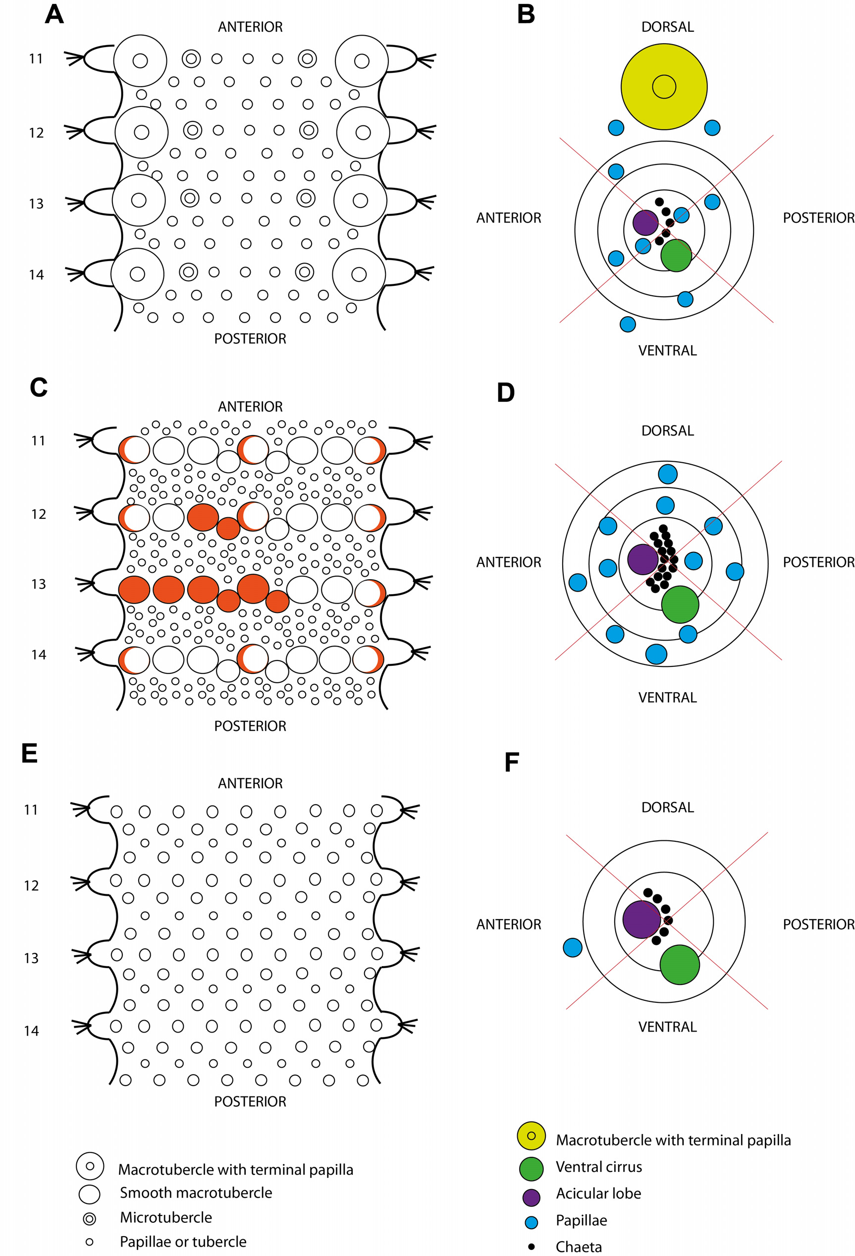
Diagnosis. Body long and slender. Two longitudinal rows of macrotubercles, one pair per segment, may be absent in first chaetiger. Macrotubercles spherical, sessile, with terminal papillae. Two longitudinal rows of microtubercles, with a collar and a terminal papillae, one pair per segment, running parallel between macrotubercles. Additionally, papillae arranged in three transverse rows on dorsum and in 4–5 transverse rows on ventrum. Head appendages digitiform. Mid-body parapodia with six semi-spherical papillae, all similar in size: one anterior dorsal, one anterior, near the acicular lobe, one anterior ventral two posterior-dorsal, one of the latter distal. Parapodia from chaetiger 2 with compound or semi-compound, shaft provided with a distal spine; hooks not observed.
Description. (of Lizard Island specimen) Body elongate, measuring 4.2 mm long, 0.4 mm wide, with 63 segments, slightly narrowing along posterior segments ( Fig. 1 View FIGURE 1 A). Sub-quadrangular in section, with convex dorsum ( Fig. 1 View FIGURE 1 A). Tegument with transverse wrinkles and segmentation inconspicuous ( Fig. 1 View FIGURE 1 A). Preserved material lacking pigmentation. Head externally indistinct. Anterior end bluntly rounded and damaged. Prostomium with five appendages; paired palps and lateral antennae digitiform and three times longer than wide; median antenna, shorter, hemispherical. Prostomial papillae, present but numbers uncertain. Tentacular cirri slightly shorter than palps and lateral antennae. First chaetiger with two sessile and spherical macrotubercles, each provided with a digitiform terminal papillae; microtubercles absent. Rest of chaetigers with two macrotubercles each, arranged in two longitudinal dorso-lateral rows, shape and size of all macrotubercles similar, slightly increasing in size in first segments, and decreasing posteriorly ( Fig. 1 View FIGURE 1 A–D). A pair of microtubercles on each segment from chaetiger to until last segment, running in two longitudinal rows inside macrotubercles; with terminal papillae longer than collar ( Fig. 1 View FIGURE 1 C). Spherical and elliptical papillae present over dorsum, arranged in three transverse rows per segment, with around 15–20 papillae per segment on mid body region, including 2–3 papillae between macrotubercles and parapodia ( Figs 1 View FIGURE 1 A, 2B). Ventral surface with spherical papillae, arranged in 4–5 transverse rows (seemingly a zig-zag in anterio-posterior view), with a total of 20–25 per segment, in mid-body; numbers decreasing towards posterior end. Parapodia sub-conical, increasing in size towards chaetiger 3 and as longer as wide ( Fig. 1 View FIGURE 1 C–E). Acicular lobe projecting distally anterior to chaetae, resembling other parapodial papillae or slightly longer ( Fig. 1 View FIGURE 1 E). Ventral cirri bottle-shaped similar in length to acicular lobe ( Fig. 1 View FIGURE 1 E). Anterior parapodia with four hemispherical papillae: one anterior, one anterio-ventral, one anterior-dorsal and one posterior, in addition to the acicular lobe. Mid-body parapodia with six semi-spherical papillae, all similar in size: one anterior dorsal, one anterior, near the acicular lobe, one anterior ventral, two posterior-dorsal, one of the latter distal ( Figs 1 View FIGURE 1 D–E, 2B). Compound chaetae, appearing as semi-compound in some chaetigers, arranged in a curved transverse rows around acicular lobe and numbering 4–6 per fascicle ( Fig. 1 View FIGURE 1 G–I). First chaetiger with blades 5–6 times longer than wide and conspicuous serration along cutting edge. Hooks not seen. Chaetae from chaetiger 3 with shaft distally enlarged and serrated with a main distal tooth; blades showing a slight dorso-ventral gradation (1–2 times longer than wide; except for anterior chaetiger with longer blades) recurved and with smooth or few serration of the cutting edge ( Fig. 1 View FIGURE 1 G–I). Pygidium terminal, with mid-ventral digitiform anal cirrus and a pair of dorsal anal cirri, similar in shape but slightly smaller than macrotubercles ( Fig. 1 View FIGURE 1 F). Eyes, muscular pharynx, gonads, gametes or copulatory organs not observed.
Remarks. Ephesiella is a cosmopolitan genus that requires revision. It currently groups 15 species supposedly sharing the presence of only compound chaetae from chaetiger 2. Nevertheless, some species have been described as bearing semi-compound chaetae (e.g., Moore 1909, and description above), a feature half way to members of Sphaerodorum (with only simple chaetae) and Ephesiopsis (with both simple and compound chaetae in all parapodia). Moreover, several species have been described from a single or few specimens (e.g., Desbruyères 1980; Hartmann-Schröder 1982) and consequently, the intraspecific variation has not been addressed. Most specific diagnostic characters rely on the absence or presence of hooks, the number of prostomial appendages, the number and arrangement of parapodial papillae. The types and specimens examined for this study (cited above) all appear to: i) lack hooks and antenniform papillae; ii) have the median antenna distinctly shorter than other prostomial appendages; iii) have three transverse rows of dorsal papillae per chaetiger; iv) number of parapodial papillae range between 4–7; v) bear compound, or semi-compound, chaetae with short blades (2–3 times longer than wide), most of them provided with a distinct spine on the tip of the shaft (not observed in the types, due to their fragile condition for manipulation). Most specimens examined from other Australian localities do share these morphological traits. However, some specimens showed different chaetal morphology (longer blades and/or distal tooth in shaft absent), or different relative position of the macro and microtubercles (Capa & Bakken 2015). Ephesiella australiensis is represented elsewhere in Queensland, but only reported from Townsville (Capa & Bakken 2015).
| ZMH |
Zoologisches Museum Hamburg |
No known copyright restrictions apply. See Agosti, D., Egloff, W., 2009. Taxonomic information exchange and copyright: the Plazi approach. BMC Research Notes 2009, 2:53 for further explanation.
|
Kingdom |
|
|
Phylum |
|
|
Class |
|
|
Order |
|
|
Family |
|
|
Genus |
Ephesiella australiensis Hartmann-Schröder, 1982
| Capa, María & Rouse, Greg W. 2015 |
Ephesiella australiensis Hartmann-Schröder, 1982 : 80
| Hartmann-Schroder 1982: 80 |