
Gymnesigobius medits, Kovačić & Ordines & Ramirez-Amaro & Schliewen, 2019
|
publication ID |
https://doi.org/ 10.11646/zootaxa.4651.3.6 |
|
publication LSID |
lsid:zoobank.org:pub:4F58CBEE-A382-4E48-9363-569FC7437587 |
|
persistent identifier |
https://treatment.plazi.org/id/F36C87BE-FF9D-4457-FF6F-FB08DDFD0E46 |
|
treatment provided by |
Plazi |
|
scientific name |
Gymnesigobius medits |
| status |
sp. nov. |
Gymnesigobius medits sp. nov.
Holotype ( Fig. 1 View FIGURE 1 ). Male, 42.6 + 11.1 mm, PMR VP4649 View Materials , west of Mallorca , 11 June 2018, 39º17’59”N, 002º25’52”E, depth of the bottom trawl sampling ranging between 344 and 364 m (mean 354 m), collector F. Ordines. GoogleMaps
Diagnosis. As for genus.
Description. General morphology ( Fig. 1 View FIGURE 1 ): Body proportions are given in Table 3 View TABLE 3 . Body elongate, laterally more compressed towards caudal peduncle. Head of moderate size and depressed (head depth 70.5% of head width). Snout gently oblique, shorter than eye (snout length 61.9% of eye diameter). Anterior nostril long, tubular, without process on rim; posterior nostril short tube smaller of diameter than anterior nostril. Eyes large, eye diameter about 2/5 length between mouth tip and preopercular edge, dorsolateral, distinctly elevated above dorsal profile with narrow interorbital space. Mouth terminal, lips end frontally equal, mouth oblique, with anterior tip above horizontal level of lower eye edge, posterior angle of jaws below pupil. Upper lip width uniform. Chin without fold or barbels. Ventrolateral head ridge broad, extends in flap reaching lateral midline and nearly touching fellow at the midline ( Fig 2a View FIGURE 2 ). Branchiostegal membrane attached along entire lateral margin of isthmus from immediately anterior to pectoral margin. Cranial roof covered by dorsal axial musculature. No spines on preopercle. Pectoral girdle without dermal flaps on anterior edge. Tongue well developed, anteriorly truncated. Teeth in upper and lower jaws erect, caniniform, in several rows; teeth in the first row larger in both jaws.
Fins. D 1 VI; D 2 I/11; A I/10; C segmented rays 17, branched 14; P 17; V I/5 + 5/I. Fin morphometrics in proportion to standard body length given in Table 3. D View TABLE 3 1 View TABLE 1 spines not elongate or filamentous, II spine longest, from III to V spines slowly decreasing in length and all barely reaching D2, VI spine shorter. D2 originates above anus, the longest D2 rays not reaching base of uppermost caudal-fin rays, ending on caudal peduncle before caudal fin. A originates at vertical through D2 second soft ray. P posteriorly barely reaching vertical of D2 base anterior begin- ning. No free tips on upper P rays, P rays all branched except uppermost and lowermost rays unbranched. Pectoral girdle without flaps on anterior edge. V with fifth ray longest and the visible remaining of broken membrane con- necting fifth rays at least to the half of the pelvic fin i.e. pelvic fins not divided but the degree of fin emargination is unknown due to damaged membrane. V anterior pelvic membrane (frenum) well-developed, without lateral lobes (anterior membrane in midline depth about 1/3 of spinous ray) ( Fig. 2b View FIGURE 2 ). C rounded, shorter than head (caudal fin length 86% of head length).
Scales. Body scaled, scales lost on both specimens, counts made from stained scale pouches. Head with cheek and opercle naked. Predorsal area and D1 base naked, with upper edge of scaled area extending from behind upper part of pectoral axilla backwards and up to D 2 I. Prepectoral naked, breast scaled with two scale pouches visible, belly scaled. No trace of scales or scale pouches on caudal fin. LL 28, TR 6, CP 8.
Lateral line system ( Figs. 2a View FIGURE 2 and 3 View FIGURE 3 ). Head with anterior oculoscapular and preopercular canals, with pores σ, λ, κ, ω, α, β, ρ and γ, δ, ε respectively; posterior oculoscapular canal absent. Pores of head canals enlarged e.g. pores α and ρ larger or about same size as interspaces to pore β. Rows with range of number of sensory papillae in parentheses (counted on both sides of the holotype) as follows: (1) preorbital: snout with three median preorbital series, row r (2 and 3), horizontal row s 1 (1) below pore σ, a single papilla visible inside pore σ, horizontal row s 2 not visible and vertical s 3 (1) more medially above upper lip. Lateral series c in four parts: superior (c 2) as two papillae above posterior nostril and middle (c 1) (3) below anterior nostril; inferior rows: upper horizontal c 2 (3 and 4) above upper lip and lower horizontal c 1 (2) between upper lip and row 1. (2) suborbital: six transverse suborbital rows (1-6) of sensory papillae, four continuous suborbital rows in front of row b, row 5 divided in three parts but in front of row b, lowermost segment of row 5 at the level of row d, row 6 just as superior part above row b and below pore α, rows 2 to 4 short, length less than half distance from eye orbit to row d, and distant from orbit and row d, (1: 5 and 6, 2: 2 and 3, 3: 3 and 4, 4: 4 and 5, 5: 2 and 3 + 2 and 3 + 4, 6: 2). A single papilla visible inside pore α. Longitudinal row b (5 and 6) barely reaching forward to the vertical from posterior edge of eye, ending before row 5. Longitudinal row d (20 and 21) continuous with supralabial and cheek horizontal part joined. (3) preoperculomandibular ( Fig. 2a View FIGURE 2 ): external row e (20 + 20) and internal row i (7 + 7) divided into anterior and posterior sections; longitudinal row f (6 and 7). (4) oculoscapular: anterior longitudinal row x 1 (6 and 7) above pore ρ, not extending forwards to pore β, posterior longitudinal row x 2 as two longitudinally arranged papillae above opercular edge; row z (2 and 4) above pore γ, row q (3) longitudinal at pore ρ, row u as single larger papilla behind pore ρ and two more papillae backwards on the place of the missing posterior oculoscapular canal in longitudinal arrangement (marked POC on Fig. 3 View FIGURE 3 ); transversal row y (2) behind row x 2. Axillary vertical rows as 1 (5) and as 2 (3) present, rows as 3, la 1, la 2 not visible. (5) opercular: transverse row ot (22 and 25); superior longitudinal row os (7 and 8); and interior longitudinal row oi (4 and 5). (6) anterior dorsal: anterior transverse row n (5 and 7) behind pore ω, transverse row o (1 and 3) distant at the dorsal midline from the equivalent row of the other lateral side; longitudinal row g (3) short, not reaching anteriorly to row o, longitudinal row m single papilla behind below row g, longitudinal row h (6 and 7) continuous. Interorbital papillae absent.
Osteology ( Fig. 1c View FIGURE 1 ). A total of 28 vertebrae including the urostyle (10 precaudal and 18 caudal vertebrae). Dorsal-fin pterygiophore insertion pattern: 3-22110. Two anal-fin pterygiophores before haemal spine of the 1st caudal vertebra. Total number of branched C rays: hypurals 1 + 2 (fused) with 5 branched and segmented caudal-fin rays attached to it, hypurals 3 + 4 (fused) with 6 branched and segmented rays, hypural 5 with one branched and one unbranched ray and parhypural with one branched ray; two additional C rays, one branched and one unbranched, inserting at end of haemal spine of 27th vertebra. 10 procurrent rays above epural and epural cartilage, 9 below haemal spines of 26th and 27th vertebra.
Coloration. Color of freshly collected material from photographs ( Fig. 1a View FIGURE 1 ). The scales had already been lost on the freshly collected specimen, so coloration may have been influenced. Head and body whitish to beige, with scattered melanophores and gentle yellow orange pattern. The pattern of melanophores concentrated at posterior edges of scales, resulting in an imbricated pattern, more intensively on upper body. Melanophores also present in five poorly defined blotches along lateral side below D1 and D2, and one more intensive vertical dark band on the posterior end of caudal peduncle and base of caudal fin. The remaining part of caudal peduncle paler than the rest of the body. Traces of yellow orange pattern visible above lateral midline roughly following imbricated pattern, more intensive small yellow-orange marks arranged longitudinally along lateral midline inside poorly defined grey blotches and also dorsally along dorsal fin bases. Upper head more or less covered with same dotted melanophore pattern as body, about six irregular curved yellow orange stripes going laterally forward and downward, two from eye to lips and chin and four from predorsal over preopercle and opercle, continuing on predorsal area as four poorly defined transversal stripes. Preopercle and opercle with reddish tone visible, possibly from gills. Underside of head, prepelvic and belly whitish, except melanophore presence at chin and prepelvic area. Prepectoral base same as preopercle and opercle. Fins mostly transparent and partially pigmented. D1 and D2 with melanophores mostly in upper fin part and with yellowish orange pigments below it. C at base contingent with posterior edge of caudal peduncle sharing dark vertical stripe; main part of C gently yellow orange and poorly dotted with melanophores; more intensive melanophore pigmentation at the C fin edges and the posterior part of the fin. A and V pigmented with melanophores. P transparent, only lower rays gently yellow orange and poorly dotted with melanophores.
Color of preserved specimens in alcohol ( Fig. 1b View FIGURE 1 ). Coloration pattern very similar to that of live specimens. Head and body yellowish white, with scattered dark brown dots. In preservation the yellow orange pattern has been lost. The dark brown imbricated pattern follows posterior edges of scales, more intensive on upper body. The dark brown dots also concentrated as five poorly defined blotches along lateral side below D1 and D2 and as vertical band on the posterior end of caudal peduncle. The remaining part of caudal peduncle paler than the rest of the body. Upper head more or less covered with same brown dotted pattern as body. At lateral side of head poorly defined irregular stripes of brown dots going downward across cheek, preopercle and opercle. Underside of head, prepelvic and belly whitish, except dark brown dotted area at chin and rarely scattered brown dots at prepelvic area. Fins mostly transparent and partially pigmented. The fin membranes mostly destroyed in D1, D2 and A, with dark pigment in upper fin part of all three fins still visible. C at base contingent with posterior edge of caudal peduncle sharing dark brown vertical stripe. The main part of C transparent and poorly dotted, the fin edges and the posterior fin part dark. V transparent at anterior part of the fin, the most of the fin dark pigmented. P transparent, only lower rays poorly dotted forming dark lower edge.
Etymology. The generic name refers to the Greek name for the Balearic Islands (Gymnesian Islands). The species name is derived from the MEDITS surveys that made the discovery of the new species possible. The name is treated here as an arbitrary combination of letters in the sense of the International Code of Zoological Nomenclature Articles 30.2.2 and is treated as a noun in apposition. The name is an appreciation of all the people making the MEDITS surveys possible.
Distribution and ecology. The species is only known from the sampling station of the holotype. During the 2018 survey a total of 50 sampling stations where performed (42 between 50 and 450 m depth) but no other individual was collected. Moreover, in these stations catches have been thoroughly searched for gobies since 2008 with no other evidence of the new species. For the moment the new species should be considered as a very rare one, restricted to the upper slope bathyal bottoms of the Balearic Islands, and only known from the west coast of Mallorca.
At the sampling station of the finding a beam trawl was performed during the 2016 MEDITS survey, recovering bathyal mud with very few individuals of epibenthic species, most belonging to the decapod crustacean genus Munida Leach 1820 (mainly Munida intermedia A Milne Edwards & Bouvier 1899 ). In 2018, the bottom trawl collected a higher diversity of accompanying species, including epibenthic species, such as the decapod crustaceans, Dardanus arrosor (Herbst 1796) and Macropipus tuberculatus (Roux 1830) , and nektobenthic species such as the decapod crusteceans Plesionika heterocarpus (A. Costa 1871) , P. edwarsii (Brandt 1851) , P. antigai Zariquiey Álvarez 1955 and Parapenaeus longirostris (Lucas 1846) , the chondrichthyan fishes Galeus melastomus Rafinesque 1810 and Scyliorhinus canicula (Linnaeus 1758) and the osteichthyan fishes Gadiculus argenteus Guichenot 1850 , Lepidotrigla dieuzeidei Blanc & Hureau 1973 , Helicolenus dactylopterus (Delaroche 1809) , Synchiropus phaeton (Günther 1861) , Lepidorhombus boscii (Risso 1810) and Peristedion cataphractum (Linnaeus 1758) . The sampling station was located in the Balearic Islands upper slope bottom trawl fishery, targeting at those depths P. longirostris . In the Balearic Islands the upper slope is the bathymetric stratum exposed to a comparatively lower fishing effort ( Quetglas et al., 2012). Particularly, the sampling station of the finding is among the fishing grounds subjected to the lowest bottom trawl fishing effort (Farriols et. al., 2017).
Genetics and phylogeny. Two concatenated mitochondrial gene fragments resulted in a sequence alignment of 984 bp (COI: 570 bp; Cytb: 414 bp) comprising haplotypes of the new species, as well as for the remaining goby species. Interspecific distances between concatenated sequences ranged from 11.8 to 22.6% ( Table 4 View TABLE 4 ). Specimen PMR VP4649 (holotype) revealed to be most similar to Vanneaugobius dollfusi with an average genetic p -distance of 15.47%. This genetic distance was larger than intrageneric distance found for Wheelerigobius and Gobius genera, 11.8 and 13.1%, respectively, while it was closer to the genetic distance between Vanneaugobius and Odondebuenia genera averaging 15.9% ( Table 4 View TABLE 4 ). Genetic distances of the specimen PMR VP4649 to any other genus in the analysis ranged between 15.47 and 19.7%. Bayesian and Maximum-Likelihood phylogenetic reconstructions placed specimen PMR VP4649 within the clade including Wheelerigobius , Odondebuenia and Vanneaugobius ( Figure 4 View FIGURE 4 ).
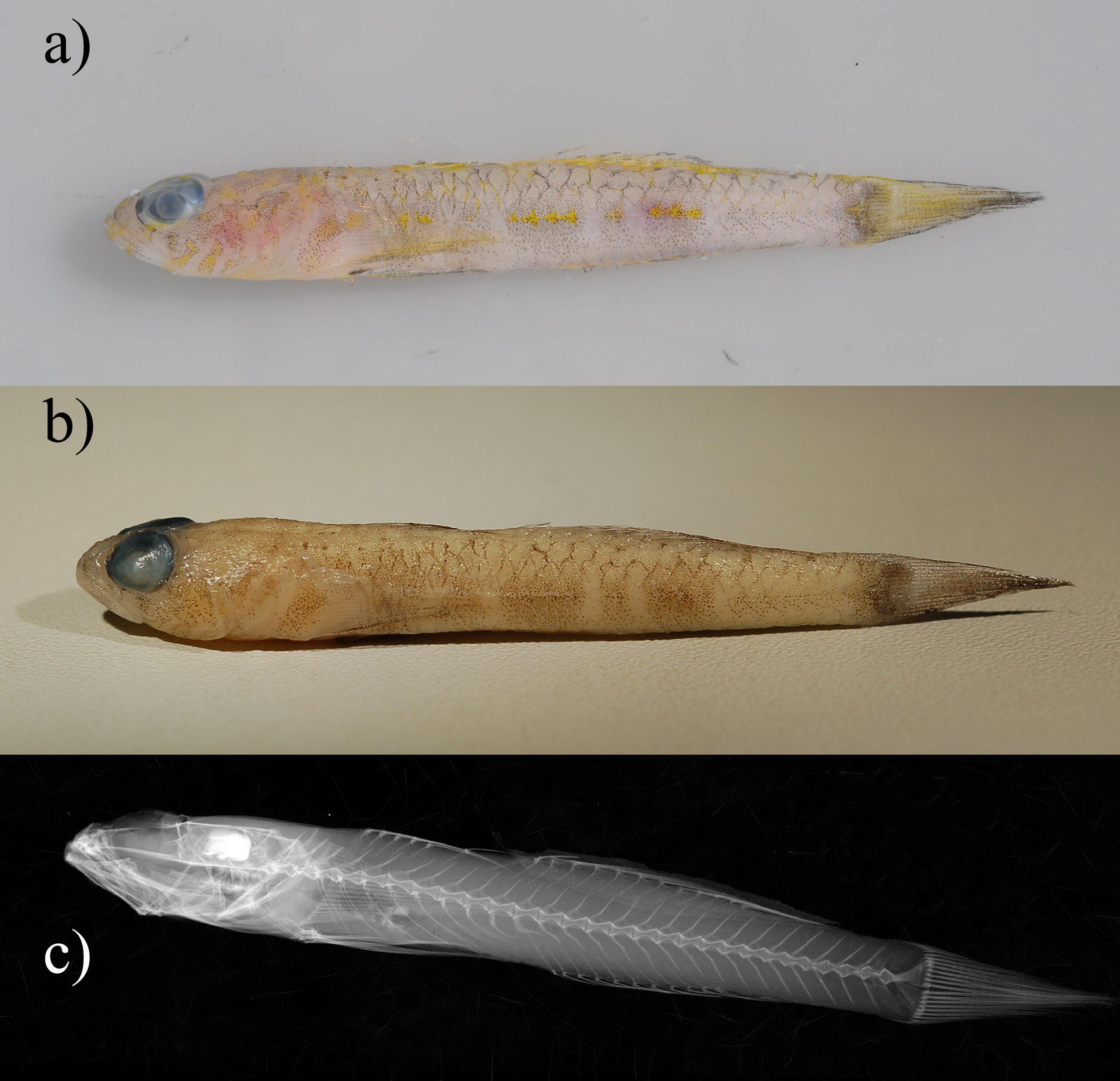
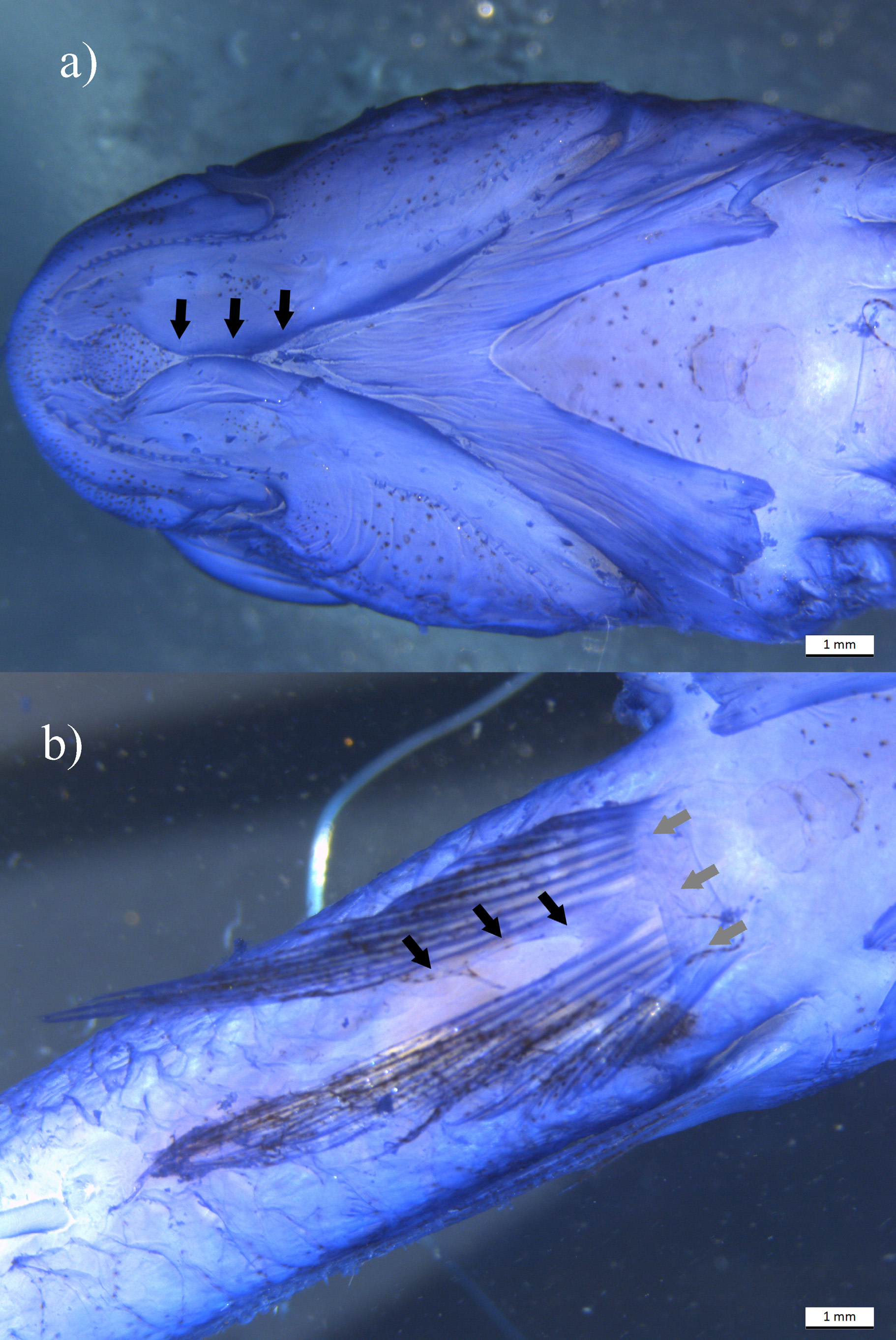
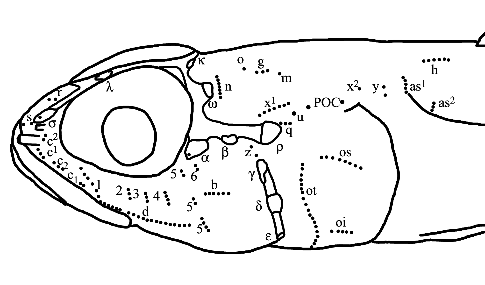
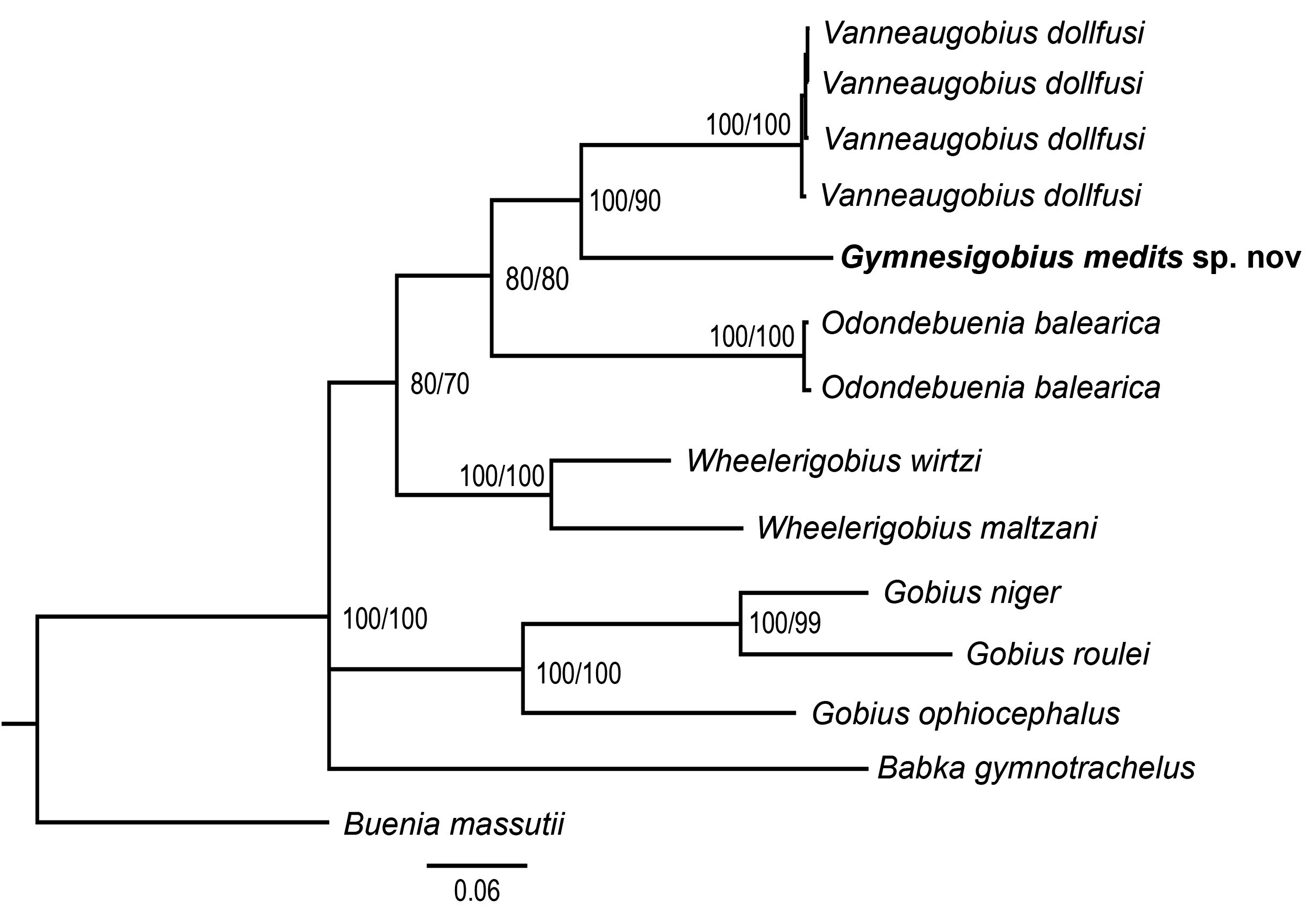
TABLE 3. Morphometric characters (as proportional measurements in %) of Gymnesigobius medits sp. nov.
| Specimen | PMR VP4649 |
|---|---|
| Sex | male |
| Standard length (Sl) in mm | 42.6 |
| % of standard length | |
| 1st spine length of first dorsal fin | 14.3% |
| 2nd spine length of first dorsal fin | 16.2% |
| 3rd spine length of first dorsal fin | 14.6% |
| 4th spine length of first dorsal fin | 14.3% |
| 5th spine length of first dorsal fin | 12.4% |
| 6th spine length of first dorsal fin | 8.2% |
| Anal fin base | 19.7% |
| Anal fin spine length | 7.3% |
| Body depth at anal fin origin | 13.1% |
| Body depth at pelvic fin origin | 14.1% |
| Body width at anal fin origin | 8.9% |
| Body width at pelvic fin origin | 9.9% |
| Caudal fin length | 26.1% |
| Caudal peduncle depth | 9.2% |
| Caudal peduncle length | 22.1% |
| Eye diameter | 9.9% |
| First dorsal fin base | 10.8% |
| Head length | 30.3% |
| Head width | 18.3% |
| Pectoral fin length | 26.8% |
| Pelvic fin length | 24.2% |
| Pelvic to anus | 24.2% |
| Second dorsal fin base | 24.2% |
| Second dorsal fin spine length | 9.6% |
| Snout to origin of first dorsal fin | 36.2% |
| Snout to origin of second dorsal fin | 55.2% |
| Snout to vertical of anal fin origin | 58.2% |
| Snout to vertical of anus | 54.0% |
| Snout to vertical of pelvic fin origin | 31.0% |
| % of caudal peduncle: | |
| Caudal peduncle depth | 41.5% |
| % of head length | |
| Cheek depth | 4.7% |
| Eye diameter | 32.6% |
| Head depth | 42.6% |
......continued on the next page
TABLE 4. Genetic distance and number of base differences for mitochondrial concatenated fragments (COI + Cyt b) of gobies species below and above the diagonal, respectively.
| Num | Species | 1 | 2 | 3 | 4 | 5 | 6 | 7 | 8 | 9 | 10 | 11 | 12 | 13 | 14 |
|---|---|---|---|---|---|---|---|---|---|---|---|---|---|---|---|
| 1 | Buenia massutii | 190 | 188 | 189 | 188 | 186 | 187 | 194 | 199 | 193 | 203 | 222 | 199 | 179 | |
| 2 | Vanneaugobius dollfusi | 19.3 | 7 | 1 | 4 | 159 | 156 | 181 | 178 | 153 | 180 | 181 | 187 | 182 | |
| 3 | Vanneaugobius dollfusi | 19.1 | 0.71 | 6 | 7 | 158 | 155 | 179 | 174 | 151 | 179 | 179 | 183 | 178 | |
| 4 | Vanneaugobius dollfusi | 19.2 | 0.1 | 0.61 | 3 | 158 | 155 | 180 | 177 | 152 | 181 | 182 | 186 | 181 | |
| 5 | Vanneaugobius dollfusi | 19.1 | 0.41 | 0.71 | 0.3 | 158 | 155 | 179 | 176 | 153 | 181 | 182 | 184 | 180 | |
| 6 | Odondebuenia balearica | 18.9 | 16.2 | 16.1 | 16.1 | 16.1 | 9 | 158 | 167 | 170 | 190 | 195 | 202 | 204 | |
| 7 | Odondebuenia balearica | 19 | 15.9 | 15.8 | 15.8 | 15.8 | 0.91 | 159 | 169 | 171 | 194 | 199 | 206 | 202 | |
| 8 | Wheelerigobius wirtzi | 19.7 | 18.4 | 18.2 | 18.3 | 18.2 | 16.1 | 16.2 | 116 | 171 | 181 | 196 | 175 | 184 | |
| 9 | Wheelerigobius maltzani | 20.2 | 18.1 | 17.7 | 18 | 17.9 | 17 | 17.2 | 11.8 | 176 | 180 | 189 | 205 | 186 | |
| 10 | Gymnesigobius medits sp. nov. | 19.6 | 15.5 | 15.3 | 15.4 | 15.5 | 17.3 | 17.4 | 17.4 | 17.9 | 190 | 194 | 191 | 182 | |
| 11 | Gobius niger | 20.6 | 18.3 | 18.2 | 18.4 | 18.4 | 19.3 | 19.7 | 18.4 | 18.3 | 19.3 | 129 | 192 | 167 | |
| 12 | Gobius roulei | 22.6 | 18.4 | 18.2 | 18.5 | 18.5 | 19.8 | 20.2 | 19.9 | 19.2 | 19.7 | 13.1 | 207 | 175 | |
| 13 | Babka gymnotrachelus | 20.2 | 19 | 18.6 | 18.9 | 18.7 | 20.5 | 20.9 | 17.8 | 20.8 | 19.4 | 19.5 | 21 | 198 | |
| 14 | Zosterisessor ophiocephalus | 18.2 | 18.5 | 18.1 | 18.4 | 18.3 | 20.7 | 20.5 | 18.7 | 18.9 | 18.5 | 17 | 17.8 | 20.1 |
| PMR |
Prirodoslovni muzej Rijeka |
No known copyright restrictions apply. See Agosti, D., Egloff, W., 2009. Taxonomic information exchange and copyright: the Plazi approach. BMC Research Notes 2009, 2:53 for further explanation.
|
Kingdom |
|
|
Phylum |
|
|
Class |
|
|
Order |
|
|
Family |
|
|
Genus |