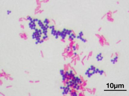
The terms Gram positive and Gram negative are commonly used to describe bacteria. The main difference between the two is the structure of their cell wall which changes their susceptibility to different antibiotics. The separation also loosely fits the location of these organisms in the body – Gram negative organisms predominate in the bowel (eg. E. coli and other coliforms, Bacteroides species etc) whilst Gram positives (Staphs. and streptococci) predominate on the skin, upper respiratory tract and oropharynx. Gram staining (see example Gram stains) was invented in the 1880s by Danish scientist Hans Christian Gram and takes several steps. A Gram positive organism holds onto the first stain and appears a purple-blue colour, whilst a Gram negative organism holds the second stain and appears pink. A bacterium’s ability to hold onto a stain is dependent on the structure of their cell wall.
A Gram positive organism lacks an outer (LPS) membrane but has a thick layer of peptidoglycan and no LPS outer membrane. This facilitates access of cell-wall active antibiotics (eg. penicillin/betalactam or vancomycin-type antibiotics) to their site of action (the peptidoglycan).
In coliform-type Gram negative bacteria such as E. coli, these antibiotics have to traverse the LPS layer via porin (channel) proteins. The ability to transfer through these channels is dependent on the charge and shape of the antibiotic. For instance benzylpenicillin cannot cross whereas ampicillin can, causing intrinsic antibiotic resistance to the former drug. Porins may be modified or removed via genetic changes (via mutation usually) that occur which then potentially alters the resistance character of the organism. Efflux proteins may also be present in Gram negative cell walls. These are relevant to antibiotics that act intracellularly and pump them out of the cell, lowering the intracellular antibiotic concentration to a level where the antibiotic becomes inactive.

Image from: http://static.diffen.com/uploadz/0/0e/cell-wall-gram-bacteria.png
Image from: https://en.wikipedia.org/wiki/File:Gram_stain_01.jpg : Microscopic image of a Gram stain of mixed Gram-positive cocci (Staphylococcus aureus ATCC 25923, purple) and Gram-negative bacilli (Escherichia coli ATCC 11775, red). Magnification:1,000.

I was asked to explain this to a nursing colleague last week. I have now forwarded to her for reinforcement
LikeLike
[…] This posting may be of interest – it contrasts why Gram negative and positive bacteria exhibit different antimicrobial susceptibilities. […]
LikeLike