Biology LibreTexts

2.5.2.3: Bryophyta

  • Page ID
    37011
  • \( \newcommand{\vecs}[1]{\overset { \scriptstyle \rightharpoonup} {\mathbf{#1}} } \) \( \newcommand{\vecd}[1]{\overset{-\!-\!\rightharpoonup}{\vphantom{a}\smash {#1}}} \)\(\newcommand{\id}{\mathrm{id}}\) \( \newcommand{\Span}{\mathrm{span}}\) \( \newcommand{\kernel}{\mathrm{null}\,}\) \( \newcommand{\range}{\mathrm{range}\,}\) \( \newcommand{\RealPart}{\mathrm{Re}}\) \( \newcommand{\ImaginaryPart}{\mathrm{Im}}\) \( \newcommand{\Argument}{\mathrm{Arg}}\) \( \newcommand{\norm}[1]{\| #1 \|}\) \( \newcommand{\inner}[2]{\langle #1, #2 \rangle}\) \( \newcommand{\Span}{\mathrm{span}}\) \(\newcommand{\id}{\mathrm{id}}\) \( \newcommand{\Span}{\mathrm{span}}\) \( \newcommand{\kernel}{\mathrm{null}\,}\) \( \newcommand{\range}{\mathrm{range}\,}\) \( \newcommand{\RealPart}{\mathrm{Re}}\) \( \newcommand{\ImaginaryPart}{\mathrm{Im}}\) \( \newcommand{\Argument}{\mathrm{Arg}}\) \( \newcommand{\norm}[1]{\| #1 \|}\) \( \newcommand{\inner}[2]{\langle #1, #2 \rangle}\) \( \newcommand{\Span}{\mathrm{span}}\)\(\newcommand{\AA}{\unicode[.8,0]{x212B}}\)

    Learning Objectives
    • Use morphological traits and cellular components to distinguish between mosses and other bryophytes.
    • Identify structures and phases in the moss life cycle; know their ploidy.
    • Label a moss sporophyte and describe its development.

    Mosses, Phylum Bryophyta

    Most described bryophyte species diversity (around 13,000 species) belongs to the mosses. Unlike other bryophytes, mosses are exclusively leafy. Sporophytes in most species form complex capsules, involving multiple layers of structures. Members of the mosses have defied many of the typical bryophyte descriptors. For example, some mosses have evolved vascular tissue analogs called leptoids (analogous to phloem) and hydroids (analogous to xylem). Other mosses can grow quite tall: Dawsonia superba, the world's tallest moss, can grow up to be nearly 2 feet tall (50-60 cm).

    Tall moss gametophytes with spirally arranged leaves
    Figure \(\PageIndex{1}\): Gametophytes of Dawsonia superba, the world's tallest moss species, growing in New Zealand. Photo by Jacqui-nz, CC-BY-NC.

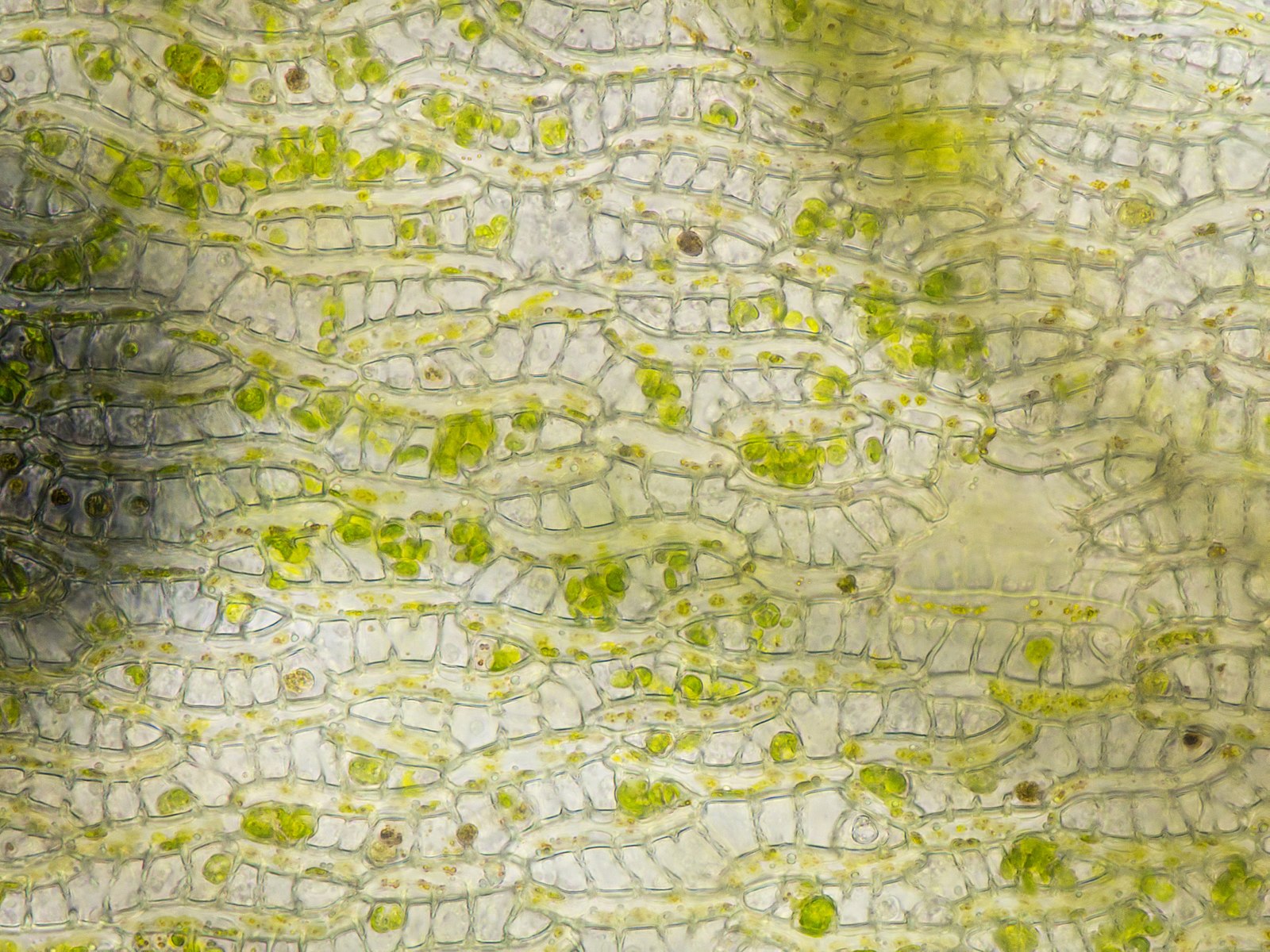
    Sphagnum is an ancestral genus of mosses that grow in bogs and are commonly referred to as peat moss or peat, in its compressed form. They have large, empty cells (Figure \(\PageIndex{2}\)) capable of absorbing around 20 times their dry weight in water and can exude compounds to make their environment more acidic. Because of this, and the compounds in their cell walls, the bogs where they grow have slow decomposition, allowing plant matter to accumulate and compress over time. This makes peat, a common source of fuel in higher latitudes. It's use as fuel, flavoring (the smoke), and in horticulture for its water holding capacity have contributed to its commercial importance. Many peat bogs are frozen over for most of the year, halting metabolic activity and slowing the release of methane from anaerobic decomposition. As the climate in northern latitudes warms, so do the peat bogs, increasing the release of methane.

    A clump of Sphagnum gametophytes with densely packed leaves grows surrounded by waterMicroscopic view of Sphagnum leaf cells reveals that they are mostly empty. Some large green cells can be seen within the leaf cells.
    Figure \(\PageIndex{2}\): Sphagnum gametophytes and cells. The gametophytes in the first photo are growing submerged in water. In the second photo, the strange, empty cells of Sphagnum leaves have algae growing within them! These large, empty cells give Sphagnum their immense water holding capacity. First photo by Tarpinian, CC-BY-NC. Second photo by Казаков Александр Павлович, CC BY 4.0, via Wikimedia Commons.

    Gametophyte Generation

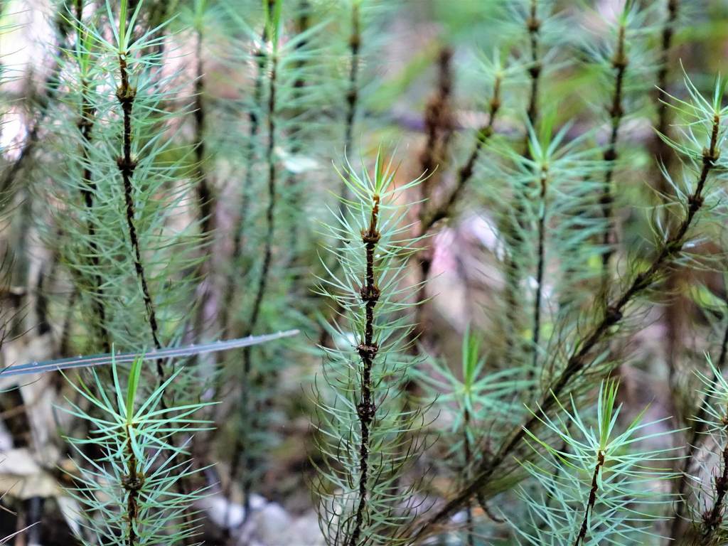
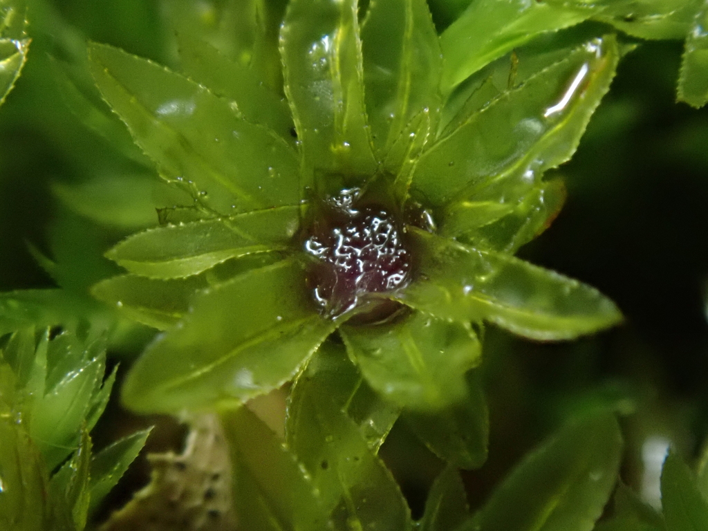
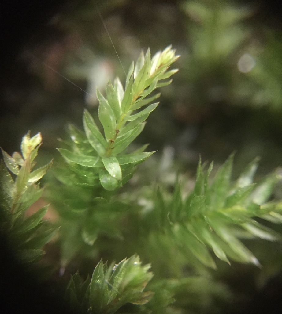
    Moss gametophytes have spirally arranged leaves that emerge from all sides of the stem (Figure \(\PageIndex{3}\)). In most mosses, the leaves have a central line of tissue called a costa that looks a bit like a midrib (though it is not, as bryophytes don't have lignified vascular tissue) and are only 1-2 cell layers thick. Rhizoids are produced at the base of the gametophytes. In the common haircap moss, Polytrichum commune, there are three kinds of gametophytes:

    • Female, which develop archegonia at their tip. A single egg forms in each archegonium. (see Figure \(\PageIndex{6}\))
    • Male, which develop antheridia at their tip. Multiple swimming sperm form in each antheridium. (see Figure \(\PageIndex{5}\))
    • Sterile, which do not form sex organs.
    Clustered green, leafy gametophytes. Thin leaves emerge at all angles from each gametophyte, making them look like tiny trees.
    Figure \(\PageIndex{3}\): A closer look at spiral leaf arrangement, this time with Polytrichum commune, the haircap moss. Note that leaves emerge from all directions of each gametophyte. Photo by Mizuki Maeda, CC-BY-NC.
    A cluster of moss gametophytes. These have densely packed leaves emerging at all angles.
    Figure \(\PageIndex{4}\): These moss gametophytes show spiral leaf arrangement and a distinct, tan costa that emerges on each leaf. Can you spot them? Photo by Geoffrey Cox, CC-BY-NC.
    Labeled cross section of a Mnium antheridial head
    Mnium male gametophyte, looking down at the splash cup
    Figure \(\PageIndex{5}\): On the left is a labeled cross section of the antheridial head of a Mnium male gametophyte. On the right is an actual image of the splash cup (antheridial head) from a bird's eye view. The cross section is labeled as follows: A) An antheridium, B) paraphyses, C) sterile jacket of the antheridium, D) sporogenous tissue, E) male gametophyte. First photo by Maria Morrow, CC-BY-NC. Second photo by Bosmon, CC-BY-NC.
    Labeled Mnium female gametophyte long section
    Mnium female gametophyte, with no obvious structures visible
    Figure \(\PageIndex{6}\): On the left is a labeled cross section of the archegonial head of a Mnium female gametophyte. On the right is an actual image of a female gametophyte. The cross section is labeled as follows: A) An archegonium, B) neck of the archegonium, C) venter, D) egg, E) paraphyses, F) female gametophyte. First photo by Maria Morrow, CC-BY-NC. Second photo by Tisrel, CC-BY-NC.

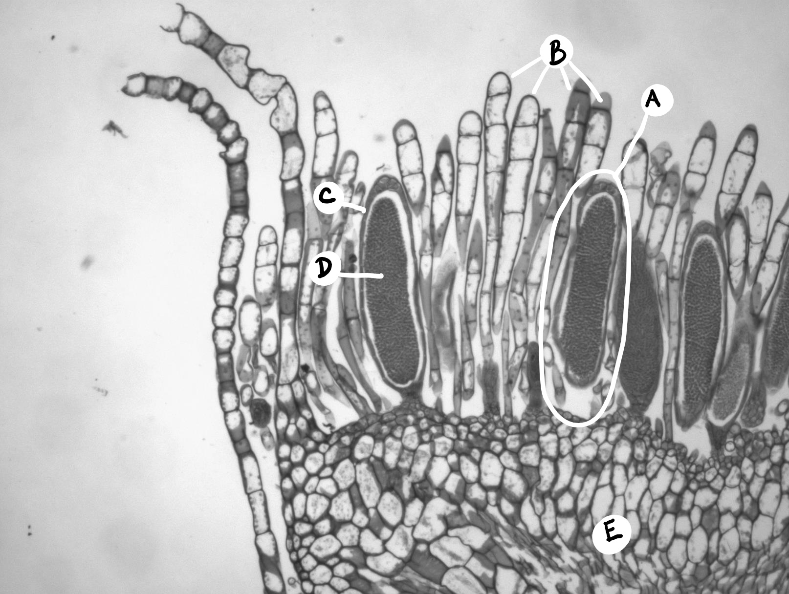
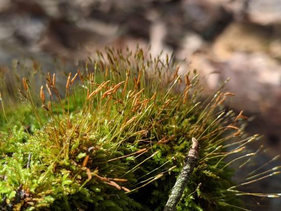
    Sporophyte Generation
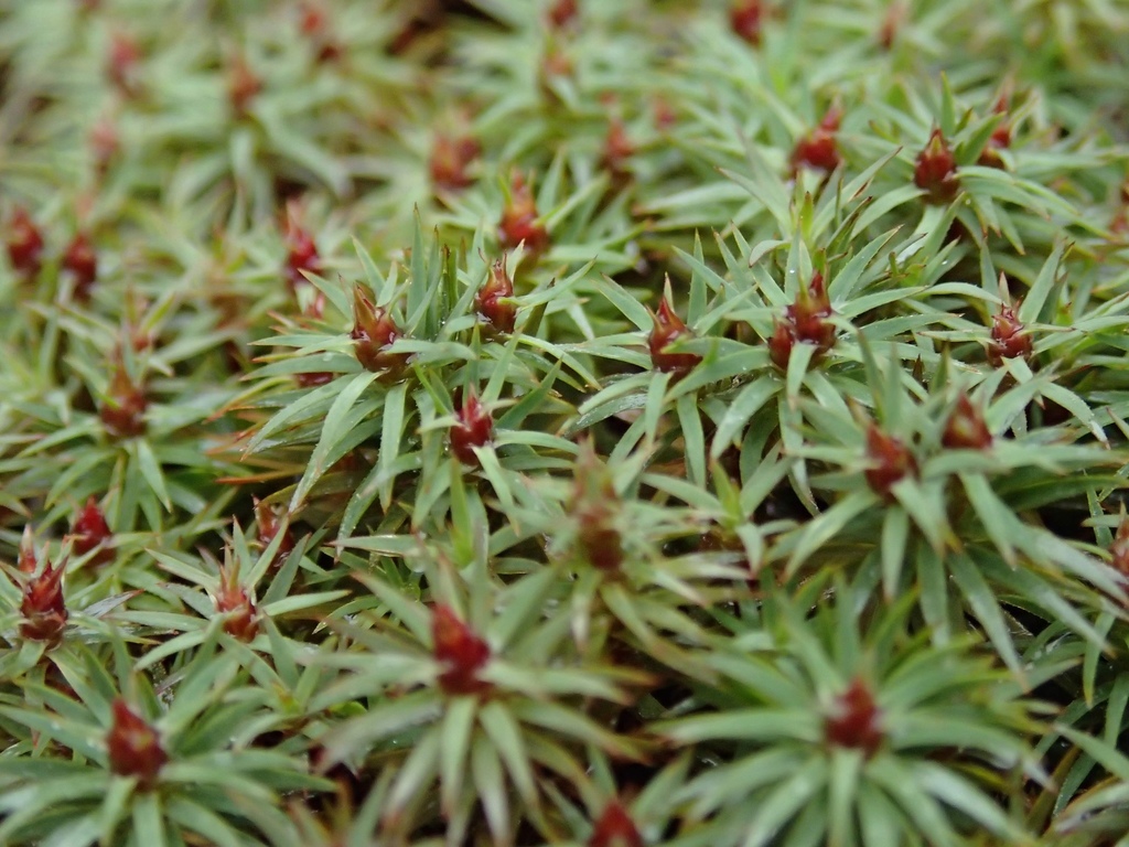
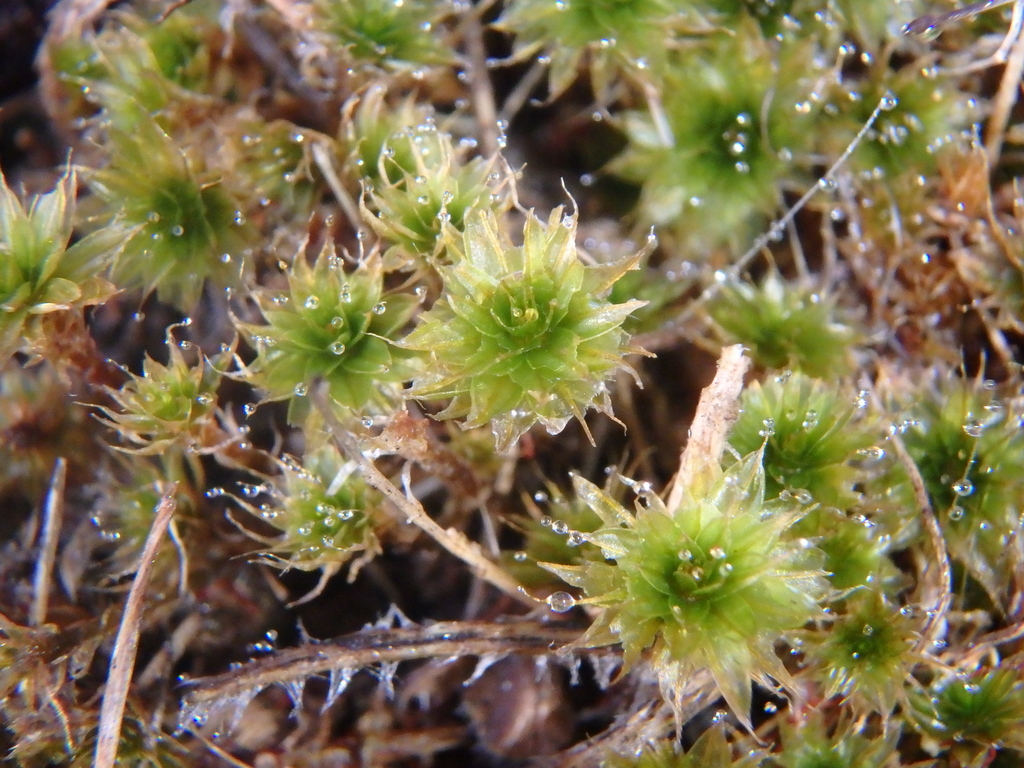
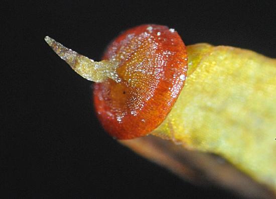
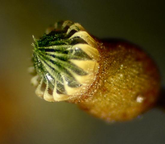
    Moss sporophytes (with calyptras) emerging from gametophytes
    Figure \(\PageIndex{7}\): Moss sporophytes are composed of a seta and capsule, much like liverworts. When the sporophyte emerges from the archegonium, it brings a piece of that archegonium with it. This gametophyte remnant sits on the capsule like a cap, which is why it is called a calyptra. Photo by Maria Morrow, CC-BY-NC.

    When the calyptra falls off, another feature of the sporophyte is visible: the operculum. This is a lid-like structure on the capsule that pops off when the spores are mature. Most mosses have a structure under the operculum that lines the capsule opening, called the peristome. The peristome is a series of cellular flaps (peristome teeth) that line the edge of the capsule opening and aids in spore dispersal via hygroscopic movements.

    A red, cap-like structure on the end of a moss capsule
    Two rows of pointed, cellular flaps line the mouth of a moss capsule.
    Figure \(\PageIndex{8}\): Moss sporophyte structures. In the first photo, the operculum of a Polytrichum sp. is visible. It is red and has a long, conical beak. In the second photo, the operculum of this moss has already popped off, revealing the peristome. There are two ranks of teeth in the peristome of this moss, the outer rank is bent and the tips are reaching inward. Green spores can bee seen clustered under the inner rank of teeth. Photos by George Shepherd, CC BY-NC-SA.

    Tetraphis moss (Tetraphis pellucida) produces both asexual propagules (gemmae) and spores, which result from sexual reproduction (Figure \(\PageIndex{9}\)). In 1991, Robin Wall Kimmerer found that spores were dispersed an average of 20 times farther from the parent plant than gemmae were, but gemmae established more easily. Each reproductive strategy thus served a different purpose: gemmae helped the plant spread locally, and spores were important for colonization of an entirely new location.

    Tetraphis moss has long, brown sporophytes with oval capsules at the tip. The rest of the plant is the green, cushiony gametophyte.
    Figure \(\PageIndex{9}\): Sporophytes extend from the lush, green gametophyte in Tetraphis pellucida. The spores produced in the capsules of the sporophyte will function in long-distance dispersal. Photo by Nate Marineau, CC-BY-NC.

    Mnium Life Cycle

    Male gametophytes form antheridia at the top of the gametophyte in a structure called the perigonium or antheridial head (Figure \(\PageIndex{10}\)). These are cup-shaped, and commonly referred to as splash cups. In early spring, raindrops splash sperm from male to female plants. These swim down the canal in the archegonium to the chamber containing the egg. The resulting zygote begins the sporophyte generation.

    Mitosis of the zygote produces an embryo that grows into the mature sporophyte generation. It consists of:

    • A foot, which absorbs water, minerals, and food from the parent gametophyte
    • A stalk (seta), at the tip of which is formed a sporangium (the brownish objects in the photo).

    The sporangium is

    • filled with spore mother cells
    • in most cases, ined by a peristome around the opening
    • sealed by an operculum
    • covered with a calyptra. The calyptra develops from the wall of the old archegonium and so is actually a part of the gametophyte generation. It is responsible for the common name ("haircap moss") of this species.

    During the summer, each spore mother cell undergoes meiosis, producing four haploid spores - the start of the new gametophyte generation. Late in the summer, the calyptra and operculum become detached from the sporangium allowing the spores to be released. These tiny spores are dispersed so effectively by the wind that many mosses are worldwide in their distribution. If a spore reaches a suitable habitat, it germinates to form a filament of cells called a protonema. Soon buds appear and develop into the mature leafy shoots.

    Thus the gametophyte generation is responsible for sexual reproduction. The sporophyte generation is responsible for dispersal.

    Mnium life cycle diagram
    Figure \(\PageIndex{10}\): The Mnium life cycle. Starting with meiosis (left, center): Meiosis produces haploid spores which will grow by mitosis into either male or female gametophytes. Moss spores germinate and begin to grow into a chain of cells called a protonema. Male gametophytes are flat topped, called splash cups. Many antheridia are produced within the splash cup, surrounded by sterile cells called paraphyses. Antheridia produce biflagellate sperm by mitosis. Female gametophytes produce multiple archegonia at the top of the gametophyte. These are also surrounded by paraphyses. Each archegonium produces a single egg by mitosis. When water hits the splash cup, it can splash sperm onto a female gametophyte. If the sperm are able to swim through the water to an egg, fertilization occurs producing a diploid zygote. The zygote grows into a sporophyte from within the archegonium. The remaining archegonial tissue is called the calyptra and will remain on top of the sporophyte as a cap-like structure. A mature sporophyte will have a sterile stalk called a seta. Underneath the calyptra, there is a capsule containing cells that will undergo meiosis to make haploid spores. The capsule has a lid-like operculum that will pop off when the spores have matured. Peristome teeth along the opening of the capsule aid in spore dispersal. Drawing by Nikki Harris, CC-BY-NC, with colors and labels added by Maria Morrow.

    Attribution

    Content by Maria Morrow, CC BY-NC, except the following:

    • Life cycle text from 16.3B Moss Life Cycle from Biology by John. W. Kimball (licensed CC-BY)
    • Paragraph that goes with Figure \(\PageIndex{9}\) by Melissa Ha (licensed CC BY-NC)


    This page titled 2.5.2.3: Bryophyta is shared under a CC BY-NC-SA 4.0 license and was authored, remixed, and/or curated by Melissa Ha, Maria Morrow, & Kammy Algiers (ASCCC Open Educational Resources Initiative) .