


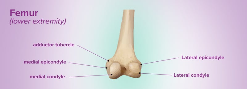
Condyles are rounded bony features that have articular surfaces to mate with other bones to form joints. The femoral condyles are at the distal end of the femur. The medial condyle is the larger of the two and it lies toward the midline of the body. The lateral condyle is towards the outside of the body. A recent biomechanical study has shown almost equal loading between the medial and lateral condyles during common activities.
Epicondyles are smaller bony prominences that are found on the larger condyles and serve as attachment sites for tendons of muscles and ligaments. The medial epicondyle is an attachment point for the medial head of the gastrocnemius (calf) muscle, a large muscle that provides push-off force during walking, running, and jumping. The lateral epicondyle provides an attachment point for the lateral head of this muscle.
The femoral condyles articulate with the relatively flat surface of the proximal tibia called the tibial plateau. The condyles and the tibial plateau are covered with articular cartilage. Between the femoral condyles and the tibial plateau are two thin cartilage pads called the medial and lateral meniscus. The menisci (plural) are anchored to the tibia and deepen the concavity of the tibial plateau. This helps to improve stability and dampen shearing forces at the joint by preventing anterior, posterior (rearward), and side-to-side slipping of the rounded condyles off the otherwise flat tibial plateau. The menisci work with the articular cartilage to absorb compressive forces.
The knee joint, like the hip joint, is a synovial joint. Synovium fills the joint cavity and bathes the articular surfaces, providing lubrication that significantly reduces friction. The patella (knee cap) is positioned on the front of the knee and rests against the femoral condyles (not shown in image). The patella connects the large quadriceps of the thigh to the tibia and significantly increases the lever arm of the muscle group. Several ligaments keep the joint together, including the collateral ligaments, anterior cruciate ligament (ACL), and posterior cruciate ligament (PCL).