A Positron Emission Tomography (PET) Scan uses a tracer to provide your cardiologist with images to diagnoses disease in your body. The tracer is a minorly radioactive substance that shows itself on the imaging device. A PET scan gives your doctor a clear idea of how well your organs and tissue are functioning. This is not the same as an MRI and CT scans which show structure and b
Related Positron Emission Tomography (PET) Scan tests include:
- Brain PET scan
- Breast PET scan
- Heart PET scan
- Lung PET scan
How is a Positron Emission Tomography (PET) Performed?
The tracer is inserted into an IV, located on the inside of your elbow. It flows with your blood and will pool or collect inside organs and tissues. This will allow a radiologist to see the issue area clearly.
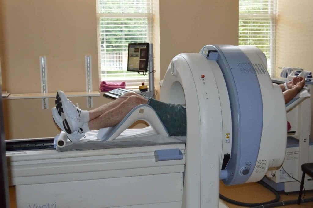
After the tracer is inserted, you will wait and hour for it to be absorbed and flow through your body. Then your body will be placed inside of a large scanner. The Positron Emission Tomography (PET)will detect signals from the tracer. And the images will be generated by a computer for your doctor to interpret.
During the test, it is vital that you remain still to protect the images. The length of time varies for each case.
How do you prepare for a PET scan?
You may be asked not to eat anything for 4 – 6 hours before the scan. You will be able to drink water.
Tell your health care provider if:
- You are afraid of close spaces (have claustrophobia). You may be given a medicine to help you feel sleepy and less anxious.
- You are pregnant or think you might be pregnant.
- You have any allergies to injected dye (contrast).
Always tell your health care provider about the medicines you are taking, including those bought without a prescription. Sometimes, medications may interfere with the test results.