File:Anatomy, descriptive and surgical (1897) (14761736601).jpg
From Wikimedia Commons, the free media repository
Jump to navigation Jump to search

Original file(1,440 × 1,400 pixels, file size: 520 KB, MIME type: image/jpeg)

Captions

Captions

Add a one-line explanation of what this file represents

Summary[edit]


Description
English:

Identifier: anatomydescripti1897gray (find matches)
Title: Anatomy, descriptive and surgical
Year: 1897 (1890s)
Authors: Gray, Henry, 1825-1861 Carter, H. V. (Henry Vandyke), 1831-1897 Pick, T. Pickering (Thomas Pickering), 1841-1919
Subjects: Anatomy Human anatomy Anatomy, Surgical and topographical
Publisher: London : Longmans, Green
Contributing Library: Francis A. Countway Library of Medicine
Digitizing Sponsor: Open Knowledge Commons and Harvard Medical School

View Book Page: Book Viewer
About This Book: Catalog Entry
View All Images: All Images From Book
Click here to view book online to see this illustration in context in a browseable online version of this book.

Text Appearing Before Image:
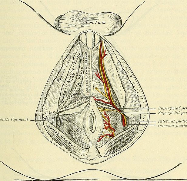
erted into the central tendinous point of the perinaeum, joining inthis situation with the muscle of the opposite side, the External sphincter anibehind, and the Accelerator urinte in front. Nerve-supply.—The perineal branch of the internal pudic. Actions.—By their contraction they serve to j&x the central tendinous pointof the perinseum. The Accelerator urinse (Ejaculator seminis, or Bidbo-cavernosus) is placed inthe middle line of the perinseum, immediately in front of the anus. In consistsof two symmetrical halves, united along the median line by a tendinous raphe.It arises from the central tendon of the perinaeum, and from the median raphein front. From this point its fibres diverge like the plumes of a pen ; the mostposterior form a thin layer, which are lost on the anterior surface of the triangularligament; the middle fibres encircle the bulb and adjacent parts of the corpus Fig. 336.—The superficial muscles and vessels of the perinseum. Gi~eat aacro-seiatic Ugatneiii
Text Appearing After Image:
Ji SuperflciaC_perineal arleri -U . Superficial perineal nerve Internal pudic nerveInternal pudic artery spongiosum, and join with the fibres of the opposite side, on the upper part ofthe corpus spongiosum, in a strong aponeurosis; the anterior fibres, the longestand most distinct, spread out over the sides of the corpus cavernosum, to beinserted partly into that body, anterior to the Erector penis, occasionally extend-ing to the OS pubis; partly terminating in a tendinous expansion, which coversthe dorsal vessels of the penis. The latter fibres are best seen by dividing themuscle longitudinally, and dissecting it outwards from the surface of the urethra.Action.—This muscle serves to empty the canal of the urethra, after thebladder has expelled its contents ; during the greater part of the act of micturitionits fibres are relaxed, and it only comes into action at the end of the process.The middle fibres are supposed, by Krause, to assist in the erection of the corpusspongiosum, by c

Note About Images

Please note that these images are extracted from scanned page images that may have been digitally enhanced for readability - coloration and appearance of these illustrations may not perfectly resemble the original work.
Date
Source

https://www.flickr.com/photos/internetarchivebookimages/14761736601/

Author Internet Archive Book Images
Permission
(Reusing this file)
At the time of upload, the image license was automatically confirmed using the Flickr API. For more information see Flickr API detail.
Flickr tags
InfoField
Flickr posted date
InfoField
28 July 2014

Licensing[edit]

This image was taken from Flickr's The Commons. The uploading organization may have various reasons for determining that no known copyright restrictions exist, such as:
  1. The copyright is in the public domain because it has expired;
  2. The copyright was injected into the public domain for other reasons, such as failure to adhere to required formalities or conditions;
  3. The institution owns the copyright but is not interested in exercising control; or
  4. The institution has legal rights sufficient to authorize others to use the work without restrictions.

More information can be found at https://flickr.com/commons/usage/.


Please add additional copyright tags to this image if more specific information about copyright status can be determined. See Commons:Licensing for more information.
This image was originally posted to Flickr by Internet Archive Book Images at https://flickr.com/photos/126377022@N07/14761736601. It was reviewed on 17 September 2015 by FlickreviewR and was confirmed to be licensed under the terms of the No known copyright restrictions.

17 September 2015

File history

Click on a date/time to view the file as it appeared at that time.

Date/TimeThumbnailDimensionsUserComment
current16:27, 17 September 2015Thumbnail for version as of 16:27, 17 September 20151,440 × 1,400 (520 KB) (talk | contribs)== {{int:filedesc}} == {{subst:chc}} {{information |description={{en|1=<br> '''Identifier''': anatomydescripti1897gray ([https://commons.wikimedia.org/w/index.php?title=Special%3ASearch&profile=default&fulltext=Search&search=insource%3A%2Fanatomydescri...

There are no pages that use this file.