


Filters

Stock royalty-free photos and images of Ecg monitoring
Discover unlimited high resolution images of Ecg monitoring and stock visuals for commercial use.
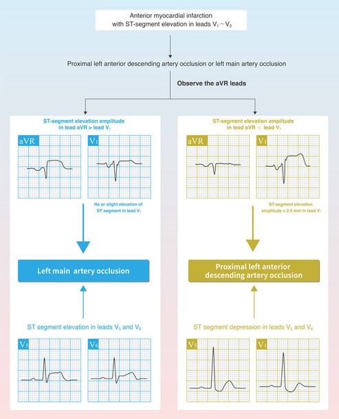
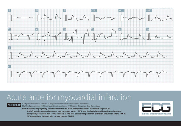
In case of acute anterior myocardial infarction, the characteristics of ST segment elevation in ECG can be used to deduce whether the culprit vessel system is the left main trunk or the proximal LAD.
When the middle - distal segments of the LAD were occluded, the ischemic potential was toward the anterior, inferior and left, and the infarct spread to the anterior lower part of the left ventricle.
ECG monitoring, monitoring with a spirometer and a dermatoscope, telemedicine concept

In imaging medicine, although the myocardium is divided into many segments, in anatomy, these myocardium are continuous rather than completely divided.

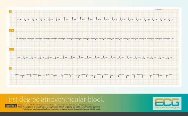
A normal ECG is an ECG that does not have any abnormal changes and can be seen in healthy people as well as in people with heart disease.
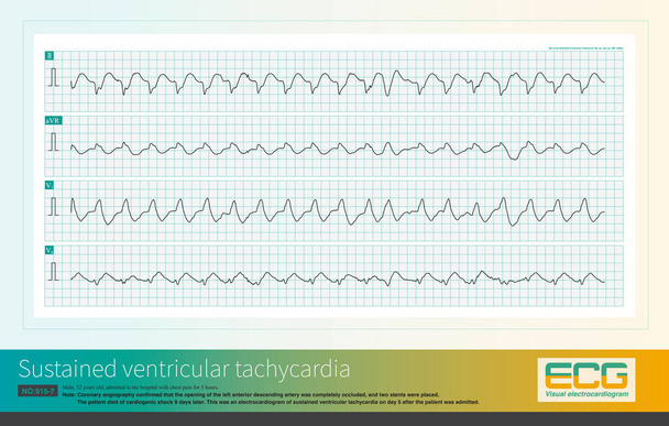
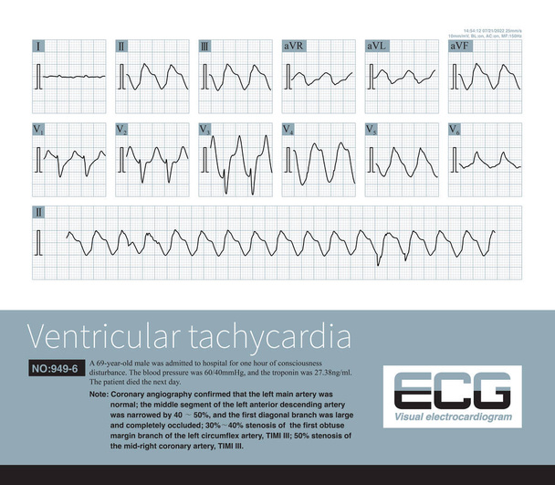
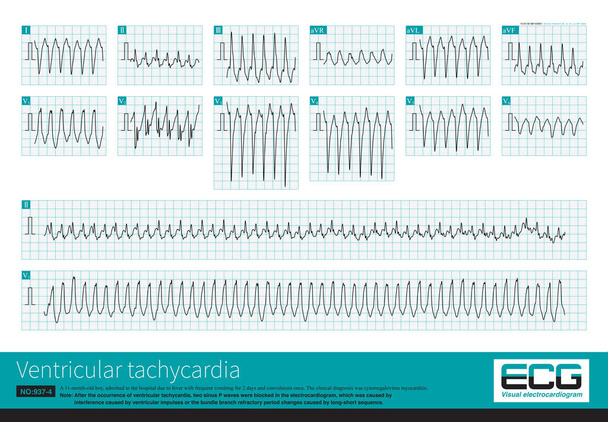
Male, 52 years old, diagnosed with acute extensive anterior wall myocardial infarction. The patient repeatedly experienced ventricular tachycardia and eventually died of cardiogenic shock.

When the frontal axis is +62 , the maximum QRS depolarization potential is closest to the axis of lead so the R-wave amplitude of lead is the highest.

A 1-year-old child developed atrioventricular reentrant tachycardia. When tachycardia occurred, retrograde P waves can be detected each QRS complex.
When the frontal axis is +43 , the maximum QRS depolarization potential is closest to the axis of lead so the R-wave amplitude of lead is the highest
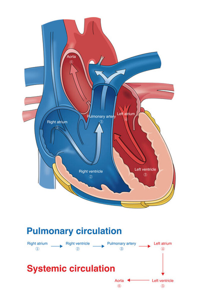
After venous blood enters the pulmonary circulation, oxygenated blood returns to the left atrium and is pumped through the left ventricle into the aorta to complete systemic circulation.
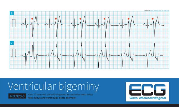
A 17 year old male was clinically diagnosed as ventricular septal defect and his ECG was ventricular bigeminy.Ventricular premature contraction originated in the left posterior fascicular region.

Male, 75 years old, clinically diagnosed as acute anterior septal myocardial infarction, prolonged QT interval, complicated with torsade de pointes.
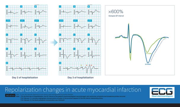
In acute myocardial infarction, the dynamic change of repolarization and prolongation of QT interval are the hallmarks of torsade de pointes.

Acute left main artery occlusion can cause both ST segment elevation and non ST segment elevation myocardial infarction, regardless of which type, the risk of death is high.

The absent septal q wave in ECG may be a normal variant, and the pathological reasons are ventricular septal fibrosis, intraventricular block and myocardial disease.

Under physiological conditions, because the initial part of the frontal vector loop can be projected on the negative side of some lead axes, physiological Q waves are formed.
A is idiopathic right ventricular outflow tract ventricular tachycardia, and B is arrhythmogenic right ventricular cardiomyopathy.The former is benign, while the latter is malignant.
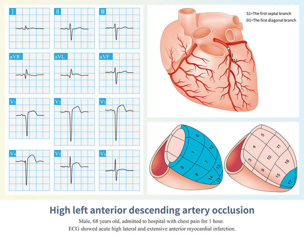
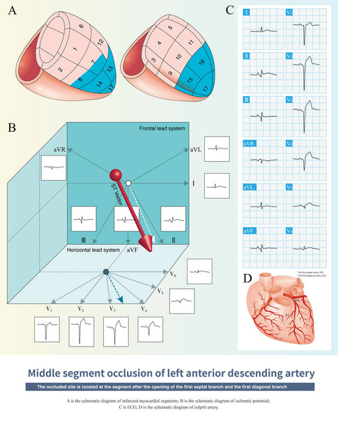
When the occlusion of the LAD is located in the common trunk above the opening of the first septal branch and the first diagonal branch, a large area of left ventricular infarction is caused.
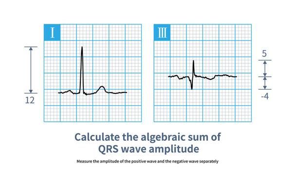
The calculation of algebraic sum of QRS wave is to measure the amplitude of positive wave (positive number) and negative wave (negative number) respectively, and then calculate the algebraic sum.

The proximal occlusion of the left anterior descending artery leads to a large area of anterior myocardial infarction, which belongs to the high occlusion of the left coronary artery.
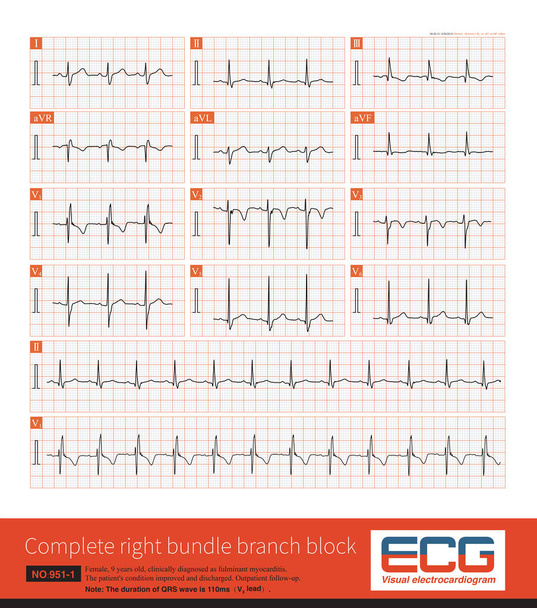
In 2009, American AHA ECG guidelines defined children aged 4 to 16 years old, and QRS duration 110ms can be diagnosed as complete right bundle branch block.
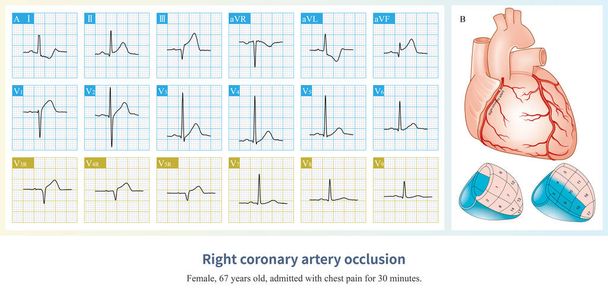
Acute occlusion of the proximal to middle segment of the right coronary artery can clinically cause myocardial infarction in the right ventricle, inferior and posterior wall.

Male, 68 years old, chest pain for 7 hours. Coronary angiography suggests occlusion of the distal right coronary artery. The patient was successfully placed with a coronary stent.

When the frontal axis is + 19 , the maximum QRS depolarization potential is closest to the axis of lead , so the R-wave amplitude of lead , is the highest.
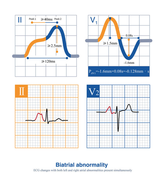
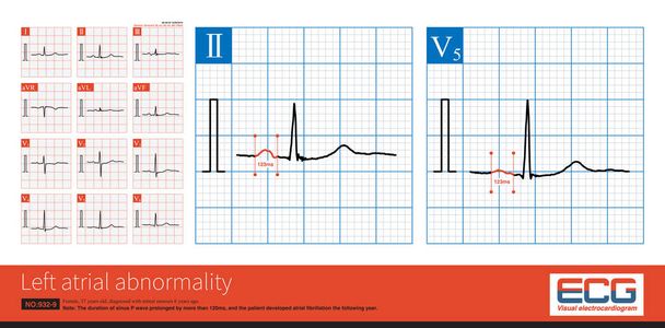
When the duration of the P wave exceeds 120 ms, the amplitude of the limb leads exceeds 2.5 mm, and the amplitude of the thoracic leads exceeds 1.5 mm, it is interpreted as a biatrial abnormality.
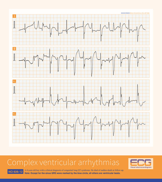
A 4-year-old boy with a clinical diagnosis of long QT syndrome. No genetic testing was done during hospitalization. The child died suddenly during follow-up.

The retrograde P wave of the inferior leads can come from the lower part of the atrium, the lower part of the atrial septum, and the atrioventricular junctional region.
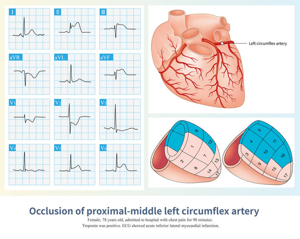
The occlusion of the proximal and middle left circumflex artery can lead to myocardial infarction in the lateral, inferior and posterior walls, with a large infarction area.
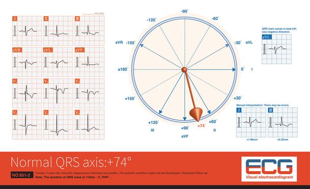
When the frontal QRS axis is located at +74, the QRS main wave in lead aVL is negative and the highest amplitude of the QRS wave in the limb leads is the lead.

On ECG, U wave is a low wave after T wave, and its mechanism is still unclear. It may be an electromechanical feedback wave.It is easy to recognize when the heart rate is slow.
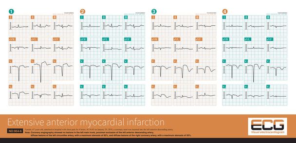
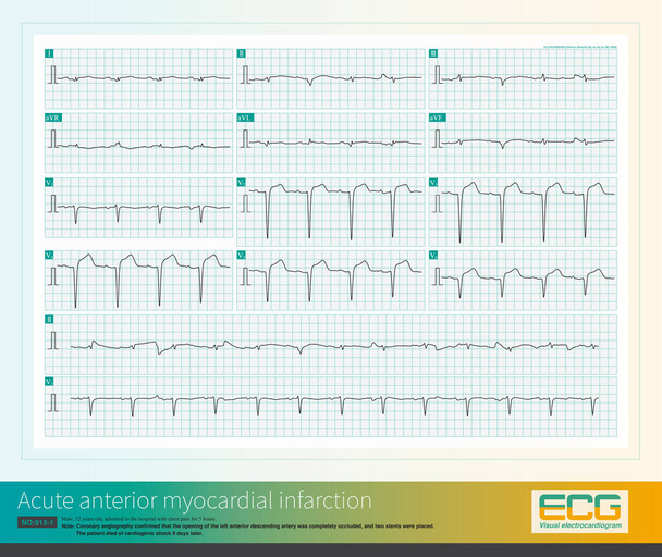
A 67-year-old patient with acute extensive anterior myocardial infarction showed obvious ST-T evolution on the sequence electrocardiogram during hospitalization.
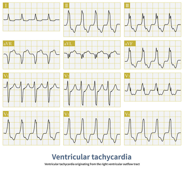
Ventricular tachycardia originating from the right ventricular outflow tract is a benign ventricular tachycardia.It usually does not cause sudden death.Radiofrequency ablation can cure this arrhythmia.
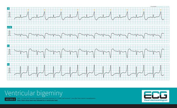
Some ventricular bigeminy are precursors of ventricular tachycardia, a sign of clinical deterioration and severe impairment of cardiac function, and predisposition to ventricular fibrillation and death.
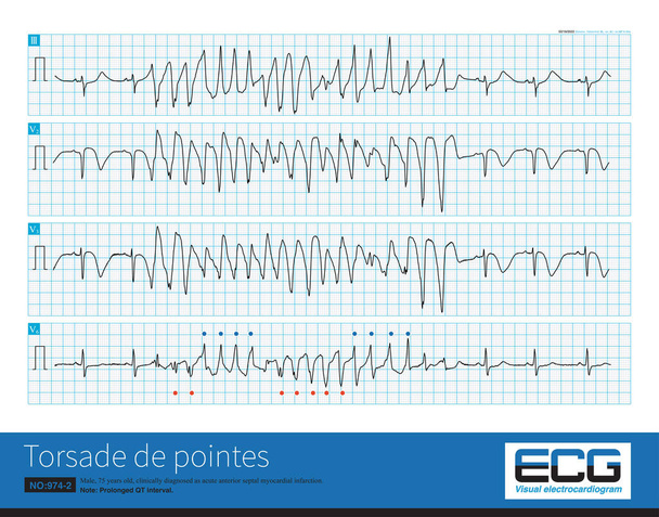
Male, 75 years old, clinically diagnosed as acute anterior septal myocardial infarction.It is best to use 12 leads to diagnose torsade de pointes because some leads may not have torsade characteristic

A 17 year old male was clinically diagnosed as ventricular septal defect and his ECG was ventricular bigeminy.Sinus and ventricular beats alternated.This is a long V1 lead.
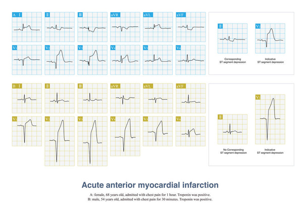
In some patients with acute anterior myocardial infarction, the inferior leads of the electrocardiogram will have ST segment depression, which is called corresponding ST segment depression.
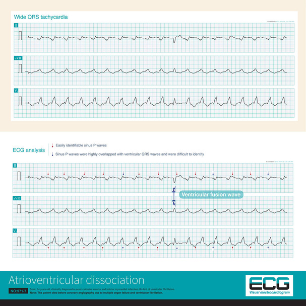
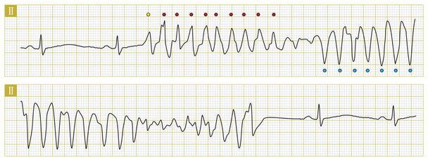
If atrioventricular dissociation and ventricular fusion wave can be found in wide QRS wave tachycardia, the diagnosis of ventricular tachycardia is highly supported.
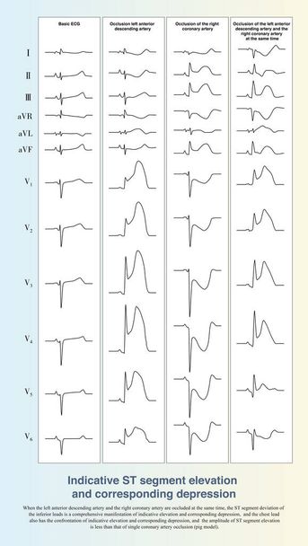
When the two coronary arteries are occluded at the same time, the ST segment elevation of the ischemic myocardium on one side is countered by the corresponding ST segment depression on the other side.

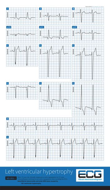
Sometimes, because the QRS axis is in the upper left quadrant, the high-amplitude R wave of left ventricular hypertrophy occurs in the limb leads, and left chest leads is normal.
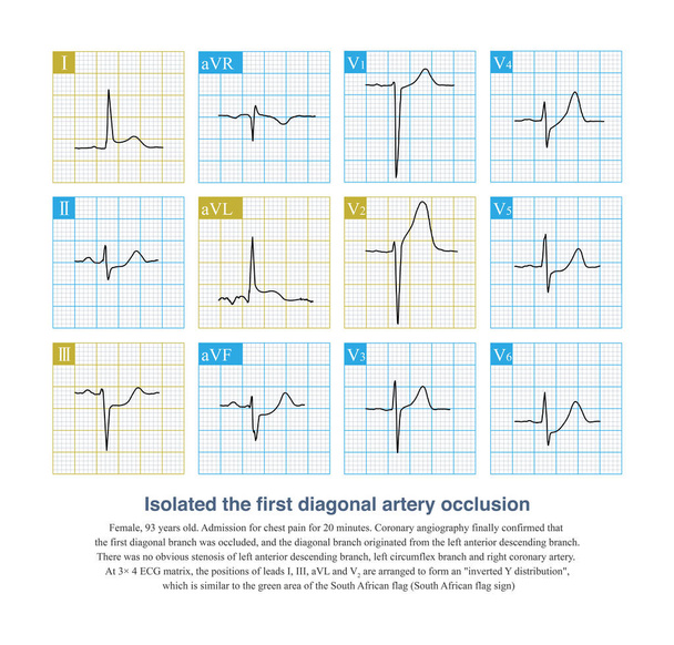
18-When the isolated first diagonal artery is occluded, ECG can show ST segment elevation in leads I, aVL, and V2, and ST segment depression in lead III, and the layout resembles the South African flag.
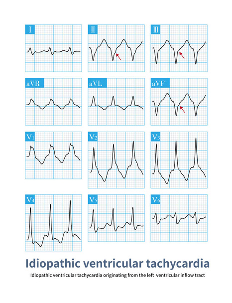
This case of ventricular tachycardia originated from the left ventricular inflow tract, which is a benign idiopathic ventricular tachycardia.
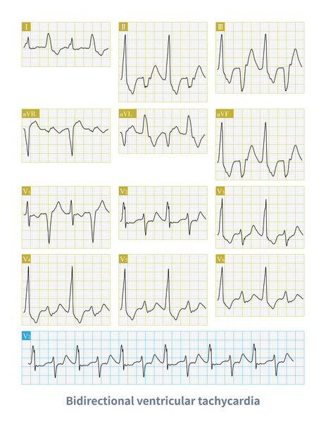
Bidirectional ventricular tachycardia is a kind of malignant arrhythmia. The polarity of QRS main wave alternates from beat to beat, and it is easy to degenerate into ventricular fibrillation.

When acute inferior myocardial infarction occurs, the ST segment offset of the electrocardiogram can be used to derive the culprit vessel, commonly known as the Fiol process.

When the frontal axis is + 19 , the maximum QRS depolarization potential is closest to the axis of lead , so the R-wave amplitude of lead is the highest.

Male, 76 years old, clinically diagnosed with coronary heart disease. ECG showed that sinus rhythm and rapid junctional rhythm alternate, with the latter being the predominant.
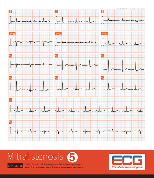
Female, 55 years old, diagnosed with mitral stenosis 5 years ago. When this ECG was taken, the patient still maintained sinus rhythm.Note that the P wave duration was widened.
Female, 65 years old, clinically diagnosed with advanced ovarian cancer. She repeatedly experienced rapid ventricular arrhythmias during her hospitalization and ultimately died.
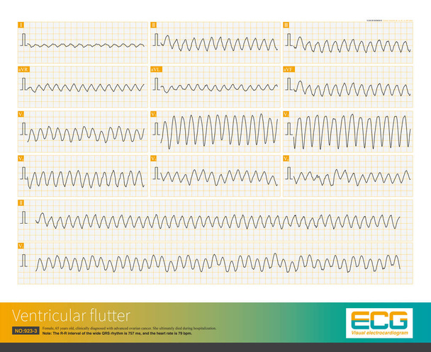
Female, 65 years old, clinically diagnosed with advanced ovarian cancer. She repeatedly experienced rapid ventricular arrhythmias during her hospitalization and ultimately died.

Female, 58 years old, clinically diagnosed with aortic stenosis. Electrocardiogram showed left ventricular hypertrophy and ST-T changes.

A and B are the normal S wave and R wave amplitude; C and D are the S wave and R wave of a case of left ventricular hypertrophy with a significant increase in amplitude.
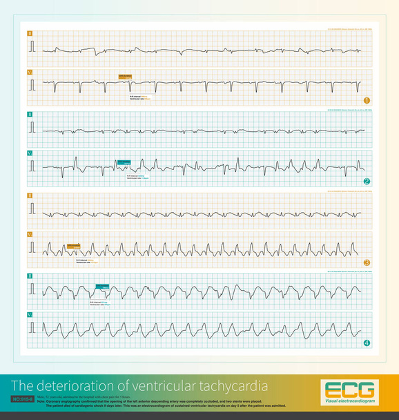
Male, 52 years old, diagnosed with acute extensive anterior myocardial infarction. The patient suffered recurrent ventricular tachycardia, worsened and died of cardiogenic shock.

Male, 84 years old, admitted to hospital with chest pain for 1 day. ECG showed acute inferior and posterior MI and possibly right MI. The patient died of ventricular fibrillation the next day.

QRS complex in lead V1 presents rSR shape with high amplitude R wave.The electrocardiogram of a patient with atrial septal defect showed right ventricular hypertrophy with complete RBBB.
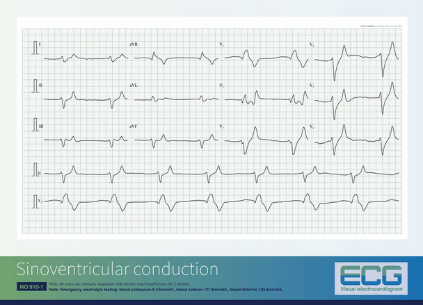
When the blood potassium concentration significantly increases, it can inhibit ventricular conduction, cause diffuse intraventricular block, and generate wide QRS waves and sinoventricular conduction.

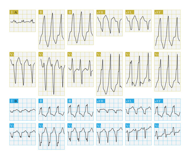
A middle-aged man sought medical attention due to palpitations. The electrocardiogram indicates arrhythmia. Can you correctly diagnose this electrocardiogram?
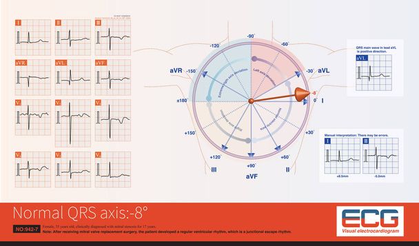
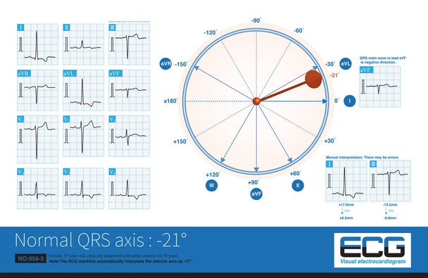
When the frontal QRS axis is located at -8, the maximum ventricular depolarization potential is oriented towards lead I, and the R wave amplitude of lead I is the highest.

When the rhythm of the atria originates in the lower part of the atria, the whole atria are excited from inferior to superior, producing negative P waves in the inferior leads.
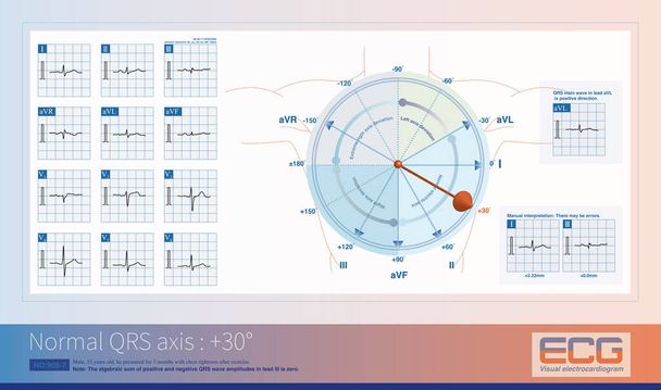
When the frontal QRS axis is+30 and perpendicular to the lead III axis, the algebraic sum of positive and negative QRS wave amplitudes in lead III is zero.
When the opening of left anterior descending artery is occluded, it can cause a large area of anterior myocardial infarction (some patients with inferior wall), and the prognosis is poor.
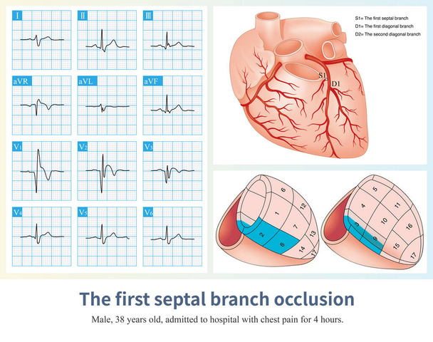
When isolated first septal branch occlusion occurs, it causes acute anteroseptal myocardial infarction and ST segment elevation in leads V1 to V2-V3.
A 69 year old patient with acute anterior and inferior myocardial infarction developed a series of electrocardiograms of ventricular tachycardia, and ultimately died in the hospital.

Cardiac surgery is one of the common causes of iatrogenic atrioventricular block. Once atrial fibrillation exhibits a slow and regular ventricular rhythm, it is important to be alert to 3 AVB.
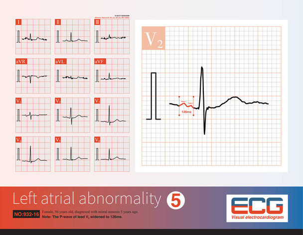
Female, 56 years old, diagnosed with mitral stenosis 5 years ago. When this ECG was taken, the patient still maintained sinus rhythm.Note that the P wave duration was widened.
A 69 year old patient with acute myocardial infarction, whose infarction affected the anterior wall, high lateral wall, and inferior wall, ultimately died during hospitalization.

A 4-year-old boy with a clinical diagnosis of long QT syndrome. No genetic testing was done during hospitalization. The child died suddenly during follow-up.

Only type 1 Brugada phenotype ECGs can directly diagnose Brugada syndrome, an inherited cardiac ion channel disorder associated with risk of sudden death.
Torsades de pointes is a polymorphic ventricular tachycardia that occurs in patients with long QT intervals and can degenerate into ventricular fibrillation.
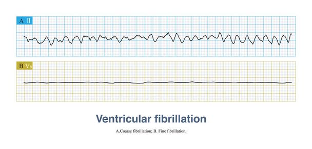
Ventricular fibrillation is a fatal arrhythmia and also a cardiac arrest rhythm. It can be divided into coarse fibrillation and fine fibrillation according to the amplitude of the fibrillation wave.
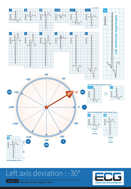
Left ventricular hypertrophy is a common cause of left axis deviation, and on the frontal lead system, the maximum ventricular depolarization potential is facing the upper left quadrant, and the QRS main wave in lead is negative.
When the frontal QRS axis is between 0 and - 30 , it is located at the negative side of the aVF lead axis, so the main QRS wave of the aVF lead is negative.
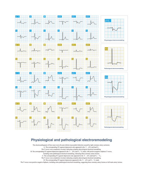
In acute inferior myocardial infarction, physiologically corresponding ST-segment depression is characterized by aVL and V2-V4 leads, complete inversion of T waves.

When the first diagonal branch of the LAD is occluded, the ischemic potential is toward the left, upper and antirior and the ST segment of the corresponding limb leads and chest leads is elevated.
When aortic valve regurgitation occurs, in diastole, blood returns from the aorta to the left ventricle, and the volume of return blood from the left atrium causes left ventricular dilation.
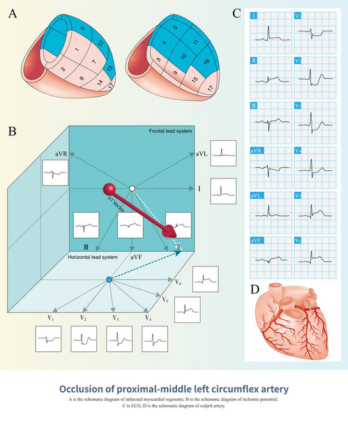
When the proximal and middle segments of the LCA were occluded, the ischemic potential was toward the rear, left and lower, and the corresponding ECG leads showed ST segment elevation.

SQT sign is an electrocardiogram change of right heart disease, which is not only seen in acute pulmonary embolism, but also in other diseases affecting the right heart.
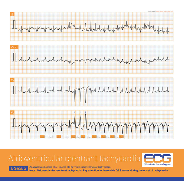
A 1-month-old child developed atrioventricular reentrant tachycardia. At the beginning of the attack of tachycardia, three wide QRS waves appeared successively, which was complete LBBB.
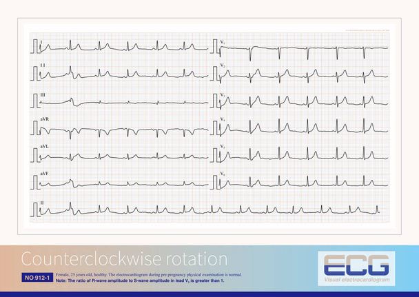
Counterclockwise rotation is a common ECG phenomenon, with an RS amplitude ratio greater than 1 occurring in leads V2 and V3, which is poorly correlated with anatomical transposition.

A 69 year old patient with acute myocardial infarction, whose infarction affected the anterior wall, high lateral wall, and inferior wall, ultimately died during hospitalization.
An 11-month-old boy was clinically diagnosed with cytomegalovirus myocarditis. During hospitalization, the child had repeated episodes of VT and was eventually discharged from the hospital.

Male, 48 years old, clinically diagnosed with dilated cardiomyopathy. Significant high-amplitude R waves in lead V5 of the ECG suggest left ventricular hypertrophy.
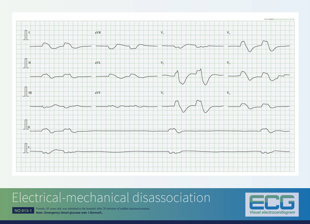
Female, 85 years old, was admitted to the hospital after 20 minutes of sudden unconsciousness. The admission ECG indicated electrical mechanical disassociation, which is a type of cardiac arrest.

Medical and healthcare concept for background. A real chart of an ECG. Electrocardiogram registered on paper. Hearts electrical activity. Selective focus. Close up. Piece of ECG. Free space to write.

Cardiogram. ECG shows the heart beat

Electrocardiogram, ecg background
Red heartbeat icon. Vector illustration. Heartbeat sign horizon banner in flat design. Smooth thick and thin lines, template set

Multicolored EKG Tracings - ICU Monitor
Vital signs portable monitoring equipment in intensive care unit.

Image of a dramatic electrocardiogram in red and orange with heart symbol on peak

Pulse line ilustration vector template red

Electrocardiogram drawn in white on a black background

Man doctor in cardiology telemedicine concept

Heartbeat Medical Cardiogram electrocardiogram pulse graph

Ecg with stethoscope suggesting the periodical monitorization of heart
Purple Computer monitor with cardiogram icon isolated on white background. Monitoring icon. ECG monitor with heart beat hand drawn. Minimalism concept. 3d illustration 3D render
Flatline blip on a medical heart monitor ECG - EKG (electrocardiogram) with white background

Heart Monitor Red Lines

Electrocardiogram, ecg background

Medical monitor isolated on white background

Monitoring patients vital sign in operating room. Cardiogram monitor during surgery in operation room.

ECG Electrocardiography, medical and healthcare background