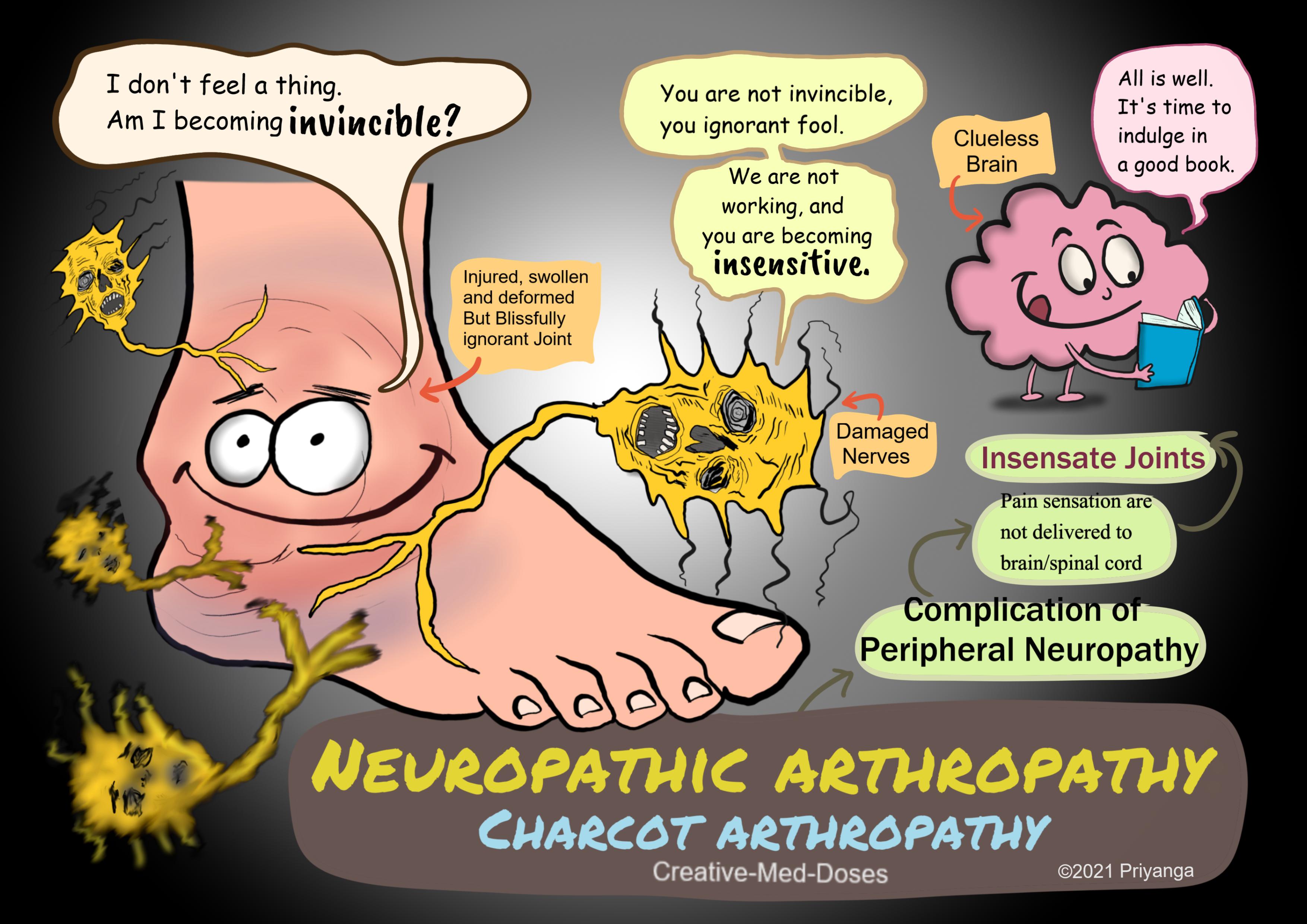
Neuropathic arthropathy: Charcot arthropathy
Neuropathic arthropathy (Charcot arthropathy) is a complication of peripheral neuropathy that results in fractures, dislocations, subluxations. It has an increased risk of progressive deformity of the affected joint. Sometimes, the resulting joint deformity increases the risk of amputation.
Charcot arthropathy is a specific manifestation of peripheral neuropathy, also known as Neuropathic Joint Disease. It is named after Jean-Martin Charcot, who recognized that peripheral neuropathy could lead to neuropathic joints. Any condition resulting in decreased peripheral sensation, proprioception, and fine motor control can cause Charcot arthropathy. There is a progressive degeneration of a weight-bearing joint. The affected joint has bony destruction, bone resorption, and eventual deformity.
...
...
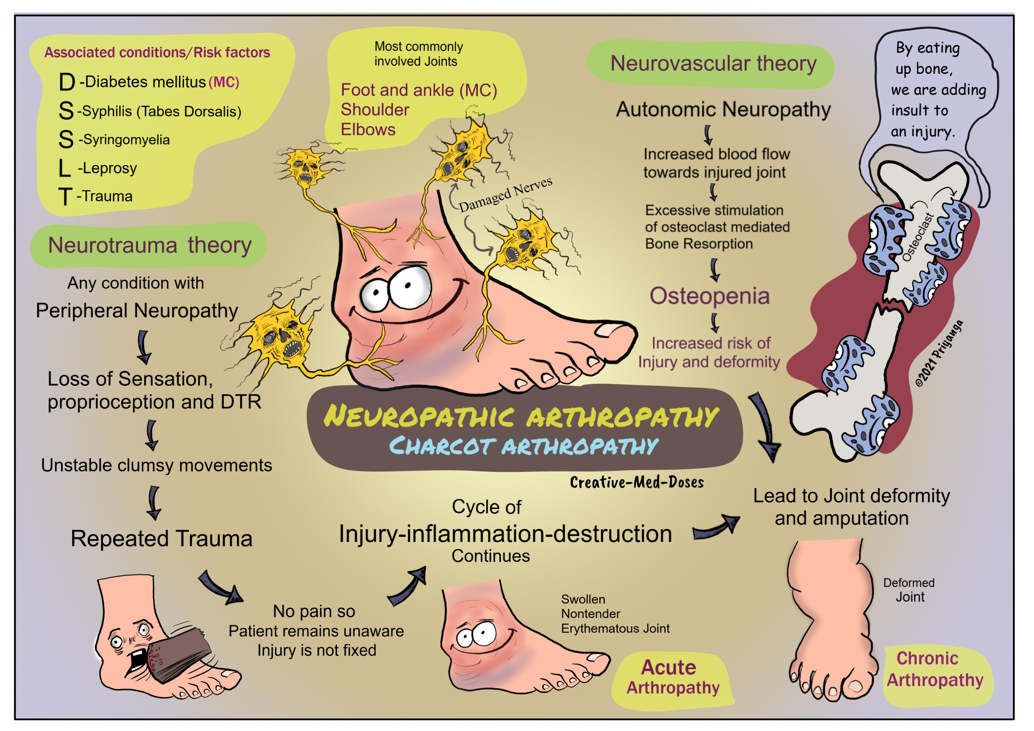
Most common locations
- Foot and ankle (most commonly affected joint)
- Shoulder
- Elbows
Conditions causing neuropathy and Charcot arthropathy
- Diabetes mellitus- foot and ankle are most affected
- Tertiary syphilis (tabes dorsalis) - the knee is the most affected joint
- Syringomyelia – shoulder is the most affected joint
- Trauma
- Leprosy
- Spinal cord tumor
- Subacute combined degeneration (Vit B12 deficiency)
Pathophysiology (underlying mechanisms responsible for the neuropathic joint)
Mechanisms are responsible for joint destruction in these cases are-
Neurotrauma
Peripheral neuropathy → loss of sensation, proprioception, and deep tendon reflexes → imbalanced and clumsy joints with poor fine motor control → repetitive trauma → no pain since joint is insensate → patient remains unaware of injury and doesn’t fix it → prolonged inflammation, destruction and Injury continues → swollen, red and nontender joint in beginning → deformed joint later in course of the disease → may lead to amputation
Neurovascular
Repetitive injury → Joint inflammation → increased blood flow towards the joint by dilating vessels → Autonomic Neuropathy → reduced ability to vasoconstrict → vessels fails to constrict → hyperemia in injured and inflamed joint → stimulates osteoclasts → increased osteoclast-mediated bone resorption→ decreased bone mineral density→ osteopenia→ increased risk of fractures and the deformity
Clinical presentation
Acute arthropathy – patient presents with swollen, erythematous but nontender joint. Physical examination shows loss of sensation and absent deep tendon reflexes (DTR)
Chronic arthropathy- patient presents with deformed joint, bony prominences, and foot ulcers. The most common deformity is a collapse of the tarsometatarsal joint, with valgus angulation.
...
...
Diagnosis
The diagnosis of neuropathic arthritis is based on the clinical features and characteristic radiographic findings in a patient with underlying sensory neuropathy.
Joint fluid (synovial fluid) test
It has a normal white blood cell count and markers of inflammation. In contrast to this, osteomyelitis has increased inflammatory marker levels and increased leucocyte count.
X-ray
bony consolidation with fractures and bone destruction. Chronic cases show bony sclerosis, eburnation, and Bone deformity.
Bone scintigraphy
It is used to distinguish between neuropathic arthropathy from osteomyelitis.
- Negative/cold for neuropathic arthropathy
- Positive /hot for osteomyelitis
Treatment
Acute arthropathy
- Rest and elevation
- Immobilization of affected joint
Chronic arthropathy
- Accommodative shoes
- braces and splints to support unstable joint
- Surgical repair in severe deformity- Fusion of an unstable joint may improve function and reduce pain
- Amputation (only in rare cases)
Case scenario
A 56-year-old man presents to his physician for painless swelling of the ankle. Physical exam reveals a non-tender, swollen, and erythematous ankle joint. The joint also has decreased range of motion. The foot has lost sensation, and the deep tendon reflex is absent. Laboratory studies show a normal leukocyte count, and inflammatory markers are also under normal levels.
All of the following can be associated with his condition except-
A. Diabetes
B. Syringomyelia
C. Syphilis
D. Osteomyelitis
The answer is D. osteomyelitis
Clue 1. Painless swelling
Clue 2. Normal leukocyte count and inflammatory markers
Clue 3. All other options cause neuropathic joint disease except osteomyelitis it causes inflammatory joint disease.
Revision for today Hydronephrosis: Dilated Pelvis and calyx - Creative Med Doses
Buy fun review books here (these are kindle eBook’s you can download kindle on any digital device and log in with Amazon accounts to read them). Have fun and please leave a review.
https://creativemeddoses.com/books/