Some Ventricular Bigeminy Precursors Ventricular Tachycardia Sign Clinical Deterioration Severe — Stock Photo, Image

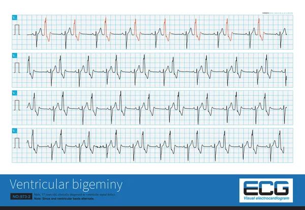
Some ventricular bigeminy are precursors of ventricular tachycardia, a sign of clinical deterioration and severe impairment of cardiac function, and predisposition to ventricular fibrillation and death. — Photo

Some ventricular bigeminy are precursors of ventricular tachycardia, a sign of clinical deterioration and severe impairment of cardiac function, and predisposition to ventricular fibrillation and death.

 — Photo by asia11m

Same Series:

One patient with acute myocardial infarction had three ventricular beats on ECG, which should not be misdiagnosed as common ventricular premature contractions. They were ventricular parasystole.
One Patient Acute Myocardial Infarction Had Three Ventricular Beats Ecg — Stock Photo, Image
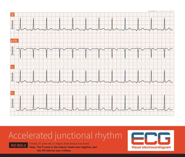
The retrograde P wave of the inferior leads can come from the lower part of the atrium, the lower part of the atrial septum, and the atrioventricular junctional region.
Retrograde Wave Inferior Leads Can Come Lower Part Atrium Lower — Stock Photo, Image
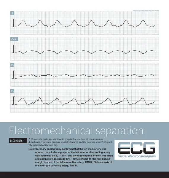
Electromechanical separation is a kind of terminal ECG. The patient's ECG has electrical signals, the ECG wave is widened with morphological abnormalities, and the ventricle has no contraction.
Electromechanical Separation Kind Terminal Ecg Patient Ecg Has Electrical Signals — Stock Photo, Image
A 17 year old male was clinically diagnosed as ventricular septal defect and his ECG was ventricular bigeminy.Sinus and ventricular beats alternated.This is a long V1 lead.
Year Old Male Clinically Diagnosed Ventricular Septal Defect His Ecg — Stock Photo, Image
A 4-year-old male was admitted to hospital with recurrent amaurosis and syncope. Electrocardiogram showed long QT syndrome. Note the widening of the base of the T wave.He was at risk of sudden death.
Year Old Male Admitted Hospital Recurrent Amaurosis Syncope Electrocardiogram Showed — Stock Photo, Image
During acute transmural myocardial ischemia, the repolarization waves of ECG show beat to beat alternating changes, which is an ECG sign of impending malignant ventricular arrhythmia.
Acute Transmural Myocardial Ischemia Repolarization Waves Ecg Show Beat Beat — Stock Photo, Image
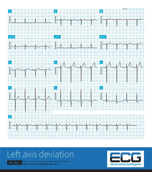
Male, 45 years old, suffering from hypertension. The frontal QRS axis was -38 , which was not enough to diagnose left anterior fascicular block.Lead II QRS main wave is negative.
Male Years Old Suffering Hypertension Frontal Qrs Axis Which Enough — Stock Photo, Image
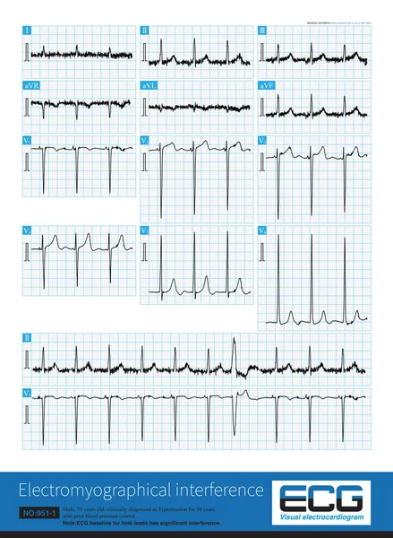
When the baseline of ECG is interfered by its own or external electrical signal, it will affect the shape and measured value of ECG wave, especially ST segment offset amplitude.
Baseline Ecg Interfered Its Own External Electrical Signal Affect Shape — Stock Photo, Image
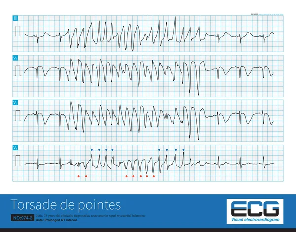
Male, 75 years old, clinically diagnosed as acute anterior septal myocardial infarction, prolonged QT interval, complicated with torsade de pointes.
Male Years Old Clinically Diagnosed Acute Anterior Septal Myocardial Infarction — Stock Photo, Image
Male, 75 years old, clinically diagnosed as acute anterior septal myocardial infarction.It is best to use 12 leads to diagnose torsade de pointes because some leads may not have torsade characteristic
Male Years Old Clinically Diagnosed Acute Anterior Septal Myocardial Infarction — Stock Photo, Image
Acute myocardial ischemia can lead to secondary long QT syndrome, R on T ventricular premature contraction and torsade de pointes.
Acute Myocardial Ischemia Can Lead Secondary Long Syndrome Ventricular Premature — Stock Photo, Image
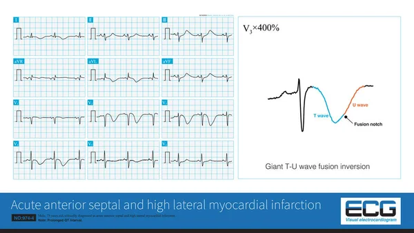
Male, 75 years old, clinically diagnosed as acute anterior septal and high lateral myocardial infarction.The 12 lead ECG showed a large inversion of T wave with prolonged QT interval.
Male Years Old Clinically Diagnosed Acute Anterior Septal High Lateral — Stock Photo, Image
When the frequency of sinus impulses is less than 60 times per minute, it is called sinus bradycardia. Sinus bradycardia is a common bradyarrhythmia.
Frequency Sinus Impulses Less Times Minute Called Sinus Bradycardia Sinus — Stock Photo, Image
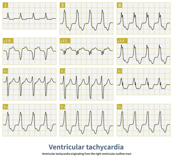
Ventricular tachycardia originating from the right ventricular outflow tract is a benign ventricular tachycardia.It usually does not cause sudden death.Radiofrequency ablation can cure this arrhythmia.
Ventricular Tachycardia Originating Right Ventricular Outflow Tract Benign Ventricular Tachycardia — Stock Photo, Image
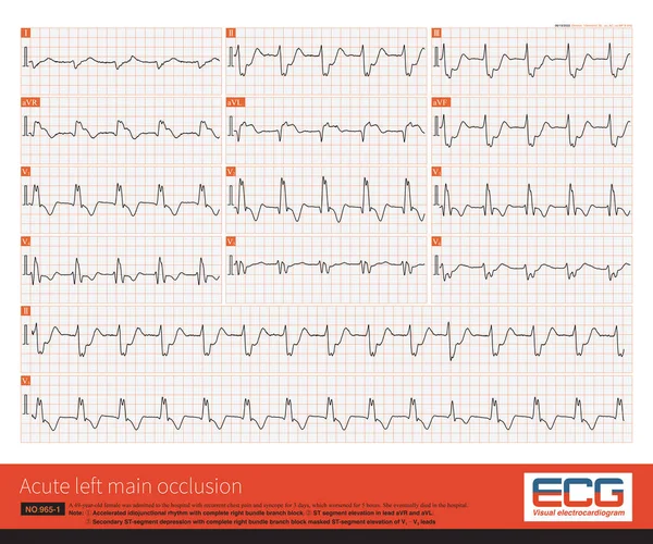
The coronary angiography showed complete occlusion of the left main artery, TIMI 0 and no stenosis in the right coronary artery with TIMI 3, and a lateral circulation to left coronary artery.
Coronary Angiography Showed Complete Occlusion Left Main Artery Timi Stenosis — Stock Photo, Image

Usage Information

You can use this royalty-free photo "Some ventricular bigeminy are precursors of ventricular tachycardia, a sign of clinical deterioration and severe impairment of cardiac function, and predisposition to ventricular fibrillation and death." for personal and commercial purposes according to the Standard or Extended License. The Standard License covers most use cases, including advertising, UI designs, and product packaging, and allows up to 500,000 print copies. The Extended License permits all use cases under the Standard License with unlimited print rights and allows you to use the downloaded stock images for merchandise, product resale, or free distribution.

You can buy this stock photo and download it in high resolution up to 10000x6107. Upload Date: Feb 24, 2023