Tag Archives: Neodermata

Friday Fellow: Common Horse Tapeworm

by Piter Kehoma Boll

Flatworms are the fourth largest animal phylum after arthropods, mollusks and chordates and most species known to date belong to the clade Neodermata, which includes parasitic species such as flukes and tapeworms, some of which infect humans. Among tapeworms, the species that infect humans and belong to the genus Taenia are certainly the most popular, but it is expected that all vertebrates can have at least one tapeworm parasite, so that it is only a matter of time and opportunity for us to discover them all.

Among the species known to parasitize horses, the most widespread is Anoplocephala perfoliata, which I decided to call the common horse tapeworm. As tapeworms in general, the adult common horse tapeworm lives in the intestine of its definitive host, in this case a horse.

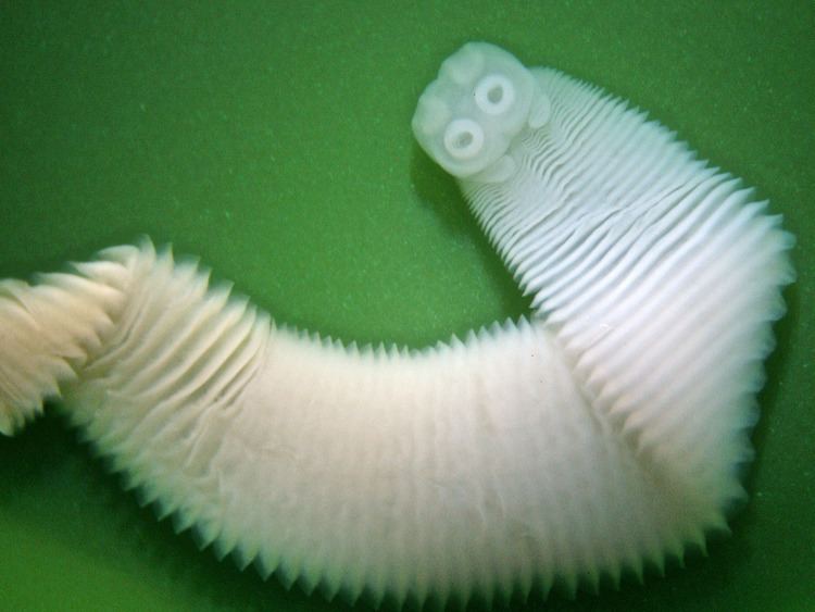
Different from species of Taenia, which can grow up to several meters in length, species of Anoplocephala are much smaller. The whole body of adult specimens measures about 5 to 8 cm in length and 1 to 2 cm in width and is divided into the same parts seen in other tapeworms. The anteriormost part of the body includes the scolex, which has 4 large suckers. Although the scolex of most tapeworms measures less than 1 mm, in the common horse tapeworm it reaches up to 3 mm.

A preserved specimen. Photo extracted from alchetron.com

After the scolex there is a small neck of undifferentiated tissue that grows constantly to build new proglottids, which form the rest of the body. Proglottids are connected to each other in a chain fashion and the posteriormost ones are continuouly lost and released into the environment. Each proglotid contains male and female sexual organs and is released when it contains mature eggs.

Mature proglottids are released in the environment through the horse’s feces and release their eggs on the ground and the vegetation. The eggs can survive outside a host for as long as 9 months. During this time, they hope to be accidentally ingested by oribatid mites that live in pastures. If this happens, the egg hatches inside the mite due to the mechanical action of the mite’s mouth parts and releases the first-stage larva called the oncosphere.

An egg of Anoplocephala perfoliata. The small 20-µm-diamter sphere is the oncosphere waiting to be released. Photo by Martin K. Nielsen, extracted from msdvetmanual.com

When the oncosphere reaches the mite’s gut, it is activated, probably via ions present in this environment, and uses a group of hooks to penetrate the mite’s tissues. After about 8 to 20 weeks, the oncosphere develops into a cysticercoid. This stage looks like an inverted miniaturized tapeworm inside a bladder-like vesicle, having already a protoscolex inside it.

While horses are grazing, they always ingest some invertebrates together with the grass. It they happen to ingest an infected mite, the cysticercoids are released during digestion, evert the protoscolex and attach to the intestinal walls of the horse. There, the tapeworm develops into an adult, restarting the cycle.

Attached to the intestine of their hosts, tapeworms do not feed on blood or other tissues as many parasites do. Instead of that, they collect nutrients directly from the host’s gut by absorbing them via the worm’s body surface.

For a long time its has been thought that the common horse tapeworm was a harmless parasite since most horses did not seem to have any symptom and the tapeworms were often only discovered during dissection after the horse’s death by other causes. The preferred area for the common horse tapeworm to attach is the caecum and the ileocaecal junction but in heavily infected animals some individuals can be found in suboptimal sites throughout the small and large intestines. In such heavily infected horses, the tapeworms can cause colics and even intestinal obstruction.

Large number of adult tapeworms in a heavily infected horse. Credits to Tomczuk et al. (2014).*

The common horse tapeworm can infect other equids as well, such as donkeys and zebras. Ironically, domesticated horses seem to be the most infected individuals exactly because horse owners treat them with anthelmintics. Most modern anthelmintics do not affect tapeworms and only remove other parasites, such as roundworms, which reduces competition and allows tapeworms to thrive.

– – –

More parasitic flatworms:

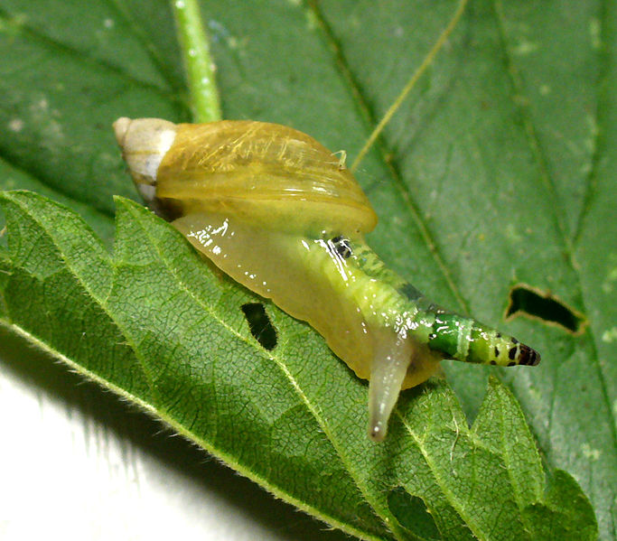
Friday Fellow: Green-banded broodsac (on 09 March, 2018)

Friday Fellow: Salmon Fluke (on 11 January, 2019)

– – –

Like us on Facebook!

Follow us on Twitter!

– – –

References:

Gasser RB, Williamson RMC, Beveridge I (2005) Anoplocephala perfoliata of horses – significant scope for further research, improved diagnosis and control. Parasitology 131(1): 1–13. https://doi.org/10.1017/S0031182004007127

Tomczuk K, Kostro K, Szczepaniak KO, Grzybek M, Studzińska M, Demkowska-Kutrzepa M, Roczeń-Karczmarz M (2014) Comparison of the sensitivity of coprological methods in detecting Anoplocephala perfoliata invasions. Parasitology Research 113(6): 2401–2406. doi: 10.1007/s00436-014-3919-4

Wikipedia. Anoplocephala perfoliata. Available at < https://en.wikipedia.org/wiki/Anoplocephala_perfoliata >. Access on 11 June 2020.

– – –

*Creative Commons License This work is licensed under a Creative Commons Attribution 4.0 International License.

1 Comment

Filed under flatworms, Friday Fellow, Parasites, worms

Friday Fellow: Salmon Fluke

by Piter Kehoma Boll

Leia em Português

Everybody knows salmons, especially the Atlantic salmon, Salmo salar, and many of us love to eat this fish species as well. However, I’m not here to talk about the Atlantic salmon itself, but to talk about one of its closes companions and antagonists, the salmon fluke.

Scientifically known as Gyrodactylus salaris, the salmon fluke is a flatworm of the clade Monogenea, a group of ectoparasites that infect mainly fish. As its name suggests, the salmon fluke infects salmons, such as the Atlantic salmon, and closely related species, such as the rainbow trout Onchorhynchus mykiss.

Several salmon flukes on a host. Photo by Tora Bardal. Extracted from https://www.drivaregionen.no/no/Gyrodactylus-salaris/

The salmon fluke was first discovered in 1952 in salmons from a Baltic population that were kept in a Swedish laboratory. Measuring about 0.5 mm in length, the salmon fluke attaches to the skin of the fish and is too small to be seen with the naked eye. The attachment happens using a specialized organ full of tiny hooks, called haptor, located at the posterior end of the body. When feeding, the salmon fluke attaches its mouth to the surface of the fish using special glands in its head and everts its pharynx through the mouth, releasing digestive enzymes on the fish, dissolving its skin, which is then ingested. The wounds caused by the parasite’s feeding activity can lead to secondary infections that can seriously affect the salmon’s health.

Artificially colored SEM micrograph of five specimens of Gyrodactylus salaris. Credits to Jannicke Wiik Nielsen. Extracted from https://www.vetinst.no/nyheter/kan-gyrodactylus-salaris-utryddes-i-drammensregionen

Different from most parasitic flatworms, monogeneans such as the salmon fluke have a single host. During reproduction, the hermanophrodite adults release a ciliated larva called oncomiracidium that infects new fish. A single fluke can originate an entire population because it is able to self fertilize.

During the 1970’s, a massive infection by the salmon fluke occurred in Norway following the introduction of infected salmon strains. This led to a catastrophic decrease in the salmon populations in the country, affecting many rivers. Due to this evident threat to such a commercially important species, several techniques have been developed to control and kill the parasite. The first developed methods included the use of pesticides in the rivers, but those ended up having a negative effect on many species, including the salmons themselves. Currently, newer and less aggressive methods have been used.

– – –

Like us on Facebook!

Follow us on Twitter!

– – –

References:

Jansen, P. A., & Bakke, T. A. (1991). Temperature-dependent reproduction and survival of Gyrodactylus salaris Malmberg, 1957 (Platyhelminthes: Monogenea) on Atlantic salmon (Salmo salar L.). Parasitology, 102(01), 105. doi:10.1017/s0031182000060406

Johnsen, B. O., & Jenser, A. J. (1991). The Gyrodactylus story in Norway. Aquaculture, 98(1-3), 289–302. doi:10.1016/0044-8486(91)90393-l

Meinilä, M., Kuusela, J., Ziętara, M. S., & Lumme, J. (2004). Initial steps of speciation by geographic isolation and host switch in salmonid pathogen Gyrodactylus salaris (Monogenea: Gyrodactylidae). International Journal for Parasitology, 34(4), 515–526. doi:10.1016/j.ijpara.2003.12.002

Wikipedia. Gyrodactylus salaris. Available at < https://en.wikipedia.org/wiki/Gyrodactylus_salaris >. Access on December 26, 2018.

2 Comments

Filed under flatworms, Friday Fellow, Parasites, Zoology

Friday Fellow: Green-Banded Broodsac

by Piter Kehoma Boll

Parasites are very speciose, and I often feel that I’m not giving enough space for them here, especially when I bring you a flatworm, which is likely the group with the largest number of parasite species. So let’s talk about one today at last.

The first parasitic flatworm I am introducing to you is Leucochloridium paradoxum, the green-banded broodsac. It is a member of the flatworm group Trematoda, commonly known as flukes and, as all flukes, it has a complex life cycle.

Adults of the green-banded broodsac live in the intestine of various passerine birds of North America and Europe. The eggs they lay reach the environment through the bird’s feces and are eventually ingested by land snails of the genus Succinea.

145

Adult individual of Leucochloridium paradoxum (left), an infected intermediate hose, a sail of the genus Succinea (center) and the sporocysts along the snail’s internal organs (right). Images not to scale. Extracted from http://medbiol.ru/medbiol/dog/0011a975.htm

Adults of Leucochloridium paradoxum are very similar to the adults of other species of the genus Leucochloridium, the main differences being seen in the larval stages. Inside the body of the snail, the eggs hatch into the first larval stage, the miracidium, which inside the snail’s digestive system develops into the next stage, the sporocyst.

The sporocyst has the form of a long and swollen sac (the broodsac, hence the common name) that is filled with many cercariae, which are the next larval stage. The sporocyst than migrates towards the snail’s eye tentacles, invading them and turning them into a swollen, colorful and pulsating structure that resembles a caterpillar. In this stage of infection, the poor snail is most likely blind and cannot avoid light as it normally does. As a result, it becomes exposed to birds that mistake it for a juicy caterpillar, eating it eagerly.

683px-succinea_mit_leucocholoridium

A poor snail of the species Succinea putris with a broodsac in its left eye stalk. There is only one terrible fate for this creature. Photo by Thomas Hahmann.*

When the snail is eaten, the sporocyst burst and the several cercariae are released. In the bird’s intestine, they develop into adults and restart the nightmarish cycle.

– – –

Like us on Facebook!

Follow us on Twitter!

– – –

References:

Rząd, I.; Hofsoe, R.; Panicz, R.; Nowakowski, J. K. (2014) Morphological and molecular characterization of adult worms of Leucochloridium paradoxum Carus, 1835 and L. perturbatum Pojmańska, 1969 (Digenea: Leucochloridiidae) from the great tit, Parus major L., 1758 and similarity with the sporocyst stages. Journal of Helminthology 88(4): 506-510. DOI: 10.1017/S0022149X13000291

Wikipedia. Leucochloridium paradoxum. Available at < https://en.wikipedia.org/wiki/Leucochloridium_paradoxum >. Access on March 8, 2018.

– – –

*Creative Commons License
This work is licensed under a Creative Commons Attribution-ShareAlike 3.0 Unported License.

1 Comment

Filed under flatworms, Friday Fellow, Parasites, worms, Zoology