History
A patient's presenting symptoms are determined by the site of initial and subsequent infection and by his or her immune status. Factors favoring primary cutaneous infection include a solitary lesion, lesions on uncovered parts, a history of primary inoculation, and regional lymph node involvement. Factors favoring secondary cutaneous infection include the presence of systemic involvement, lesions on covered parts, multicentric skin involvement, and deep dermal or subcutaneous lesions. Cryptococcal meningitis with skin involvement in a 20-year-old woman has been reported and indicates that immunosuppression is not necessary for cryptococcal infection to occur and that skin manifestations can give clues to meningitis. [19]
Several reports in 2011 have noted that C gattii can affect immunocompetent hosts, so this must be considered when examining otherwise-well patients. [20] C gattii type VGII resulted in primary infection after trauma in one patient. [21]
C laurentii in a renal transplant recipient resulted in primary cutaneous cryptococcosis. [22] An immunosuppressed psoriatic patient with palmopustular involvement experienced generalized Cryptococcus albidus infection manifesting as generalized hemorrhagic plaques. [23]
Primary cutaneous infection findings are as follows:
-
History of trauma and direct inoculation
-
Most often occurs in immunosuppressed patients but may also occur in immunocompetent patients
-
Single lesion develops at the site of infection
-
Solitary nodules that may ulcerate
-
Cellulitis
-
Ulcers
-
Abscesses
-
Sporotrichoid-pattern, capsule-deficient cutaneous cryptococcosis [26]
Disseminated cutaneous Cryptococcus infection findings are as follows:
-
Occurs in 10-15% of patients with disseminated infection
-
Cutaneous signs possibly preceding other symptoms of disseminated disease by 2-8 months
-
Lesions mostly found on head and neck
-
First present as painless papules or pustules, which then become nodules that may ulcerate (eg, pustular eruption in a cardiac transplant patient with cryptococcal meningitis and headache [27] )
-
Numerous other cutaneous manifestations (eg, acneiform papules or pustules, warty or vegetating crusted plaques and ulcers, hard infiltrated plaques or nodules, subcutaneous swellings, abscesses, blisters, tumorlike masses, eczematous plaques, granulomas, vasculitic lesions resembling palpable purpura [especially in transplantation patients])
Primary pulmonary infection findings are as follows:
-
May be limited to fever, cough, and pleuritic chest pain
-
Can progress to acute respiratory distress syndrome
CNS Cryptococcus infection findings are as follows:
-
Nuchal rigidity
-
Headache
-
Vertigo
-
Nausea and vomiting
-
Changes in consciousness
-
Mental changes
-
Nerve palsies
Immunosuppressive medications are as follows:
-
Treatment with methotrexate and adalimumab [28]
Other possible developments are as follows:
-
Hepatitis
-
Osteomyelitis
-
Prostatitis
-
Pyelonephritis
-
Peritonitis
Physical Examination
Primary cutaneous Cryptococcus infection findings
The patient may have solitary nodules that break down and ulcerate (eg, facial ulceration [31] ).
Cutaneous Cryptococcus infection has been reported to mimic leishmaniasis (ulcerated nodule on the face). [8]
Other signs include local lymphadenopathy, cellulitis, confluent erythematous plaques, ulcerated tumor mass, and abscesses. [32, 33, 34, 35]
Disseminated cutaneous Cryptococcus infection findings
Numerous manifestations may be seen, but seldom are they pathognomonic.
Lesions are most often found on the head, neck, mouth, and nose.
Xanthomalike lesions (crusted yellowish-brown plaques, which were accompanied by smaller papules with telangiectasia) histologically and clinically have been noted. [36]
The most common manifestations are molluscum contagiosum–like umbilicated papules of varying sizes (approximately 50% of patients with HIV infection), as seen in the image below, which may have a gelatinous quality and develop a central hemorrhagic crust.
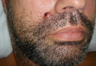 Umbilicated papule of cutaneous Cryptococcus infection on the face of a male.
Cellulitis is seen most commonly in transplantation patients. In one report, 72% of organ transplantation patients with cutaneous Cryptococcus infection presented with cellulitis. Of these, 94% had cellulitis on the extremities. A particularly high rate was found among patients treated with tacrolimus. Primary cryptococcal cellulitis caused by C neoformans var gattii has also been reported in an immunocompetent host.
Patients may have acneiform papules or pustules and/or warty or vegetating crusted plaques and ulcers.
Other signs include hard infiltrated plaques or nodules, subcutaneous swellings, abscesses, blisters, tumorlike masses, eczematous plaques, granulomas, and/or vasculitic lesions resembling palpable purpura (especially in transplantation patients).
Disseminated cryptococcosis initially presenting as cellulitis in a rheumatoid arthritis patient was noted by Lu et al. [37] Cryptococcal cellulitis should be suspected in any immunosuppressed patient with cellulitis that is unresponsive to antibiotics.
Hoang and Burruss [38] described localized cutaneous Cryptococcus albidus infection in a 14-year-old boy on etanercept therapy.
Dilhuydy et al [39] reported on a patient who had cutaneous cryptococcosis with alemtuzumab; this patient was being treated for chronic lymphocytic leukemia.
Van Grieken et al [40] described a case of primary cryptococcal cellulitis in a lung transplant recipient.
Cutaneous cryptococcosis can resemble molluscum. [41]
Complications
In a person with AIDs, cutaneous plasmablastic lymphoma with disseminated cryptococcosis has been reported, [42] stressing that persons with HIV cryptococcal disease can have other significant pathology.
-
Umbilicated papule of cutaneous Cryptococcus infection on the face of a male.


