Approach Considerations
Treatment of Buruli ulcers relies on timely and accurate diagnoses. When treated early, antibiotics alone are adequate. If treatment is delayed, surgical debridement, skin grafts, extensive wound care, and physical therapy may be needed to attenuate debilitating sequelae.
Medical Care
Antibiotics
Since 2004, the medical management of Buruli ulcers has become an active area of research. Through the use of the mouse footpad model developed by Fenner, rifampicin, rifabutin, amikacin, and streptomycin have demonstrated bactericidal activity and azithromycin, clarithromycin, and moxifloxacin to have bacteriostatic activity. [55, 56] Antibiotics not only destroy or inhibit the causative mycobacteria, they reverse the immunosuppression of the mycolactone. [43, 55] Antibiotics have reduced the rate of infection recurrence and reduced the need for surgical intervention. Unfortunately, the antibiotic treatment courses are prolonged and may lead to permanent sequelae.
In 2004, the WHO recommended a treatment protocol that divided lesions into three categories (see Table 2 below). The WHO recommended that all categories receive an eight-week course of rifampicin (10 mg/kg once daily) and streptomycin (15 mg/kg once daily) as the standard of care. [57] Recurrence rates after antibiotic treatment are reported to be 2-3%. [10, 58, 59] This regimen has been reported to heal lesions without requiring surgery in 47-95% of patients. [58, 60] The median healing times for category I, II, and III were 8, 10, and 20 weeks, respectively. [60]
Table 2. Categories of Treatment [57] (Open Table in a new window)
Category |
Form of Disease |
Treatment |
Primary Aim |
Secondary Aim |
Level of Health Care System |
Diagnosis |
I |
Small, early lesion (eg, nodules, papules, plaques, ulcers < 5 cm in diameter) |
Complete antibiotics If at or near a joint, maintain same movement as on unaffected side If surgery is needed in noncritical areas, consider this after 8 weeks of antibiotic treatment |
Cure without surgery Cure without movement limitations |
Reduce or prevent recurrence |
Community health centers and district hospitals |
Strong clinical diagnosis (with or without laboratory confirmation) |
II |
Nonulcerative and ulcerative plaque and edematous forms Single, large ulcerative lesion 5-15 cm in diameter |
Complete antibiotics, before surgery (if possible) If at or near a joint, maintain same movement as on unaffected side |
Cure without surgery Reduce extent of the surgical debridement when needed Cure without movement limitations |
Reduce or prevent recurrence |
Health centers, district and tertiary hospitals |
Strong clinical diagnosis (with or without laboratory confirmation) |
III |
Lesions in the head and neck region, particularly the face Disseminated/mixed forms (eg, osteitis, osteomyelitis, joint involvement) Multiple lesions and osteomyelitis Extensive lesion >15 cm |
Complete antibiotics, before surgery (if possible) If at or near a joint, maintain same movement as on unaffected side |
Cure without surgery Cure without movement limitations |
Reduce or prevent recurrence |
District and tertiary hospitals |
Strong clinical diagnosis (with or without laboratory confirmation) |
Streptomycin use is limited by patient compliance, as it has poor bioavailability and requires daily intramuscular administration. Additionally, it has ototoxic, nephrotoxic, and neurotoxic effects, which may be permanent. It is pregnancy risk factor category D and may cause bilateral congenital deafness by crossing the placenta. [61] Therefore, alternative antibiotics are currently being researched.
Nienhuis et al compared the efficacy of two antibiotic regimens for Mycobacterium ulcerans infection. In Ghana, patients aged 5 years or older were randomly assigned to receive streptomycin (15 mg/kg IM daily) plus rifampicin (10 mg/kg PO daily) for 8 weeks (n = 76) or streptomycin and rifampin for 4 weeks followed by rifampin and clarithromycin (7.5 mg/kg PO daily) for 4 weeks (n = 75). No significant difference was observed for each treatment regimen (healed lesions at 1 y were 96% for 8-wk streptomycin vs 91% for 4-wk streptomycin); however, the number of streptomycin injections was able to be reduced by switching to oral clarithromycin after 4 weeks. [62]
In 2007, the Australian Victorian Department of Human Services recommended the combination of rifampicin and clarithromycin or ciprofloxacin or moxifloxacin for 3 months. [63] It has proven effective in small case series. [51] . Friedman et al demonstrated a successful treatment in 42 of 43 patients using rifampin (10 mg/kg PO daily) combined with either ciprofloxacin (500 mg twice daily) or clarithromycin (500 mg twice daily). The majority of these patients had WHO category I lesions. [64] In severe disease, oral rifampicin with intravenous amikacin is the treatment of choice.
Converse et al, in 2015, demonstrated on the mouse footpad model that rifampin plus clofazimine has potential as a continuation-phase regimen for treatment of M ulcerans infection. [65]
While further research is needed to determine the optimal duration of treatment of an all-oral antibiotic regimen, the existing research has led the WHO to summarize its stance on treatment as follows [57] :
"In summary, there is now overwhelming evidence that 8 weeks of streptomycin–rifampicin or 4 weeks of rifampicin–streptomycin followed by 4 weeks of rifampicin–clarithromycin or 8 weeks of other oral regimens all achieve recurrence-free healing with an acceptable level of side-effects. This is true for ulcers of all sizes, even without additional surgery to remove necrosis or skin grafting to accelerate healing."
While awaiting further confirmation of efficacy, the WHO has stated that an alternative regimen based on vast clinical practice is as follows [57] :
-
Rifampicin (10 mg/kg PO daily) for 8 weeks and clarithromycin (7.5 mg/kg PO twice daily) for 8 weeks OR
-
Rifampicin (10 mg/kg PO daily) for 8 weeks and moxifloxacin (400 mg PO daily) for 8 weeks (for adults only)
Wound care
While traditionally Buruli ulcers are thought to be painless as a result of the neurotoxic effects of mycolactone, many patients experience pain during wound care, which must be addressed. Currently, the only dressing used for wound care is gauze, which results in pain and bleeding when removed. More research needs to be performed to determine the best dressings to be used for these patients. [66] Additionally, the availability of clean water and good hygiene are important for the management of Buruli ulcers and prevention of secondary infections.
Other supportive treatments
Other treatment modalities are currently being explored.
Given the sensitivity of M ulcerans to temperatures greater than 37°C, hyperthermia with a 40°C water bath, such as a circulating water jacket, and local heat application devices have shown some success. [67, 68, 17]
Hyperbaric oxygen also has been reported as effective in a small number of patients. [69]
Ozone therapy is being explored after initially encouraging results. [70]
Negative-pressure wound therapy (NPWT) has also been shown to be effective. [71]
One report suggested that acid-oxidizing solutions can help kill M ulcerans and possibly can be used in support of antibiotic therapy, as well as to prevent cross-contamination. [72]
Surgical Care
Prior to 2004, excision was the treatment of choice for Buruli ulcers. Most patients required multiple staged surgeries and extensive skin grafts, which resulted in prolonged hospitalizations, averaging around three months. [57] Recurrence rates after surgery alone were 16-28%. [57] With the use of antibiotics, 40% of patients do not require surgery. In conjunction with antibiotics, surgery is used to remove devitalized tissue, cover open wounds with skin grafts, and correct or minimize deformities. [73] Note the images below.
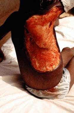 An edematous Buruli ulcer in a 9-year-old Togolese girl. Courtesy of Wayne M Meyers, MD.
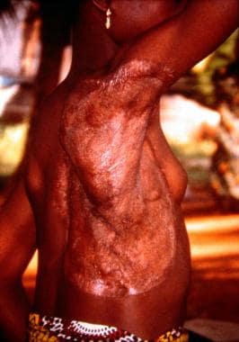 Photo of Togolese girl taken 5 years after the Buruli ulcer had been excised and repaired with autologous split-skin graft by GB Priuli, MD. Courtesy of Wayne M Meyers, MD.
For those who refuse or cannot tolerate antibiotics, surgeries can have excellent cure rates in certain cases. Small subcutaneous nodules or small ulcerations younger than 6 months and smaller than 10 cm in diameter may be excised en bloc with primary closure. Risk factors for recurrence in surgery-alone patients include immunosuppression, positive histologic margins for inflammation or infection, patients older than 60 years, and clinical symptoms present longer than 75 days. [39] Use of PCR to evaluate surgical margins may reduce recurrences. [74]
Complications
Scarring, contractures, and lymphedema may result after healing of Buruli ulcers. Scarring and contractures can have significant social, psychological, physical, and economic impact on patients. Osteomyelitis, metastatic lesions, and secondary infections are additional potential complications. Squamous cell carcinomas have been reported in Buruli ulcers. [75]
Debilitation
The most common complication from Buruli ulcers is physical disability. In a 2015 study from Togo, of 199 patients with confirmed cases of M ulcerans infection, 109 patients (84.5%) healed completely without any complications, 5 patients (3.9%) had secondary lesions, and 15 patients (11.6%) had functional limitations. [76] Risks factors for complications included edema, ulcers larger than 15 cm, healing times longer than 180 days, and a limitation of movement at the time of discharge. [76] A study from Benin in 2014 showed that 55.6% of patients who present with M ulcerans osteomyelitis had long-term crippling sequelae. [77]
Paradoxical reactions
Antibiotic treatment leads to a reversal of the immune suppression, which can lead to a brisk inflammatory response and release mycobacterial antigens from dead organisms. Clinically, this is apparent as clinical deterioration of lesions after initial improvement on antibiotics or the appearance of new lesions. This phenomenon, known as a paradoxical reaction, was first described in Buruli ulcers by O'Brien et al. [78] A similar process has been well documented in Mycobacterium tuberculosis infection, Mycobacterium leprae infection, and HIV infection among patients who are undergoing antiviral treatment.
This immune response can be misinterpreted as treatment failure or secondary infection and lead to unnecessary medical or surgical intervention if a paradoxical reaction is not considered. Paradoxical reactions occur in 9-23% of patients treated with antibiotics. [64, 39, 79, 80] Most occur within 3-10 weeks of initiating antibiotic treatment, but they can occur anywhere from the first week of treatment to 6 months after antibiotic treatment has been completed. Paradoxical reactions may mimic treatment failure, and they do not necessitate restarting antibiotics.
Patients who are at an increased risk of paradoxical reactions include those with an edematous lesion, patients treated with amikacin, patients with polymorphisms in the SLC11A1 gene, larger ulcers, or ulcers located on the trunk. [79, 81]
If a paradoxical reaction is suspected, a specimen should be sent for histopathological examination and culture. It should be cautioned that PCR and AFB stains can be positive because of the detection of nonviable M ulcerans.
Treatment strategies include clinical observation, needle aspiration of fluctuant lesions, minimal debridement if necessary, and adjunctive corticosteroid administration to settle inflammation in severe reactions. Prednisone may be used at a dose of 0.5-1 mg/kg for 2-3 weeks with gradual tapering for a course of 4-8 weeks. [80] Prolonging the antibiotic course to 12 weeks can be considered.
Coinfections
Secondary infections are common with Buruli ulcers, as clean water sources for wound care are frequently limited. A staggering 23% of patients diagnosed with Buruli ulcers in Ghana between August 2010 and December 2012 were coinfected with Mansonella perstans nematodes. [82] Proper wound care hygiene should be emphasized with these patients.
In coendemic countries, M ulcerans infections in HIV-positive patients lead to rapidly spreading osteomyelitis. Additionally, a severe paradoxical reaction may occur after starting antibiotic treatment for Buruli ulcers combined with antiretroviral therapy. Wanda et al reported successful treatment of a severe paradoxical reaction in a patient with HIV and M ulcerans coinfection. [83]
Aminoglycoside toxicity
As discussed in Treatment/Medical Care, prolonged streptomycin use is limited by systemic toxicity. In a long-term follow-up study from the BURULICO Drug Trial, Klis et al found that ototoxicity was present in 29% of adults and 25% of children, especially in the high-frequency range. In contrast, nephrotoxicity that had been detected in 14% of adults and in 13% of children during treatment was present in only 2.4% of patients at long-term follow-up. [84] Further research needs to be done to find safer alternatives for long-term treatments. In the meantime, streptomycin should be given with caution in patients at risk for renal dysfunction or hearing loss.
Prevention
Buruli ulcers disproportionally affect children and may result in functional limitations. Accurate and timely laboratory diagnosis is difficult in endemic rural areas. Standard treatment requires a minimum of 8 weeks of intramuscular injections, which may leave the patients with permanent adverse effects. Prevention of M ulcerans infections is crucial, but difficult, as the exact mode of transmission has yet to be identified. Further research is needed to better understand this neglected emerging infectious disease.
The regular use of insect repellent, wearing protective clothing, avoiding exposure to stagnant natural water sources, and prompt treatment of minor wounds with alcohol can reduce the incidence of infection. [43, 55] Using bed nets has also demonstrated a slight decrease in the incidence of disease. [23, 25]
Vaccine
A vaccine to prevent M ulcerans infections would be ideal. Unfortunately, studies of the effectiveness of the BCG vaccine against M ulcerans have been disappointing, with most studies demonstrating no lasting benefit. The BCG vaccine may provide some protection against the onset of disease, although this effect does not last more than one year. [85, 86] Individuals who were previously immunized were less likely to have ulcers that cause osteomyelitis. Despite the disappointing results with the BCG vaccine, a new vaccine is an active area of research. See BuruliVac for more information.
Public health efforts
In highly endemic areas, public health officials have trained community-based volunteers to aid in education and early detection of Buruli ulcers. As a result, in Ghana, the percentage of cases being reported in the earliest WHO category I-stage of the disease has increased from 32% to nearly 70%. [87] Public health efforts are necessary for patient education and assistance with early detection to minimize associated morbidity. Although these programs have had encouraging results with improved clinical outcomes and decreased morbidity, studies have shown that continued refinement of these programs is needed. [88, 89, 90]
Long-Term Monitoring
Patients who receive extended courses of streptomycin should be monitored for kidney function and hearing loss.
The WHO recommends teaching patients basic physical therapy to minimize functional limitations. [91] This self-administered physiotherapy has been show to improve and even resolve existing functional limitations during antibiotic therapy. [60]
-
Buruli ulcer can extend to 15% of a person's skin surface and may destroy nerves and blood vessels. Metastatic bone lesions may develop.
-
An edematous Buruli ulcer in a 9-year-old Togolese girl. Courtesy of Wayne M Meyers, MD.
-
Photo of Togolese girl taken 5 years after the Buruli ulcer had been excised and repaired with autologous split-skin graft by GB Priuli, MD. Courtesy of Wayne M Meyers, MD.
-
Well-circumscribed ulceration with sharp, undermined borders on the lower leg. Courtesy of Ronald E Grimwood, Jr, MD, Baylor Scott and White Health.
Tables
Method |
Pros |
Cons |
Direct smear examination |
• Easy to perform at local level • Does not require expensive materials and equipment • Rapid results • Uses swabs, fine-needle aspiration, and biopsy samples |
• Low sensitivity (< 60%) • Needs trained personnel • Needs external quality assurance |
PCR |
• Results fairly rapid • Uses swabs, fine-needle aspiration, and biopsy samples • High sensitivity (>95%) |
• Requires a sophisticated laboratory • Expensive to perform • Needs trained personnel • Requires strict quality control |
Culture of M ulcerans |
• Uses swabs, fine-needle aspiration, and biopsy samples |
• Requires a sophisticated laboratory • Needs trained personnel • Results take >8 weeks • Low sensitivity (20-60%) • Not useful for immediate patient management |
Histopathology |
• Sensitivity is about 90% • Results fairly rapid (if services are available) • Useful in establishing differential diagnosis and monitoring unexpected response to treatment |
• Requires a sophisticated laboratory • Expensive to perform • Needs trained personnel • Requires invasive procedure (ie, biopsy) |
Category |
Form of Disease |
Treatment |
Primary Aim |
Secondary Aim |
Level of Health Care System |
Diagnosis |
I |
Small, early lesion (eg, nodules, papules, plaques, ulcers < 5 cm in diameter) |
Complete antibiotics If at or near a joint, maintain same movement as on unaffected side If surgery is needed in noncritical areas, consider this after 8 weeks of antibiotic treatment |
Cure without surgery Cure without movement limitations |
Reduce or prevent recurrence |
Community health centers and district hospitals |
Strong clinical diagnosis (with or without laboratory confirmation) |
II |
Nonulcerative and ulcerative plaque and edematous forms Single, large ulcerative lesion 5-15 cm in diameter |
Complete antibiotics, before surgery (if possible) If at or near a joint, maintain same movement as on unaffected side |
Cure without surgery Reduce extent of the surgical debridement when needed Cure without movement limitations |
Reduce or prevent recurrence |
Health centers, district and tertiary hospitals |
Strong clinical diagnosis (with or without laboratory confirmation) |
III |
Lesions in the head and neck region, particularly the face Disseminated/mixed forms (eg, osteitis, osteomyelitis, joint involvement) Multiple lesions and osteomyelitis Extensive lesion >15 cm |
Complete antibiotics, before surgery (if possible) If at or near a joint, maintain same movement as on unaffected side |
Cure without surgery Cure without movement limitations |
Reduce or prevent recurrence |
District and tertiary hospitals |
Strong clinical diagnosis (with or without laboratory confirmation) |


