Lin RL, Szepietowski JC, Schwartz RA. Tinea faciei, an often deceptive facial eruption. Int J Dermatol. 2004. 43:437-440. [QxMD MEDLINE Link].
Nenoff P, Uhrlaß S, Krüger C, Erhard M, Hipler UC, Seyfarth F, et al. Trichophyton species of Arthroderma benhamiae - a new infectious agent in dermatology. J Dtsch Dermatol Ges. 2014 Jul. 12(7):571-81. [QxMD MEDLINE Link].
Ansari S, Ahmadi B, Tabatabaeifar SN, Hedayati MT, Javidnia J, Taghizadeh Armaki M, et al. Familial Cases of Trichophyton benhamiae Infection Transmitted from a Guinea Pig in Iran. Mycopathologia. 2021 Mar. 186 (1):119-125. [QxMD MEDLINE Link].
Tan J, Liu X, Gao Z, Yang H, Yang L, Wen H. A case of Tinea Faciei caused by Trichophyton benhamiae: first report in China. BMC Infect Dis. 2020 Feb 22. 20 (1):171. [QxMD MEDLINE Link].
Atzori L, Aste N, Aste N, Pau M. Tinea Faciei Due to Microsporum canis in Children: A Survey of 46 Cases in the District of Cagliari (Italy). Pediatr Dermatol. 2011 Oct 20. [QxMD MEDLINE Link].
Patel G, Mills C. Tinea faciei due to Microsporum canis abscess formation. Clin Exp Dermatol. 2000 Nov. 25(8):608-10. [QxMD MEDLINE Link].
Singh S, Verma P, Chandra U, Tiwary NK. Risk factors for chronic and chronic-relapsing tinea corporis, tinea cruris and tinea faciei: Results of a case-control study. Indian J Dermatol Venereol Leprol. 2019 Mar-Apr. 85 (2):197-200. [QxMD MEDLINE Link].
Nenoff P, Mugge C, Hermann J, Keller U. Tinea faciei incognito due to Trichophyton rubrum as a result of autoinoculation from onychomycosis. Mycoses. 50, suppl.2. 2007:20-25.
Pustisek N, Skerlev M, Basta-Juzbasic A, Lipozencic J, Marinovic B, Bukvic-Mokos Z. Tinea incognito caused by trichophyton mentagrophytes -- a case report. Acta Dermatovenerol Croat. 2001 Dec. 9(4):283-6. [QxMD MEDLINE Link].
Cai W, Lu C, Li X, Zhang J, Zhan P, Xi L, et al. Epidemiology of Superficial Fungal Infections in Guangdong, Southern China: A Retrospective Study from 2004 to 2014. Mycopathologia. 2016 Jun. 181 (5-6):387-95. [QxMD MEDLINE Link].
Del Boz J, Crespo V, de Troya M. Pediatric Tinea Faciei in Southern Spain: A 30-Year Survey. Pediatr Dermatol. 2011 Oct 13. [QxMD MEDLINE Link].
Ansar A, Farshchian M, Nazeri H, Ghiasian SA. Clinico-epidemiological and mycological aspects of tinea incognito in Iran: A 16-year study. Med Mycol J. 2011. 52(1):25-32. [QxMD MEDLINE Link].
Nicola A, Laura A, Natalia A, Monica P. A 20-year survey of tinea faciei. Mycoses. 2010 Nov. 53(6):504-8. [QxMD MEDLINE Link].
Lari AR, Akhlaghi L, Falahati M, Alaghehbandan R. Characteristics of dermatophytoses among children in an area south of Tehran, Iran. Mycoses. 2005. 48:32-37. [QxMD MEDLINE Link].
Borges A, Brasileiro A, Galhardas C, Apetato M. Tinea faciei in a central Portuguese hospital: A 9-year survey. Mycoses. 2018 Apr. 61 (4):283-285. [QxMD MEDLINE Link].
Romano C, Ghilardi A, Massai L. Eighty-four consecutive cases of tinea faciei in Siena, a retrospective study (1989-2003). Mycoses. 2005 Sep. 48(5):343-6. [QxMD MEDLINE Link].
Alkeswani A, Duncan JR, Theos A. Tinea faciei starting at day two of life. Pediatr Dermatol. 2019 Jan. 36 (1):e20-e22. [QxMD MEDLINE Link].
Bassi A, Greco A, Galeone M, Venturini E, Bianchi L, Scarfi F, et al. Tinea Faciei in a 14-Day-Old Girl. J Pediatr. 2016 Jun. 173:259. [QxMD MEDLINE Link].
Bardazzi F, Raone B, Neri I, Patrizi A. Tinea faciei in a newborn: a new case. Pediatr Dermatol. 2000 Nov-Dec. 17(6):494-5. [QxMD MEDLINE Link].
Cohen-Abbo A. Newborn with vesicular rash. Tinea corporis (tinea faciei). Pediatr Infect Dis J. 2000 Jul. 19(7):661, 676-7. [QxMD MEDLINE Link].
Szepietowski J. Dermatomycoses in newborns. Mikol Lek. 1997. 4:41-5.
Stanes EK, Strunk T. Tinea faciei in a very preterm infant. Arch Dis Child Fetal Neonatal Ed. 2018 Jan 23. [QxMD MEDLINE Link].
Meymandi S, Wiseman MC, Crawford RI. Tinea faciei mimicking cutaneous lupus erythematosus: a histopathologic case report. J Am Acad Dermatol. 2003 Feb. 48(2 Suppl):S7-8. [QxMD MEDLINE Link].
Singh R, Bharu K, Ghazali W, Bharu K, Nor M, Kerian K. Tinea faciei mimicking lupus erythematosus. Cutis. 1994 Jun. 53(6):297-8. [QxMD MEDLINE Link].
Agarwal A, Hassanandani T, Das A, Panda M, Chakravorty S. 'Mask tinea': tinea faciei possibly potentiated by prolonged mask usage during the COVID-19 pandemic. Clin Exp Dermatol. 2021 Jan. 46 (1):190-193. [QxMD MEDLINE Link].
Berry A, Abramovici G, Chamlin SL. A 21-day-old boy with an annular eruption. Tinea faciei / Tinea capitis. Pediatr Ann. 2014 Jan 1. 43(1):e16-8. [QxMD MEDLINE Link].
Amigo M, Milani-Nejad N, Mosser-Goldfarb J. Periocular Tinea Faciei. J Pediatr. 2020 Mar 11. [QxMD MEDLINE Link].
Salimbeni L, Delfino C. Tinea faciei incognito. G Ital Dermatol Venereol. 2017 Aug. 152 (4):390-391. [QxMD MEDLINE Link].
Noguchi H, Jinnin M, Miyata K, Hiruma M, Ihn H. Clinical features of 80 cases of tinea faciei treated at a rural clinic in Japan. Drug Discov Ther. 2014 Dec. 8 (6):245-8. [QxMD MEDLINE Link].
Jasterzbski TJ, Schwartz RA. Pseudofolliculitis cutis: a vexing disorder of hair growth. Br J Dermatol. 2015 Apr. 172 (4):878-84. [QxMD MEDLINE Link].
Laureano AC, Schwartz RA, Cohen PJ. Facial bacterial infections: folliculitis. Clin Dermatol. 2014 Nov-Dec. 32 (6):711-4. [QxMD MEDLINE Link].
Kimura U, Yokoyama K, Hiruma M, Kano R, Takamori K, Suga Y. Tinea faciei caused by Trichophyton mentagrophytes (molecular type Arthroderma benhamiae ) mimics impetigo : a case report and literature review of cases in Japan. Med Mycol J. 2015. 56 (1):E1-5. [QxMD MEDLINE Link].
Alteras I, Sandbank M, David M, Segal R. 15-year survey of tinea faciei in the adult. Dermatologica. 1988. 177(2):65-9. [QxMD MEDLINE Link].
Gorani A, Oriani A, Cambiaghi S. Seborrheic dermatitis-like tinea faciei. Pediatr Dermatol. 2005 May-Jun. 22(3):243-4. [QxMD MEDLINE Link].
Nakamura S, Yamada T, Umemoto N, Nakamura T, Wakatabi K, Iida E, et al. Cheek and periorbital peculiar discoid lupus erythematosus: rare clinical presentation mimicking tinea faciei, cutaneous granulomatous disease or blepharitis. Case Rep Dermatol. 2015 Jan-Apr. 7 (1):56-60. [QxMD MEDLINE Link].
Kye H, Kim DH, Seo SH, Ahn HH, Kye YC, Choi JE. Polycyclic Annular Lesion Masquerading as Lupus Erythematosus and Emerging as Tinea Faciei Incognito. Ann Dermatol. 2015 Jun. 27 (3):322-5. [QxMD MEDLINE Link].
Dekio S, Imaoka C, Jidoi J. Corticosteroid-modified tinea faciei simulating rosacea. J Dermatol. 1987 Oct. 14(5):509-11. [QxMD MEDLINE Link].
Czaika VA. Misdiagnosed zoophile tinea faciei and tinea corporis effectively treated with isoconazole nitrate and diflucortolone valerate combination therapy. Mycoses. 2013 May. 56 Suppl 1:26-9. [QxMD MEDLINE Link].
Wilmer A, Wollina U. Oral terbinafine in the treatment of griseofulvin-resistant Tinea capitis et faciei et corporis in a 10-month-old girl. Acta Derm Venereol. 1998 Jul. 78(4):314. [QxMD MEDLINE Link].
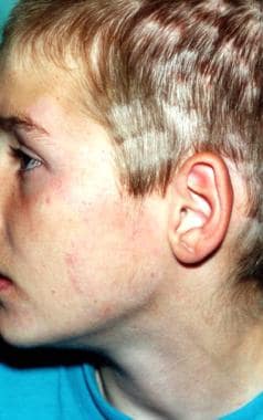
 Multiple lesions on the face caused by Microsporum canis infection in a patient who also has tinea capitis.
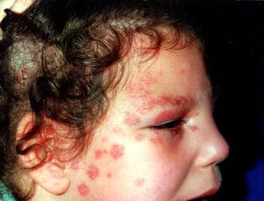