Overview
Cutaneous manifestations of human immunodeficiency virus (HIV) disease may result from HIV infection itself or from opportunistic disorders secondary to the decline in immunocompetence from the disease. [1] Cutaneous disorders may be the initial signs of HIV-related immunosuppression. Recognizing HIV-related skin changes may lead to the diagnosis of HIV infection in the early stages, allowing initiation of appropriate antiretroviral therapy. Many associated skin diseases are more severe in this group. With the use of antiretroviral therapy, the incidence of some of these skin disorders has declined, but the incidence of drug reactions and other noninfectious skin eruptions has been enhanced. [2] People living with HIV infection may not be at increased risk of catching coronavirus disease 2019 (COVID-19) and developing severe manifestations, although considerable concern remains. [3] Cutaneous manifestations of COVID-19 include vasculopathic symptoms, [4] which, in some HIV patients, may suggest the development of Kaposi sarcoma.
A variety of neoplastic, infectious, and noninfectious diseases can produce cutaneous manifestations throughout the course of HIV disease. These manifestations may occur more frequently than in persons without HIV infection and may be less responsive to usual treatment modalities.
For other discussions of HIV infection, see HIV Disease, Pediatric HIV Infection, and Antiretroviral Therapy for HIV Infection.
Manifestations by HIV Disease Stage
During acute primary HIV infection, a transient, generalized, morbilliform eruption may develop on the trunk and the arms. In the early asymptomatic stage of HIV disease, which may last from a few years to a decade or longer, no signs of infection other than lymphadenopathy are present. In a survey of 165 children in India with HIV infection, cutaneous manifestations were evident in 100 (61%) of them. Papular pruritic eruptions were evident in 16%, the most common condition, with highest prevalence in severe CD4 category. [5] In one survey of 106 West African patients, the pruritic papular eruptions of HIV infection were mainly on the exposed parts of the body, especially the upper and lower limbs. [6]
Kaposi sarcoma can occur prior to the onset of immunosuppression. With the onset of immunosuppression, nonspecific skin changes occur, such as common disorders with atypical clinical features, including recurrent varicella zoster, numerous hyperkeratotic warts, treatment-resistant seborrheic dermatitis, and oral hairy leukoplakia. However, Kaposi sarcoma of skin and/or oral cavity may develop in HIV patients with well-controlled HIV disease and may be a significant factor in their morbidity and mortality. [7]
In the later stages of HIV disease, chronic herpes simplex virus (HSV), molluscum contagiosum (MC), and cytomegalovirus (CMV) infections appear. An autopsy analysis of HIV-seropositive patients revealed that 72% had opportunistic viral infections; most patients were infected with CMV and HSV. The prevalence of clinically apparent MC infection varies from 5-18% in different series. Mycobacterial infections and mucocutaneous candidiasis occur.
A 42-month prospective study by Smith et al in 912 HIV-1–infected patients found that condylomata acuminata and verrucae are observed early, and their frequency does not increase as the disease progresses, whereas the incidence of HSV infections, MC, and oral hairy leukoplakia increases as the disease advances. [8, 9]
Verrucous herpes infection, leprosy, condylomalike molluscum contagiosum, and AIDS-associated pigmented or nonpigmented erythroderma may be seen in early HIV disease or as part of immune restoration syndrome after the initiation of antiretroviral therapy. [10, 11, 12, 13, 14] Leishmaniasis and miliary tuberculosis may be a concern in advanced HIV disease. [15, 16, 17] Diffuse or disseminated leishmaniasis may occur with HIV disease, [18, 19] including in association with the immune reconstitution inflammatory syndrome. [19]
Manifestations in HIV-Infected Children
Mucocutaneous candidiasis, including recurrent and widespread diaper dermatitis and chronic paronychia, is a common cutaneous manifestation in children with HIV infection. The most common cutaneous infections in children with HIV disease are impetigo and cellulitis caused by Staphylococcus aureus.
Other cutaneous manifestations of HIV infection in the pediatric age group are as follows:
-
Recurrent herpetic gingivostomatitis
-
Severe atopic dermatitis
Malignancies
Kaposi sarcoma (KS) was the first reported malignancy associated with HIV infection and was first documented in 1981 from reports in New York, Los Angeles, and San Francisco. [20] The worldwide prevalence of KS in patients with AIDS may approach 34%; in the United States; however, the prevalence of KS in patients with HIV disease is less than 5%. Most of the patients are homosexual men, although all patients who acquire HIV infection through sexual contact are at somewhat increased risk. Lymphedema from KS so severe as to be evident as elephantiasis nostras verrucose may occur. [21] One should also be aware of mimickers of KS, including acroangiodermatitis. [22]
Since the advent of highly active antiretroviral therapy (HAART), the incidence of non–AIDS-defining cutaneous cancers—in particular, basal cell carcinoma—among HIV-infected persons has exceeded that of AIDS-defining cutaneous cancers such as KS. In a prospective study, Crum-Cianflone et al found that 6% of HIV-infected persons developed a cutaneous malignancy over a mean follow-up period of 7.5 years. [23]
The development of cutaneous non–AIDS-defining cancers in this cohort proved to be associated with the traditional risk factors of increasing age and lighter skin color, rather than with CD4 lymphocyte counts, HIV RNA levels, or receipt of HAART. [23]
KS remains the most common HIV-associated malignancy in sub-Saharan Africa. [24] Pediatric KS is distinct, and lymph node involvement is a common manifestation.
AIDS-related B-cell non-Hodgkin lymphomas may cause skin nodules.
HIV-infected patients may also develop cutaneous T-cell lymphoma. [25] Amazingly, they had higher survival and lower mortality than those non–HIV-infected persons, possibly because of antiretroviral therapy restoring immune functions.
Anal carcinoma and cervical intraepithelial neoplasia are papillomavirus-associated tumors associated with HIV disease. These tumors tend to be more progressive and aggressive. An increase in squamous cell carcinoma of the anal mucosa has been reported, especially in young homosexual men with HIV infection.
Intraoral or multiple squamous cell carcinoma, Bowen disease, and metastatic basal cell carcinoma have occasionally been reported in patients infected with HIV.
Malignant melanoma appears to be more aggressive in patients with HIV. One study reported shorter disease-free and overall survival rates in patients with melanoma who had HIV disease, compared with those who did not have HIV. [26]
Children with AIDS have a higher risk of developing leiomyosarcoma, although the incidence is still low in this population.
Kaposi sarcoma
KS is an abnormally vascularized tumorlike lesion affecting skin, lymph nodes, and viscera. It is believed to be a proliferation of endothelial cells induced by human herpesvirus type 8. KS is promoted by various angiogenic and proinflammatory factors, including HIV-Tat. In addition, the latency-associated nuclear antigen type 1 (LANA-1) protein is highly expressed in spindle cells, which is considered important in the maintenance of human herpesvirus type 8–associated malignancies.
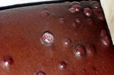
KS begins as pink macules that become disseminated and palpable. Purplish or brown macules and plaques may become nodular (see the images below). Mucosal involvement is common. KS demonstrates more aggressive clinical progression in patients infected with HIV than in other populations with the disease. It may develop in HIV patients with well-controlled HIV disease. [7]
Viral Infections
In patients infected with HIV, several viruses of the Herpesviridae family may lead to cutaneous disease, including chronic perianal and perioral herpetic ulcers caused by herpes simplex virus (HSV), recurrent typical dermatomal zoster caused by herpes zoster virus (HZV), and disseminated cytomegalovirus (CMV) infection.
Herpes simplex and herpes zoster viruses
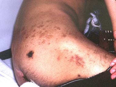
Recurrent oral and anogenital HSV infection is common in patients infected with HIV, and it may lead to chronic ulcerations. In pediatric patients, HSV stomatitis is more common than varicella-zoster virus (VZV) and may become chronic and ulcerative. Patients with VZV may develop chronic ecthymatous VZV (see the image below).
Acute disseminated HZV infection and the following atypical manifestations have also been described:
-
Hyperkeratotic papules
-
Folliculitis
-
Verrucous lesions
-
Chronic ulcerations
-
Disseminated ecthymatous lesions
According to Leibovitz et al, chronic VZV infections associated with HIV-1 infection begin as vesicles and progress into necrotic, nonhealing ulcers. [27, 28] Chronic VZV infection may mimic basal cell carcinoma. [26]
Epstein-Barr virus
Epstein-Barr virus (EBV) has been implicated in the pathogenesis of oral hairy leukoplakia, which may develop in patients infected with HIV, particularly men. Oral hairy leukoplakia is characterized by filiform white papules localized on the sides of the tongue. This condition has no malignant potential, but it may be the initial sign of progressive immunosuppression. White plaques may be confused with oral candidiasis, lichen planus, and geographic tongue.
Cytomegalovirus
CMV is a DNA virus in the Herpesviridae family. Ulcers in the perineal region are the most common presentation of CMV infection in patients infected with HIV-1. The concurrent involvement of other infectious agents, such as HSV, in the same lesions confounds the role of CMV in cutaneous lesions. HSV is proposed to be the initiating infection leading to ulcer formation, with CMV secondarily localizing in the granulation tissue.
Nonspecific maculopapular eruptions similar to those affecting patients with EBV or papulovesicular, nodular, purpuric, and ulcerative lesions of CMV infection are observed in patients who are immunocompromised. However, cutaneous lesions are rarely observed in patients infected with HIV.
CMV infection of the eccrine ducts resulting in squamous metaplasia has been described in a patient with HIV.
Diagnosing skin CMV infection in individuals infected with HIV is important. The presence of CMV infection is considered a poor prognostic sign in HIV disease.
Warts
Widespread or recalcitrant warts may be observed on the oral mucosa, the face, the perianal region, and the female genital tract in patients infected with HIV. The perianal and cervical lesions may be difficult to treat. Large plantar warts caused by human papillomoavirus–66 (HPV-66) and an epidermodysplasia verruciformislike eruption, which is believed to be associated with HPV infection, have also been reported in patients infected with HIV. [29]
Molluscum contagiosum
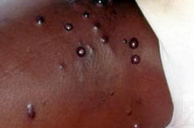
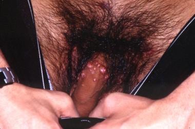
The molluscum contagiosum (MC) virus is a DNA virus in the Poxviridae family. It replicates in the cytoplasm of epidermal cells. MC lesions are small papules with central umbilication (see the images below).
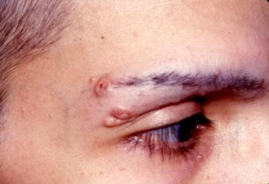 Young man with HIV disease and molluscum contagiosum on the lateral part of the eyebrow.
In HIV infection, MC may be widespread and atypical. The lesions may be observed on unusual sites, such as the face, neck, and scalp, and the lesions may be of unusual morphology and size. [30] Such unusual forms include solitary, endophytic, aggregated, inflamed, and giant MCs. MCs mimicking sebaceous nevus of Jadassohn, ecthyma, and giant condylomata acuminata have been reported.
Superficial Fungal Infections
Recurrent and persistent mucocutaneous candidiasis is common in patients with HIV infection. In the United States, recurrent vaginal candidiasis is the most common presentation of HIV infection in women.
In adults, generalized dermatophytosis, or tinea capitis, which is typically caused by Trichophyton rubrum, may suggest HIV infection. Bournerias et al reported tinea capitis from Microsporum canis in 2 patients infected with HIV. [31]
Pityriasis versicolor may be persistent and recurrent in patients with HIV infection.
Deep Fungal Infections
Rarely, cutaneous cryptococcosis may be observed in patients with HIV infection. [32] Clinical manifestations include the following:
-
Cellulitis
-
Papules
-
Plaques
-
Ulcers
-
Translucent dome-shaped papules with central umbilication, resembling MC
Cutaneous histoplasmosis may lead to red papules, a cellulitislike eruption, ulcerations, acneiform papules, or molluscumlike lesions in patients infected with HIV. [33, 34]
North American blastomycosis may present as a disseminated maculopapular eruption in HIV disease.
Talaromyces (formerly known as Penicillium) marneffei is a dimorphic fungus endemic in South and Southeast Asia that may evident cutaneously as papules with central umbilication in those with HIV disease. [35] It may become prominent as part of immune reconstitution inflammatory syndrome (IRIS) after antiretroviral therapy. Dermoscopic examination may show a round, whitish, amorphous structure.
Systemic coccidioidomycosis may disseminate to the skin, usually as hemorrhagic papules or nodules. A positive culture/staining from pus and cutaneous lesions may be used to document the cryptococcosis. [36]
Bacterial Infections
Impetigo and folliculitis may be recurrent and persistent in HIV disease, particularly in children. Disseminated furunculosis, gingivitis, gangrenous stomatitis, and abscess formation can occur in patients with HIV infection. Bacillary angiomatosis, which is caused by Bartonella henselae and rarely by Bartonella quintana, usually manifests as red papules and nodules. [37] . Antiretroviral therapy may be complicated by bacillary angiomatosis from B quintana as an immune reconstitution inflammatory syndrome and a Jarisch-Herxheimer reaction. [38]
Mycobacterial infections
Mycobacterium tuberculosis; M avium-intracellulare complex (MAC); and, rarely, M kansasii may present as acneiform papules and indurated crusted plaques.
MAC, a common opportunistic pathogen among patients with AIDS, usually causes disseminated disease involving the lungs, lymph nodes, and gastrointestinal tract. Primary cutaneous infections with MAC are extremely rare; most cutaneous lesions are caused by dissemination. Cutaneous manifestations thus far reported include the following:
-
Scaling plaques
-
Crusted ulcers
-
Ecthymalike lesions
-
Verrucous ulcers
-
Inflammatory nodules
-
Panniculitis
-
Pustular lesions
-
Draining sinuses
Localized skin involvement resembling sporotrichosis is unusual. Primary cutaneous MAC infection manifesting as sporotrichosislike lesions was described in a patient with AIDS. [39]
The possibility of coinfections, which may be multiple, should be kept in mind. Coinfection with B quintana, MAC, and CMV has been reported in an AIDS patient. [40]
In patients with HIV, M haemophilum can also present as violaceous draining nodules and superficial ulcers on the extremities, trunk, head, and genitalia.
Syphilis
Co-infection with syphilis (as well as other sexually transmitted diseases) may be found in HIV-infected patients, particularly those who are homosexual or bisexual or who use illicit drugs. Syphilitic ulcers are believed to increase HIV transmission.
Most cases of syphilis that occur in HIV disease are clinically and serologically typical, [41] but some are not. [42] A high rate of reinfection was noted in one French study, with less severe cutaneous manifestations. [41] However, syphilis seroconversion may be delayed, and standard serologic tests that aid in diagnosing syphilis may be unreliable. Also, in primary syphilis, presentation with multiple ulcers is more common in HIV-infected patients. Rapid progression of secondary syphilis to tertiary syphilis and syphilis maligna has been reported in patients infected with HIV. The Jarisch-Herxheimer reaction should be anticipated in patients neurosyphilis and coexistent HIV encephalitis. [43]
Appropriate serologic follow-up to ensure an adequate response to treatment is important in patients infected with HIV.
Staphylococcus aureus infection
Patients with HIV have been found to have increased rates of cutaneous colonization by Staphylococcus aureus, and in patients with advanced disease, sepsis and deep tissue infection can be common. Methicillin-resistant S aureus (MRSA) soft-tissue infection is an increasing problem. [44]
Parasitic Infestations
Atypical or Norwegian scabies, which is characterized by widespread hyperkeratotic, scaly maculopapular eruptions or crusted plaques, can occur in patients with HIV infection.
Atypical disseminated leishmaniasis has been reported in an HIV-infected patient. [45]
Demodex folliculorum folliculitis may lead to a pruritic papular eruption (PPE) on the face and the upper part of the trunk in patients with HIV disease.
Papulosquamous Dermatoses of AIDS
Generalized dry skin syndrome is frequently observed in patients with HIV infection. Xerosis may be the initial clinical manifestation of AIDS and is often a cause of pruritus. In the United States, pruritus has been reported in 4.5% of patients with AIDS.
Seborrheic dermatitis may be the initial cutaneous manifestation of HIV disease. According to Mathes et al, seborrheic dermatitislike eruptions are observed in 83% of patients with AIDS. [46] The eruption, which is characterized by widespread inflammatory and hyperkeratotic lesions, may progress to erythroderma in some patients. Seborrheic dermatitis may be increased in patients with AIDS-associated dementia or CNS disease. The most common mucocutaneous finding of people living with HIV in the current antiretroviral therapy era was found to be seborrheic dermatitis, with a prevalence of 28.5%. [47]
The immune alterations caused by HIV infection may lead to psoriasis and Reiter syndrome. In some instances, preexisting psoriasis may become more severe with disseminated plaques and pustules.
The typical skin lesions of pityriasis rosea may accompany HIV disease.
Acquired ichthyosis may begin on the lower extremities and disseminate in advanced HIV disease. Acquired ichthyosis may be a marker of concomitant infection with HIV-1 and human lymphotropic virus II in persons who use intravenous drugs and have profound helper T-cell depletion. [48]
Eosinophilic folliculitis manifests as an idiopathic, highly pruritic, papulopustular eruption of sterile pustules involving the face, neck, trunk, and extremities.
Pruritic papular eruption (PPE) is a common cutaneous manifestation in patients infected with HIV. It manifests as small, itchy, red or skin-colored papules on the head, neck, and upper part of the trunk. The cause is not known. According to Boonchai et al, 81.25% of patients with PPE have advanced immunosuppression. [49] (See the image below.)
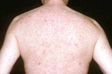 Dull red, violaceous, maculopapular lesions on the upper part of the trunk in a 49-year-old man with primary HIV-1 infection.
Hair and Nail Disorders
Diffuse alopecia or alopecia areata may be associated with HIV disease and may be inflammatory and permanent. The apoptotic follicular stem cell population in higher proportion may represent a hair cycle disturbance in patients with diffuse alopecia related to HIV-1 infection. [50]
Beau lines, telogen effluvium, and pallor of the nail beds are the general effects of the chronic illness. Elongation of the eyelashes and softening and straightening of the scalp hair may be observed in HIV disease, and proximal subungual onychomycosis is also usually a sign of HIV disease. The frequency of onychomycosis may be higher in men than in women.
Generalized alopecia can occur in patients with HIV who are treated with indinavir, an antiretroviral protease inhibitor. [51] Zidovudine is associated with longitudinal, transverse, or diffuse melanin pigmentation of the nails; however, nail pigmentation has also been observed in patients with HIV who have never taken zidovudine.
Drug Eruptions
Drug eruptions have been reported as the most common cause of erythroderma in patients infected with HIV. A study identified 177 cases of Stevens-Johnson syndrome/toxic epidermal necrolysis from 2000-2010 and found a high proportion of the patients were infected with HIV in sub-Saharan Africa, with a high frequency of antiretroviral drugs as the cause. [52] This elevated incidence of HIV patients with an adverse cutaneous drug eruption, including toxic epidermal necrolysis, may be due to a loss of skin-protective CD4+ CD25+ regulatory T cells. [53]
As many as 65-70% of patients treated with trimethoprim-sulfamethoxazole for Pneumocystis jiroveci pneumonia experience morbilliform eruptions within 7 days of starting the therapy. Reddish macules and papules may be generalized and can become permanent after the discontinuation of the therapy.
Sulfonamides may cause the following:
-
Urticaria
-
Erythema multiforme
-
Toxic epidermal necrolysis
-
Systemic reactions, including fever, leukopenia, thrombocytopenia, hepatitis, and nephritis
Toxic epidermal necrolysis has been reported with the following agents in patients with HIV:
-
Clindamycin and other antibiotics
-
Chlormezanone
Fixed drug eruption has been reported in 2 patients receiving saquinavir, an HIV-1 protease inhibitor.
Photosensitivity and Drug-Induced Pigmentation
A study by Vin-Christian et al found that photosensitivity in HIV-infected patients appears to be a manifestation of advanced disease. [54] Most of the patients in that study were sensitive to ultraviolet B (UV-B) light; however, the patients who were most severely affected were sensitive to both UV-B and UV-A light. [54]
Photo-induced lichenoid drug reactions may occur in HIV-infected patients, particularly those with dark skin. In addition, HIV-infected patients may experience drug-induced pigmentation of skin exposed to light.
Miscellaneous Dermatologic Disorders
The following dermatologic conditions may be associated with HIV disease:
-
HIV-related CD8+ cutaneous pseudolymphoma: This is an inflammatory process that results from a massive infiltration of the skin by activated, oligoclonal, HIV-specific, cytotoxic T lymphocytes and is most often seen in those that are markedly immunosuppressed. [55]
-
Severe aphthous stomatitis
-
Cutaneous vasculitis (possibly caused by CMV or parvovirus B19) [56]
-
Leukocytoclastic vasculitis with indinavir treatment [57]
Thrombocytopenic purpura, vitiligo, alopecia areata, sicca syndrome, pemphigoid, and other autoimmune blistering diseases have been reported in association with HIV disease.
Atopic disease may be reactivated by HIV disease. Atopic eczema may be severe in children infected with HIV. Increased serum IgE levels have been found in these children; however, increased IgE levels were not correlated with atopic symptoms. [58]
Urticaria may occur primarily or as a drug eruption in HIV disease. Cold urticaria has also been associated with HIV disease. [59]
Differential Diagnosis
The differential diagnosis includes the following:
-
Porphyria Cutanea Tarda: Its exacerbation in association with HIV infection is likely a result of co-infection with the hepatitis C virus rather than with HIV [60]
Other problems to be considered in HIV-infected patients with dermatologic conditions include the following:
-
Ashy dermatosis [61]
-
Normolipemic xanthomas
-
Multiple dermatofibromas [62]
-
Recurrent neutrophilic eccrine hidradenitis
-
Pemphigus vegetans
-
Lichen scrofulosorum
-
Cutaneous mucinosis
-
Papulonecrotic tuberculide
-
Kawasaki disease
-
Eruptive dysplastic nevi
-
Dissemination of vaccinia
-
Ofuji disease [63]
-
Angiomyolipomas
-
Glucan-induced keratoderma [64]
-
Disseminated superficial porokeratosis [65]
Workup
Histopathologic examination is useful to diagnose cutaneous manifestations of HIV disease with atypical clinical features and Kaposi sarcoma (KS). Routine hematoxylin and eosin and periodic acid-Schiff stainings may demonstrate multinuclear cells with intracytoplasmic inclusions of herpes simplex virus infection. Other special stains may identify additional pathogens, or they may confirm malignancies, such as KS. Histologic assessment of the CXCL12 axis may be a useful biomarker for diagnosing KS and assessing its progression. [66]
Imaging studies may be helpful in evaluating extracutaneous manifestations of KS, lymphoma, and systemic infections.
Treatment
Viral infections
For herpes simplex virus (HSV) and herpes zoster virus (HZV) infections in HIV-infected patients, the treatment of choice is acyclovir and other members of this drug class. These agents are activated by viral thymidine kinase. In some disseminated cases, the virus may be resistant to acyclovir because of the deficiency in viral thymidine kinase activity.
Prolonged therapy and chronic suppressive therapy with subtherapeutic doses have also been implicated in the development of acyclovir resistance. In the presence of acyclovir resistance, other viral therapies, including cidofovir, foscarnet, and vidarabine, may be necessary.
Treatment is usually not necessary for patients with oral hairy leukoplakia. If the patient is experiencing significant discomfort, systemic (1200 mg/day) and topical acyclovir, ganciclovir, or foscarnet may be recommended.
In most cases of molluscum contagiosum, imiquimod (Aldara) is curative. Resolution with zidovudine therapy has been reported in HIV-associated molluscum contagiosum. There are isolated reports of the efficacy of cidofovir in several viral infections (eg, molluscum contagiosum, [67] warts, [29] CMV infection) that are frequently observed in HIV-infected patients infected with HIV, but extensive studies of the safety and the efficacy of cidofovir have not been conducted to date.
Ablation and curettage may be useful in the treatment of molluscum contagiosum.
Noninfectious and nonmalignant cutaneous manifestations of HIV infection
For xerosis, emollients and dry skin care regimens are effective. For seborrheic dermatitis, coal tar, sulfur, and salicylic acid shampoos; topical corticosteroids; topical tacrolimus; and 2% ketoconazole cream may be effective.
For psoriasis and Reiter syndrome, ultraviolet B (UV-B) and psoralen with UV-A (PUVA) may be useful. Systemic corticosteroids, methotrexate, and cyclosporine may increase the immune suppression and must be considered only with careful monitoring. Zidovudine is also reported to be useful in the treatment of HIV-associated psoriasis.
For pruritic papular eruption, topical steroids, UV-B, PUVA, and pentoxifylline [68] have been reported to be effective. Eosinophilic folliculitis may respond to UV-B, [69] isotretinoin, or zidovudine treatment. For severe aphthous stomatitis, tacrolimus is reported to be effective.
Kaposi sarcoma
For patients with limited disease, local therapy with liquid nitrogen, alitretinoin, or intralesional vincristine may be effective. Surgery, radiotherapy, and systemic chemotherapy—usually with a single agent (eg, vinblastine, vincristine, bleomycin, doxorubicin, etoposide)—may be useful in the treatment of KS; however, systemic chemotherapy has not been shown to improve the long-term survival rates. Patients who recover immune response withantiretroviral therapy have had remission of KS.
Interferon (IFN)-alpha and IFN-beta photodynamic therapy and systemic hyperthermia have also been used. Cryotherapy, laser irradiation, and electrodesiccation can be useful for localized solitary lesions of KS.
Miscellaneous conditions
Mucocutaneous candidiasis is usually difficult to treat; oral azole treatment may be required. Generalized dermatophytosis may be resistant to topical antifungal creams and may require systemic antifungal therapy. Bacillary angiomatosis usually responds to oral erythromycin.
Consultations
Overall care for patients with HIV should be provided by a primary care practitioner with expertise in managing the disease. Infectious disease consultants are typically involved in the care of these patients. Patients confronting this chronic and debilitating disease may require additional support services.
The following are other specialists that may be consulted:
-
Ophthalmologist for CMV retinitis
-
Neurologist and anesthesiologist for postherpetic neuralgia and neurologic symptoms
-
Nephrologist to adjust the doses of antiviral agents
-
Psychiatrist to encourage the expression of deep emotions, such as grief, guilt, and anger
Questions & Answers
Overview
What are the cutaneous manifestations of HIV disease?
What are the cutaneous manifestations of HIV by disease stage?
What are the cutaneous manifestations of HIV disease in children?
Which malignancies cause cutaneous manifestations of HIV infection?
How are the cutaneous manifestations of Kaposi sarcoma (KS) in HIV characterized?
Which viral infections are associated with cutaneous manifestations of HIV disease?
What are atypical cutaneous manifestations of herpes zoster virus (HSV) in HIV disease?
What are the signs and symptoms of oral hairy leukoplakia in HIV disease?
How are the cutaneous manifestations of cytomegalovirus (CMV) in HIV disease characterized?
How are warts characterized in HIV disease?
What are the cutaneous manifestations of molluscum contagiosum (MC) virus in HIV disease?
What are the cutaneous manifestations of superficial fungal infections in HIV disease?
What are the cutaneous manifestations of deep fungal infections in HIV disease?
Which bacterial infections are associated with cutaneous manifestations of HIV disease?
What are the cutaneous manifestations of mycobacterial infections in HIV disease?
What are the cutaneous manifestations of syphilis in HIV disease?
What are the cutaneous manifestations of staphylococcus aureus infection in HIV disease?
Which parasitic infestations are associated with cutaneous manifestations of HIV disease?
What are the cutaneous manifestations of papulosquamous dermatoses in HIV disease?
Which hair and nail disorders are associated with HIV disease?
What is the role of drug eruptions in the development of HIV infection?
What are the cutaneous manifestations of sulfonamide use in HIV disease?
Which medications may cause toxic epidermal necrolysis in HIV disease?
What are photo-induced drug reactions in HIV disease?
Which dermatologic conditions are associated with HIV disease?
Which conditions are included in the differential diagnoses of cutaneous HIV disease?
What is the role of histology in the diagnosis of cutaneous manifestations of HIV infection?
What is the role of imaging studies in the diagnosis of cutaneous manifestations of HIV infection?
How are HSV and HZV treated in HIV disease?
How are noninfectious and nonmalignant cutaneous manifestations of HIV disease treated?
How is Kaposi sarcoma (KS) treated in HIV disease?
How are cutaneous manifestations of HIV disease treated?
-
Dull red, violaceous, maculopapular lesions on the upper part of the trunk in a 49-year-old man with primary HIV-1 infection.
-
Kaposi sarcoma in a man with HIV infection.
-
Kaposi sarcoma in a man with HIV infection.
-
Young man with HIV disease and molluscum contagiosum on the lateral part of the eyebrow.
-
Molluscum contagiosum on the penile shaft in a patient with HIV disease.
-
Old herpes zoster and Kaposi sarcoma in a patient with HIV disease.
Tables

What would you like to print?
- Overview
- Manifestations by HIV Disease Stage
- Manifestations in HIV-Infected Children
- Malignancies
- Viral Infections
- Warts
- Superficial Fungal Infections
- Deep Fungal Infections
- Bacterial Infections
- Parasitic Infestations
- Papulosquamous Dermatoses of AIDS
- Hair and Nail Disorders
- Drug Eruptions
- Photosensitivity and Drug-Induced Pigmentation
- Miscellaneous Dermatologic Disorders
- Differential Diagnosis
- Workup
- Treatment
- Consultations
- Questions & Answers
- Show All
- Media Gallery
- References


