Laboratory Studies
Hematology
Approximately three fourths of patients with an amebic liver abscess have leukocytosis. This most likely will appear if symptoms are acute or complications have developed. However, eosinophilia is rare.
Anemia may be present, but the cause usually is multifactorial.
Chemistry
Hyperbilirubinemia is present in only a small proportion of cases.
In acute liver abscess, the aspartate aminotransferase (AST) levels are high. In chronic liver abscess, the alkaline phosphatase level tends to be elevated and the AST level tends to be within the normal limits. Overall, the alkaline phosphatase level is elevated in about 70-80% of cases of amebic liver abscess. [17]
Similar complete blood cell (CBC) count and liver test abnormalities are found in patients with pyogenic liver abscesses and are not specific.
Stool Studies
Stool examination
The role of microscopic stool examination is limited. Less than 30-40% of patients with amebic liver abscess have concomitant intestinal amebiasis, and 10% of the population is infected with the nonpathogenic strain of E dispar. Hence, the microscopic examination of the stool for the identification of cysts is of little value. If positive, it may suggest the diagnosis.
Fecal findings suggestive of amebic colitis include a positive test for heme, a paucity of neutrophils, and the presence of Charcot-Leyden crystal protein. The stool examination is still of value if the serologic and antigen identification tests are not available.
Examination of the stool for hematophagous trophozoites of E histolytica must be made on at least three fresh specimens because the trophozoites are very sensitive and may be excreted intermittently. A combination of wet mount, iodine-stained concentrates, and trichrome-stained preparations is used.
Upon examination of the stool, trophozoites may be confused with neutrophils. Cysts must be differentiated morphologically from nonpathogenic Entamoeba hartmanni, Entamoeba coli, and Endolimax nana. Nonpathogenic E dispar cannot be differentiated morphologically and requires fecal antigen detection.
Stool antigen detection
Stool antigen detection facilitates early diagnosis before an antibody response occurs (< 7 d) and differentiates pathogenic from nonpathogenic Entamoeba infection. The primary drawbacks are the requirement for fresh, unpreserved stool specimens [18] and the lack of intestinal amebiasis in as many as 60% of patients with amebic liver abscess.
Stool antigen detection kits based on enzyme immunoassay (EIA) are most common and still quite sensitive compared to polymerase chain reaction (PCR)-based methods. [19]
The PCR stool test shows high sensitivity for detecting E histolytica and for distinguishing nonpathogenic amoebas. [20, 21, 22] However, this test is expensive. Real-time (rapid) PCR is sensitive but not well standardized [23] and is not widely available.
Stool culture
Stool culture for amoeba is sensitive but has limited availability.
Serologic testing
Serologic testing is the most widely used method of diagnosis for amebic liver abscess. In general, the test result should be positive, even in cases when the result of the stool test is negative (only extraintestinal disease). [24]
EIA
EIA has now largely replaced indirect hemagglutination (IHA) testing and counter immunoelectrophoresis (CIE) testing. EIA is relatively simple and easy to perform, rapid, inexpensive, and more sensitive. [25, 26]
The EIA test detects antibodies specific for E histolytica in approximately 95% of patients with extraintestinal amebiasis, in 70% of patients with active intestinal infection, and in 10% of persons who are asymptomatic cyst passers.
The EIA serology findings revert to negative in 6-12 months following eradication of infection. Even in highly endemic areas, fewer than 10% of patients who are asymptomatic have positive amebic serology findings.
Initial negative test results may appear in as many as 10% of patients with amebic liver abscess. Under these circumstances, order repeat serology testing in 1 week. This test result will usually be positive.
Serum antigen detection
E histolytica galactose lectin antigen is detectable by enzyme-linked immunosorbent assay (ELISA) in at least 75% of serum samples obtained from patients with amebic liver abscess. Studies reported an antigen seropositivity of 96% with a reversal rate of 82% after 1 week of treatment with metronidazole. This test may be useful for patients who present acutely, before an antibody response occurs. The sample needs to be obtained before starting the treatment, as the treatment leads to rapid antigen loss. This test can be used for rapid diagnosis in highly endemic areas, where serology can be misleading, but it is not widely available. [18]
Rapid antigen and antibody tests are currently being evaluated and seem very promising. [27]
Imaging Studies
None of the imaging tests can definitively differentiate between a pyogenic liver abscess, an amebic abscess, and malignant disease. [28] Clinical, epidemiologic, and serologic correlation is needed for diagnosis.
Ultrasonography
Ultrasonography is the preferable initial diagnostic test. It is rapid, inexpensive, and is only slightly less sensitive than CT scanning (75-80% sensitivity vs 88-95% for CT scan).
Ultrasonography simultaneously evaluates the gallbladder and avoids radiation exposure.
As opposed to scanning with technetium-99m, ultrasonography often can distinguish an abscess from a tumor or other solid focal lesion. The lesions tend to be round or oval, with well-defined margins, and are hypoechoic.
CT scanning
CT scanning is sensitive but the findings are not specific. The abscess typically appears low density with smooth margins and a contrast-enhancing peripheral rim.
The use of injected contrast may differentiate hepatic abscesses from vascular tumors. See the images below.
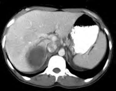 Amebic Liver/Hepatic Abscesses. CT scan of the abdomen with IV and oral contrast is shown. Note the thick-walled cavity with low attenuation center and contrast-enhanced periphery.
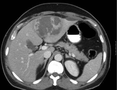 Amebic Liver/Hepatic Abscesses. CT scan of the abdomen with contrast showing a large amebic abscess with multiloculated appearance and atypical left liver lobe location. CT scan cannot differentiate amebic liver abscess from pyogenic liver abscess.
MRI
MRI is sensitive, but the findings are not specific. This imaging modality provides information comparable with less expensive imaging procedures.
Nuclear imaging studies
Technetium-99m liver scanning is useful for differentiating an amebic liver abscess from a pyogenic abscess; however, it is not used as a first-line test.
Because amebic liver abscesses do not contain leukocytes, they appear as cold lesions on hepatic nuclear scanning, with a typical hot halo or a rim of radioactivity surrounding the abscess. In contrast, pyogenic liver abscesses contain leukocytes and, therefore, typically appear as hot lesions on nuclear scanning.
Gallium scanning is helpful in differentiating a pyogenic abscess (similar to technetium-99m nuclear hepatic scanning) but requires delayed images, which makes the test less helpful.
Other imaging studies
Hepatic angiography is only useful to differentiate liver abscesses from vascular lesions.
Plain chest or abdominal films may show elevation and limitation of motion of the right diaphragm, basilar atelectasis, and right pleural effusion or gas within the abscess cavity.
Procedures
Aspiration of the abscess content is indicated only if rupture of the abscess is thought to be imminent, differentiation between amebic abscess and pyogenic abscess is critical, or there is no response to antiprotozoal therapy in 5-7 days (see Surgical Care). [29] Note the following:
-
Aspiration may be performed under computed tomography (CT) scan or sonographic guidance.
-
Send the collected specimen for Gram stain and cultures.
-
Amebae rarely are recovered from the aspirate (15%) and, often, they are present only in the peripheral parts of the abscess, invading and destroying adjacent tissue.
-
Amebic liver abscesses only rarely yield positive bacterial cultures following secondary bacterial infection of the abscess cavity.
-
Detecting E histolytica antigen in the aspirate is possible and is accomplished as previously described for stool specimens. It is highly specific. The sensitivity was only 20% using enzyme-linked immunosorbent assay (ELISA), but newer polymerase chain reaction (PCR)-based assays have a sensitivity of 83% and a specificity of 100%. [30, 31] However, currently, PCR-based detection is not widely available.
Many possible complications are associated with aspiration of the abscess, of which the most common are infection and bleeding. Other complications include amebic peritonitis or inadvertent puncture of an echinococcal cyst.
Histologic Findings
The liver involvement in amebiasis consists of necrotic abscesses and periportal inflammation. The abscess contains acellular proteinaceous debris and is surrounded by a rim of amebic trophozoites invading tissue. The abscess contains a chocolate-colored fluid that resembles anchovy paste and consists predominantly of necrotic hepatocytes. Triangular areas of hepatic necrosis, possibly due to ischemia from amebic obstruction of the portal vessels, have been observed. E histolytica can also induce hepatocyte and neutrophilic apoptosis. Some authors postulate that amebic liver abscess probably results from the coalescence of small microabscesses. Periportal fibrosis may be present, but whether this represents prior trophozoite invasion or a host reaction to amebic antigens or toxins is not known.
-
Amebic Liver/Hepatic Abscesses. CT scan of the abdomen with IV and oral contrast is shown. Note the thick-walled cavity with low attenuation center and contrast-enhanced periphery.
-
Amebic Liver/Hepatic Abscesses. CT scan of the abdomen with contrast showing a large amebic abscess with multiloculated appearance and atypical left liver lobe location. CT scan cannot differentiate amebic liver abscess from pyogenic liver abscess.


