Parker LK, Ponte C, Howell KJ, Ong VH, Denton CP, Schreiber BE. Clinical features and management of erythromelalgia: long term follow-up of 46 cases. Clin Exp Rheumatol. 2017 Jan-Feb. 35 (1):80-84. [QxMD MEDLINE Link]. [Full Text].
Jha SK, Karna B, Goodman MB. Erythromelalgia. 2020 Jan. 47 (2):91-97. [QxMD MEDLINE Link]. [Full Text].
Drenth JP, van Genderen PJ, Michiels JJ. Thrombocythemic erythromelalgia, primary erythermalgia, and secondary erythermalgia: three distinct clinicopathologic entities. Angiology. 1994 Jun. 45(6):451-3. [QxMD MEDLINE Link].
Michiels JJ, Abels J, Steketee J, et al. Erythromelalgia caused by platelet-mediated arteriolar inflammation and thrombosis in thrombocythemia. Ann Intern Med. 1985 Apr. 102(4):466-71. [QxMD MEDLINE Link].
Michiels JJ, Drenth JP, Van Genderen PJ. Classification and diagnosis of erythromelalgia and erythermalgia. Int J Dermatol. 1995 Feb. 34(2):97-100. [QxMD MEDLINE Link].
Orstavik K, Weidner C, Schmidt R, et al. Pathological C-fibres in patients with a chronic painful condition. Brain. 2003 Mar. 126(Pt 3):567-78. [QxMD MEDLINE Link].
Charkoudian N. Skin blood flow in adult human thermoregulation: how it works, when it does not, and why. Mayo Clin Proc. 2003 May. 78(5):603-12. [QxMD MEDLINE Link].
Mork C, Kalgaard OM, Kvernebo K. Impaired neurogenic control of skin perfusion in erythromelalgia. J Invest Dermatol. 2002 Apr. 118(4):699-703. [QxMD MEDLINE Link].
Waxman SG, Dib-Hajj SD. Erythromelalgia: a hereditary pain syndrome enters the molecular era. Ann Neurol. 2005 Jun. 57(6):785-8.
Drenth JP, te Morsche RH, Guillet G, et al. SCN9A mutations define primary erythermalgia as a neuropathic disorder of voltage gated sodium channels. J Invest Dermatol. 2005 Jun. 124(6):1333-8.
Michiels JJ, te Morsche RH, Jansen JB, Drenth JP. Autosomal dominant erythermalgia associated with a novel mutation in the voltage-gated sodium channel alpha subunit Nav1.7. Arch Neurol. 2005 Oct. 62(10):1587-90.
McDonnell A, Schulman B, Ali Z, Dib-Hajj SD, Brock F, Cobain S, et al. Inherited erythromelalgia due to mutations in SCN9A: natural history, clinical phenotype and somatosensory profile. Brain. 2016 Apr. 139 (Pt 4):1052-65. [QxMD MEDLINE Link].
Yang Y, Wang Y, Li S, et al. Mutations in SCN9A, encoding a sodium channel alpha subunit, in patients with primary erythermalgia. J Med Genet. 2004 Mar. 41(3):171-4. [QxMD MEDLINE Link]. [Full Text].
Eberhardt M, Nakajima J, Klinger AB, Neacsu C, Hühne K, O'Reilly AO, et al. Inherited pain: sodium channel Nav1.7 A1632T mutation causes erythromelalgia due to a shift of fast inactivation. J Biol Chem. 2014 Jan 24. 289(4):1971-80. [QxMD MEDLINE Link]. [Full Text].
Han C, Dib-Hajj SD, Lin Z, Li Y, et al. Early- and late-onset inherited erythromelalgia: genotype-phenotype correlation. Brain. 2009 Jul. 132:1711-22. [QxMD MEDLINE Link].
Davis MD, O'Fallon WM, Rogers RS 3rd, Rooke TW. Natural history of erythromelalgia: presentation and outcome in 168 patients. Arch Dermatol. 2000 Mar. 136(3):330-6. [QxMD MEDLINE Link].
Tang Z, Chen Z, Tang B, Jiang H. Primary erythromelalgia: a review. Orphanet J Rare Dis. 2015 Sep 30. 10:127. [QxMD MEDLINE Link]. [Full Text].
Zheng ZM, Zhang JH, Hu JM, et al. Poxviruses isolated from epidemic erythromelalgia in China. Lancet. 1988 Feb 6. 1(8580):296. [QxMD MEDLINE Link].
Kalgaard OM, Seem E, Kvernebo K. Erythromelalgia: a clinical study of 87 cases. J Intern Med. 1997 Sep. 242(3):191-7. [QxMD MEDLINE Link].
Dupont E, Illum F, Olivarius Bde F. Bromocriptine and erythromelalgia-like eruptions. Neurology. 1983 May. 33(5):670. [QxMD MEDLINE Link].
Perez J, Khouri C, Park S, Imbert B, Cracowski JL. Eltrombopag-associated erythromelalgia in idiopathic thrombocytopenic purpura. Br J Dermatol. 2021 Oct 26. [QxMD MEDLINE Link].
McMahon DE, Kovarik CL, Damsky W, Rosenbach M, Lipoff JB, Tyagi A, et al. Clinical and pathologic correlation of cutaneous COVID-19 vaccine reactions including V-REPP: A registry-based study. J Am Acad Dermatol. 2022 Jan. 86 (1):113-121. [QxMD MEDLINE Link]. [Full Text].
Gambichler T, Boms S, Susok L, Dickel H, Finis C, Abu Rached N, et al. Cutaneous findings following COVID-19 vaccination: review of world literature and own experience. J Eur Acad Dermatol Venereol. 2021 Oct 17. [QxMD MEDLINE Link]. [Full Text].
Zaza P, Matthieu R, Jean-Luc C, Charles K. Drug repurposing in Raynaud's phenomenon through adverse event signature matching in the World Health Organization pharmacovigilance database. Br J Clin Pharmacol. 2020 Nov. 86 (11):2217-2222. [QxMD MEDLINE Link]. [Full Text].
Drenth JP, Michiels JJ, van Joost T, Vuzevski VD. Secondary erythermalgia in systemic lupus erythematosus; [comment]. J Rheumatol. 1993 Jan. 20 (1):144-6. [QxMD MEDLINE Link].
Saviuc PF, Danel VC, Moreau PA, Guez DR, Claustre AM, Carpentier PH, et al. Erythromelalgia and mushroom poisoning. J Toxicol Clin Toxicol. 2001. 39 (4):403-7. [QxMD MEDLINE Link].
Nakajima N, Ueda M, Higashi N, Katayama Y. Erythromelalgia associated with Clitocybe acromelalga intoxication. Clin Toxicol (Phila). 2013 Jun. 51 (5):451-4. [QxMD MEDLINE Link].
Gu Y, Chen F, Liu T, Lv X, Shao Z, Lin H, et al. Early detection of an epidemic erythromelalgia outbreak using Baidu search data. Sci Rep. 2015 Jul 28. 5:12649. [QxMD MEDLINE Link]. [Full Text].
Alhadad A, Wollmer P, Svensson A, Eriksson KF. Erythromelalgia: Incidence and clinical experience in a single centre in Sweden. Vasa. 2012 Jan. 41(1):43-8. [QxMD MEDLINE Link].
Cook-Norris RH, Tollefson MM, Cruz-Inigo AE, Sandroni P, Davis MD, Davis DM. Pediatric erythromelalgia: A retrospective review of 32 cases evaluated at Mayo Clinic over a 37-year period. J Am Acad Dermatol. 2011 Jul 26. [QxMD MEDLINE Link].
Kondo T, Uehara T, Ikegami A, Ikusaka M. Paroxysmal burning pain caused by erythromelalgia. Lancet. 2014 May 10. 383(9929):1692. [QxMD MEDLINE Link].
Messeguer F, Agusti-Mejias A, Vilata Corell JJ, Requena C. Auricular erythromelalgia: report of a rare case. Dermatol Online J. 2013 Feb 15. 19(2):16. [QxMD MEDLINE Link].
Patel M, Femia AN, Eastham AB, Lin J, Canales AL, Vleugels RA. Facial erythromelalgia: a rare entity to consider in the differential diagnosis of connective tissue diseases. J Am Acad Dermatol. 2014 Dec. 71 (6):e250-1. [QxMD MEDLINE Link]. [Full Text].
Tham SW, Giles M. Current pain management strategies for patients with erythromelalgia: a critical review. J Pain Res. 2018. 11:1689-1698. [QxMD MEDLINE Link]. [Full Text].
Jha SK, Karna B, Goodman MB. Erythromelalgia. 2021 Jan. [QxMD MEDLINE Link]. [Full Text].
Hagedorn JM, Canzanello N, Lamer TJ. Dorsal Root Ganglion Stimulation for Erythromelalgia Related Foot Pain: A Case Report and Review of the Literature. Pain Pract. 2021 Jul. 21 (6):698-702. [QxMD MEDLINE Link].
Fan X, Bu H, Wen Y, Ma L, Huang C, Xu F, et al. Spinal Cord Stimulation in the Treatment of Pediatric Erythromelalgia. World Neurosurg. 2020 Oct. 142:388-390. [QxMD MEDLINE Link].
Ogawa S, Ueno H, Maruyama A, Amaya F. Extensive Lumbar Sympathetic Ganglion Block Combined With Epidural Block for Primary Erythromelalgia: A Case Report. A A Pract. 2020 Oct. 14 (12):e01325. [QxMD MEDLINE Link].
Lam CM, Zayed H, Sayed D. High frequency dorsal column spinal cord stimulation for management of erythromelalgia. BMJ Case Rep. 2021 Aug 5. 14 (8):[QxMD MEDLINE Link].
Cohen JS. High-dose oral magnesium treatment of chronic, intractable erythromelalgia. Ann Pharmacother. 2002 Feb. 36(2):255-60. [QxMD MEDLINE Link].
Skeik N, Rooke TW, Davis MD, Davis DM, Kalsi H, Kurth I, et al. Severe case and literature review of primary erythromelalgia: Novel SCN9A gene mutation. Vasc Med. 2011 Nov 9. [QxMD MEDLINE Link].
Legroux-Crespel E, Sassolas B, Guillet G, et al. [Treatment of familial erythermalgia with the association of lidocaine and mexiletine]. Ann Dermatol Venereol. 2003 Apr. 130(4):429-33. [QxMD MEDLINE Link].
Iqbal J, Bhat MI, Charoo BA, et al. Experience with oral mexiletine in primary erythromelalgia in children. Ann Saudi Med. 2009 Jul-Aug. 29(4):316-8. [QxMD MEDLINE Link]. [Full Text].
Goldberg YP, Price N, Namdari R, Cohen CJ, Lamers MH, Winters C, et al. Treatment of Na(v)1.7-mediated pain in inherited erythromelalgia using a novel sodium channel blocker. Pain. 2012 Jan. 153(1):80-5. [QxMD MEDLINE Link].
Kalgaard OM, Mork C, Kvernebo K. Prostacyclin reduces symptoms and sympathetic dysfunction in erythromelalgia in a double-blind randomized pilot study. Acta Derm Venereol. 2003. 83(6):442-4. [QxMD MEDLINE Link].
Mørk C, Salerud EG, Asker CL, Kvernebo K. The prostaglandin E1 analog misoprostol reduces symptoms and microvascular arteriovenous shunting in erythromelalgia-a double-blind, crossover, placebo-compared study. J Invest Dermatol. 2004 Mar. 122 (3):587-93. [QxMD MEDLINE Link]. [Full Text].
Davis MD, Sandroni P. Lidocaine patch for pain of erythromelalgia: follow-up of 34 patients. Arch Dermatol. 2005 Oct. 141 (10):1320-1. [QxMD MEDLINE Link].
Davis MD, Morr CS, Warndahl RA, Sandroni P. Topically Applied Midodrine, 0.2%, an α1-Agonist, for the Treatment of Erythromelalgia. JAMA Dermatol. 2015 Sep 1. 151 (9):1025-6. [QxMD MEDLINE Link].
Poterucha TJ, Weiss WT, Warndahl RA, Rho RH, Sandroni P, Davis MD, et al. Topical amitriptyline combined with ketamine for the treatment of erythromelalgia: a retrospective study of 36 patients at Mayo Clinic. J Drugs Dermatol. 2013 Mar. 12(3):308-10. [QxMD MEDLINE Link].
Pagani-Estévez GL, Sandroni P, Davis MD, Watson JC. Erythromelalgia: Identification of a corticosteroid-responsive subset. J Am Acad Dermatol. 2017 Mar. 76 (3):506-511.e1. [QxMD MEDLINE Link].
Moody S, Pacheco S, Butler IJ, Koenig MK. Secondary Erythromelalgia Successfully Treated With Intravenous Immunoglobulin. J Child Neurol. 2011 Dec 7. [QxMD MEDLINE Link].
Aruch D, Mascarenhas J. Contemporary approach to essential thrombocythemia and polycythemia vera. Curr Opin Hematol. 2016 Mar. 23 (2):150-60. [QxMD MEDLINE Link].
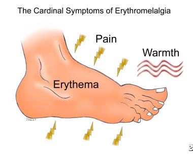 Cardinal symptoms of erythromelalgia.


