Laboratory Studies
Laboratory studies for the diagnosis of myeloproliferative disease include the following:
-
CBC counts and differential counts with microscopic examination of the peripheral smear (see example below)
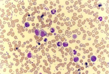 Photomicrograph of a peripheral smear of a patient with agnogenic myeloid metaplasia (myelofibrosis) shows findings of leukoerythroblastosis, giant platelets, and few teardrop cells.
-
Leukocyte alkaline phosphatase (LAP) score (to differentiate chronic myelogenous leukemia [CML] from other causes of leukocytosis)
-
Polymerase chain reaction (PCR) or fluorescent in-situ hybridization (FISH) run on peripheral blood can detect bcr-abl gene rearrangement [22] ; this helps differentiate CML from other myeloproliferative diseases
-
Red blood cell mass study (to differentiate true from spurious polycythemia)
-
Serum uric acid level

Imaging Studies
Imaging studies are not routinely required. However, a liver-spleen scan may occasionally be helpful to assess the size of these organs in the diagnosis of difficult cases.
Procedures
Bone marrow aspiration and biopsy with cytogenetic studies are required in most, but not all, patients. Cytogenetic studies detect presence or absence of the Philadelphia chromosome and help to differentiate these disorders from myelodysplastic syndrome. PCR testing on bone marrow for JAK2 is available for suspected cases of polycythemia vera, essential thrombocythemia, or myelofibrosis.
Histologic Findings
Bone marrow histology shows hypercellularity in most myeloproliferative disorders. In the case of myelofibrosis, bone marrow fibrosis is demonstrated on the reticulin stain. Bone marrow fibrosis is also detected in the spent phase of chronic myelogenous leukemia and polycythemia vera.
-
Peripheral smear of a patient with chronic myelogenous leukemia (CML) shows leukocytosis with extreme left shift and basophilia.
-
Peripheral smear of a patient with chronic myelogenous leukemia (CML) in blastic phase shows several blasts.
-
Peripheral smear of a patient with essential thrombocythemia (ET) shows markedly increased number of platelets. Some of the platelets are giant (arrow).
-
Peripheral smear of a patient with agnogenic myeloid metaplasia (myelofibrosis) shows leukoerythroblastosis. This photomicrograph also shows giant platelets.
-
Photomicrograph of a peripheral smear of a patient with agnogenic myeloid metaplasia (myelofibrosis) shows findings of leukoerythroblastosis, giant platelets, and few teardrop cells.
Tables
FAB |
WHO |
Chronic myelogenous leukemia |
Chronic myelogenous leukemia, BCR/ABL1 positive |
Polycythemia vera |
Polycythemia vera |
Essential thrombocythemia |
Essential thrombocythemia |
Agnogenic myeloid metaplasia/myelofibrosis |
Primary myelofibrosis* |
... |
Chronic neutrophilic leukemia, not otherwise specified |
... |
MPN, unclassified |
| *The 2016 WHO classification system distinguishes prefibrotic (prePMF) from overtly fibrotic PMF [4] | |