Approach Considerations
Glass slide microscopic analysis for Enterobius vermicularis may be performed looking for ova and female pinworms. A specimen is best obtained by dabbing the stretched, unwashed perianal folds in the early morning with cellophane tape and affixing the specimen onto a slide. A negative test for 5 consecutive mornings effectively rules out the diagnosis. [1, 2]
Microscopic examination shows the elongated ovoid egg distinctly compressed laterally and flattened on one side. The egg measures 50-60 µm X 20-30 µm and contains larva.
In areas where pinworms are endemic, consider analyzing any removed appendiceal stump for infestation. [9]
Patients with recurrent episodes of perianal itching should have a stool sample sent to the laboratory for analysis because different parasites, which require different treatment, may appear similar to E vermicularis using the cellophane-tape test. [2, 10]
Cellophane-tape Examination
A perianal cellophane swab or cellophane-tape examination should be used to detect Enterobius vermicularis eggs. [1, 2] Egg detection is associated with a false-positive rate of 5-11% and a false-negative rate of 70-95%. [11] One report found that a single cellophane-tape examination yielded a sensitivity of 50%, three examinations yielded a sensitivity of 90%, and five examinations yielded a sensitivity of 99%.
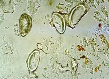 Microscopic view of Enterobius vermiculariseggs attached to cellophane tape after a perianal swab from a child in kindergarten in Seoul, Korea. Egg size was 50-60 μm X 20-30 μm. The eggs are elongated and ovoid, distinctly compressed laterally, and flattened on one side.
-
Adult female worms of Enterobius vermicularis collected from a 2-year-old girl in a Korean orphanage after treatment with pyrantel pamoate 10 mg/kg.
-
Microscopic view of Enterobius vermiculariseggs attached to cellophane tape after a perianal swab from a child in kindergarten in Seoul, Korea. Egg size was 50-60 μm X 20-30 μm. The eggs are elongated and ovoid, distinctly compressed laterally, and flattened on one side.
-
Pinworms in a young patient.


