Practice Essentials
Ependymoma is the third most common brain tumor in children, accounting for approximately 10% of primary central nervous system (CNS) neoplasms. It is a neuroepithelial tumor that arises within, or adjacent to, the ependymal lining of the ventricular system or the central canal of the spinal cord. Recent efforts have separated ependymoma into three distinct regional compartments, including the spinal column, the posterior fossa (see image below), and the supratentorium, with approximately two-thirds of pediatric ependymomas arising in the posterior fossa. Further advances have begun to delineate distinct molecular subtypes within the regional compartments, as detailed later in this article.
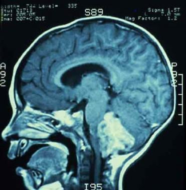 MRI showing an ependymoma of the fourth ventricle, compressing the cerebellum and brain stem.
With surgery and radiotherapy, overall 5-year survival rates have been reported to range between 50% and 81%. [1, 2, 3] Individual prognosis depends on age, extent of resection, and, increasingly, known biological variations. The role of chemotherapy for infants and patients with postoperative residual disease is currently under investigation.
Pathophysiology
Ependymomas typically arise from the ependymal lining of the ventricular system, most often the floor, roof, or lateral recesses of the fourth ventricle, as depicted below. The most recent evidence suggests that radial glia cells are the stem cells of origin for this disease. Approximately one third of ependymomas are supratentorial, arising from the surface of the lateral or third ventricles; however, they may be entirely extraventricular. They may also occur in the central canal of the spinal cord and in the filum terminale, although the latter site is uncommon in children.
Histologically, ependymoma has historically been broadly separated into three grades, although almost all pediatric ependymomas fall into one of two major subsets, grade II (cellular) and grade III (anaplastic). Grade II ependymoma is well differentiated and lacks mitosis and vascularity. Grade III ependymoma is poorly differentiated and has a high mitotic index, necrosis, calcifications, and endothelial proliferation. These display a higher frequency of ependymal rosettes clustering around a central lumen. Both histological subtypes are locally invasive into adjacent brain, and distinguishing between the grades can be very difficult. Clear cell ependymomas often exhibit cysts with characteristic clear cells with perinuclear halos and rounded nuclei, and also have high proliferation indices. Myxopapillary ependymomas are considered a distinct, grade I variant of ependymoma primarily occurring in the region of the cauda equina and are relatively rare in children. Subependymoma is another grade I ependymoma that may occur in any regional compartment, but that is likewise predominantly a disease of adult populations.
Ependymomas in the posterior fossa frequently infiltrate the brain stem, and as many as one third may project through the foramina to involve the medulla and upper spinal cord. Spread via cerebrospinal fluid (CSF) throughout the subarachnoid space is reported, primarily with the higher-grade tumors. Extraneural metastases to liver, lung, or bone are rare.
Prognosis has historically relied on age, presence of metastatic disease, extent of resection, and the appearance of significant histologic anaplasia. However, more recently, significant insight into the molecular underpinnings of ependymoma have resulted in a re-evaluation of the most important prognostic characteristics. Some molecular features of ependymoma have been previously associated with worse outcome, including chromosome 1q gain, telomerase expression and the presence of homozygous deletions of CDKN2A. [4]
As such, clinical and molecular analysis, including genomics and expression profiling, has begun to result in dividing childhood ependymoma by region (supratentorial, infratentorial, spinal) as well as by molecular phenotype. Using this categorization method, new groups of high risk disease are becoming evident.
Posterior fossa ependymoma, the most common site in childhood, may be generally divided into two groups: group A and group B. Group A tumors (also called CpG island methylator phenotype, or CIMP+) are more common, generally occur in younger children and have poorer outcomes, with roughly 20-30% 10-year progression-free survival. [5] Group A tumors have a generally balanced genome but exhibit an increased level of 1q gain, which is a marker of poor outcome. Group B tumors have better survival, with a 10-year progression free survival of almost 60%, and overall survival rates above 80%. Group B tumors occur more commonly in adults and older children, with an increased incidence ratio of female:male and have increased numbers of cytogenetic abnormalities, such as whole chromosomal gain/loss.
Supratentorial ependymomas may now be divided into groups exhibiting either YAP1 or C11orf95-RELA fusions. YAP1 fusion-positive tumors, which most typically affect younger children, have relatively good outcomes with nearly universal 10-year overall survival, although these tumors also recur. In contrast, ependymomas exhibiting RELA-fusion positivity have very poor outcomes, with rapid recurrence and progression and a 10-year progression-free survival of no better than 20%. [5] These tumors also more commonly display CDKN2A deletions, which are also postulated to be a marker of poor outcome.
Etiology
Epidemiological studies investigating parental occupational exposures, environmental exposures, and maternal nutritional intake have failed to identify linkages with any of the childhood brain tumors.
DNA sequences similar to SV40 virus and the virus-encoded large T-antigen have been found in several ependymomas, but no conclusions regarding causation have been determined. SV40 and related polyoma viruses can induce ependymoma in monkeys and other mammalian species.
Neurofibromatosis type 2 (NF2) is a genetic disorder that is known to predispose to ependymoma, typically spinal tumors.
Epidemiology
United States statistics
Approximately 200 new cases are reported annually in children. [6] Ependymoma represents the third most common brain tumor in children, following astrocytoma and medulloblastoma.
International statistics
In the United Kingdom, an estimated 30-35 new pediatric cases are diagnosed annually. More than 300 new pediatric cases per year are reported in Europe.
Race-, sex-, and age-related demographics
No specific race is predisposed to ependymoma. However, analysis of data from the Central Brain Tumor Registry of the United States shows that the incidence of ependymoma is significantly lower among African American and Native American children (by 33% and 36%, respectively), compared with non-Hispanic White children. In addition, eastern European ancestry is associated with an elevated risk of ependymoma in non-Hispanic Whites and in Hispanics. [7]
Ependymomas are more common in males.
The incidence of ependymoma peaks below 4 years of age, with almost a third of patients presenting in that time frame. The age of presentation is also related to the location of the primary disease, with spinal disease presenting at a mean age of 12.2 years, supratentorial disease at a mean of 7.8 years and infratentorial disease presenting at a mean age of 5 years. [8] Spinal cord ependymoma rarely occurs in children younger than 12 years.
Prognosis
Prognostic factors
Extent of tumor resection
Resection is the most important prognostic factor. Patients with gross total and near-total resections have reported survival rates of 51-80%, versus 0-26% in those with subtotal resections (< 90% removal of total tumor mass, visible tumor present on MRI).
Age
Very young patients (< 1 y), unrelated to radiation treatment, have a significantly worse prognosis (5-y survival rate of 25%). [9] The 5-year survival rate for children aged 1-4 years is also significantly less than for children older than 5 years (46% versus >70%). Some promising results using high-dose chemotherapy and delayed or omitted radiotherapy have been recently shown in this age group.
Other factors
Historically, anaplastic features and supratentorial location have conferred a worse prognosis. More recent reports have largely dismissed histology and tumor location as significant prognostic indicators (with the exception of better outcome observed in spinal cord tumors and myxopapillary tumors of the cauda equina). Metastatic disease is probably a poor prognostic factor; however, patient numbers are too scarce to draw a conclusion.
Morbidity/mortality
The 5-year overall survival rate has been reported to vary widely with local resection and irradiation, with a 5-year overall survival rate in the 50-60% range. However, more recently, the natural history of ependymoma has been noted to be more prolonged than previously appreciated due to the potential for late relapses and slow recurrence; therefore, survival rates do not plateau at the typically reported 5-years post-diagnosis. A more recent study noted 10-year overall survival of 50%, but only 29% progression free survival in the same timeline. [10]
Although historically age, the extent of tumor resection, and histology have been the primary predictors for survival, these characteristics are undergoing significant revision in the context of increased comprehension of the biology of ependymoma (as detailed previously).
Direct distortion and destruction of normal brain tissue by tumor, as well as increased intracranial pressure and surgical trauma, may cause some degree of irreversible neurologic impairment. Also, the volume and location of radiotherapy necessary to treat the tumor may result in cognitive impairment. The expanded use of conformal radiotherapy for localized disease has helped to ameliorate some of these effects. Radiotherapy to the hypothalamic-pituitary axis may result in deficits of growth hormone, thyroid hormone, gonadotropin, and adrenocorticotropic hormone.
Children with relapsed ependymoma often are subjected to a prolonged cycle of repeated relapses and stabilizations, with repeated surgical procedures, radiotherapy, and medical management leading to potentially significant morbidity, including organ toxicity, neurologic deterioration and cognitive/social difficulties.
Complications
Complications include the following:
-
Obstructive hydrocephaly
-
Neurologic impairment
-
Radiation-induced effects
Neurocognitive decline
Endocrinologic dysfunction
Mineralizing microangiopathy with ischemia or infarct
Secondary CNS malignancies
Transient headaches, fatigue, nausea, vomiting, and anorexia
Patient Education
The patient and his/her family members should be referred for psychosocial counseling.
The following patient education resources are available from WebMD:
-
MRI showing an ependymoma of the fourth ventricle, compressing the cerebellum and brain stem.
-
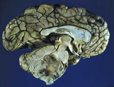
Sagittal section of an ependymoma of the fourth ventricle.
-
Section displaying typical perivascular pseudorosettes of a benign ependymoma.
-
Section displaying high cellularity, nuclear atypia, and numerous mitoses characteristic of an anaplastic ependymoma.


