Report on the Radiolaria/Plates12

From Wikisource
Jump to navigation Jump to search
1584928Report on the Radiolaria — Plates 111-120Ernst Haeckel

PLATE 111.

Legion PHÆODARIA.

Order PHÆOSPHÆRIA.

Family Aulosphærida.


PLATE 111.
Aulosphærida.
Diam. Page.
Fig. 1. Aulonia hexagonia, n. sp., × 30 1634
The complete spherical shell.
Fig. 2. Aularia ternaria, n. sp., × 300 1621
A group of six triangular meshes, with seven nodal points of radial tubes. Behind the central capsule, with its double membrane (e, outer; i, inner) and radiate operculum (o); u, the two outer parapylæ; v, vacuoles in the protoplasm. The ellipsoidal nucleus (n) contains numerous nucleoli (l).
Fig. 3. Aulastrum triceros, n. sp., × 50 1635
The complete shell.
Fig. 3a. Aulastrum triceros, n. sp., × 300 1635
A single radial tube.
Figs. 4a, 4b, 4c. Aulastrum dendroceros, n. sp., × 400 1635
Three single radial spines (taken from three different specimens).
Fig. 5a. Aulophacus lenticularis, n. sp., × 300 1631
A single radial spine.
Fig. 5b. Aulophacus amphidiscus, n. sp., × 300 1631
A single radial spine.
Fig. 6. Aulatractus fusiformis, n. sp., × 5 1632
The complete shell, five times enlarged.
Fig. 6a. Aulatractus fusiformis, n. sp., × 20 1632
Apical part of the shell.
Fig. 6b. Aulatractus fusiformis, n. sp., × 400 1632
A single radial tube.
Fig. 7. Aulatractus diploconus, n. sp., × 20 1632
Apical part of the shell.
Fig. 7a. Aulatractus diploconus, n. sp., × 400 1632
A single radial tube.
Fig. 8. Auloplegma perplexum, n. sp., × 50 1630
Half the shell.
Fig. 8a. Auloplegma perplexum, n. sp., × 400 1630
A single radial tube.
Fig. 9. Auloplegma spongiosum, n. sp., × 300 1631
A single radial tube.
Plate 111.
Plate 111.


PLATE 112.

Legion PHÆODARIA.

Orders PHÆOSPHÆRIA.

Family Cannosphærida.


PLATE 112.
Cannosphærida.
Diam. Page.
Cannosphærida.
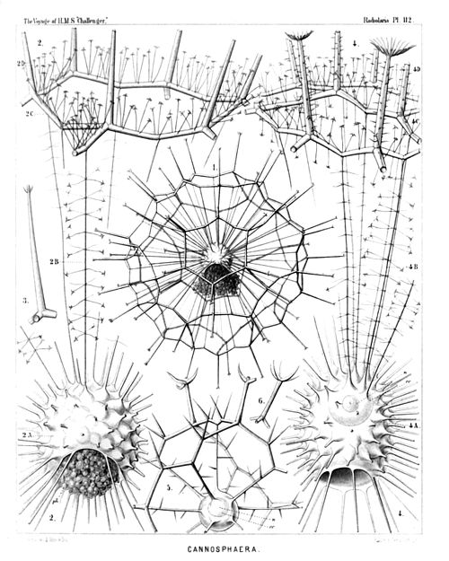
Fig. 1. Cannosphæra antarctica, n. sp., × 50 1640
The entire shell. The inner mammillate shell, from the month of which is prominent the phæodium, in connected by numerous radial beams with the outer shell.
Fig. 2. Cannosphæra antarctica, n. sp., × 200 1640
The inner shell, from the mouth of which is prominent the phæodium, and a single hexagonal mesh of the outer shell, connected with the former by thin radial threads.
Fig. 3. Cannosphæra antarctica, n. sp., × 200 1640
A single radial spine, with four terminal branches.
Fig. 4. Cannosphæra pacifica, n. sp., × 200 1641
The inner shell, exhibiting on its base the widely open mouth, and in its upper half the transparent spherical central capsule with its nucleus. Of the outer shell (which is connected with the inner by thin radial threads), only a few polygonal meshes are visible.
Fig. 5. Cannosphæra atlantica, n. sp., × 200 1640
The inner shell, connected by spiny radial beams with the outer shell, a quadrant only of which is visible.
Fig. 6. Cannosphæra atlantica, n. sp., × 200 1640
A single radial spine, with five terminal branches.
Plate 112.
Plate 112.


PLATE 113.

Legion PHÆODARIA.

Order PHÆOGROMIA.

Family Castanellida.


PLATE 113.
Castanellida.
Diam. Page.
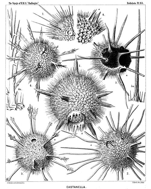
Fig. 1. Castanissa challengeri, n. sp., × 100 1686
In the lower part of the figure is visible the large corona of teeth around the mouth (a).
Fig. 2. Castanidium moseleyi, n. sp., × 80 1686
In the upper part of the figure, at left, is visible the irregular polygonal mouth (a).
Fig. 3. Castanopsis naresi, n. sp., × 80 1688
In the upper part of the figure is visible the smooth circular mouth (a).
Fig. 4. Castanura tizardi, n. sp., × 80 1689
Fig. 4a. A single main-spine of the same, × 400
Fig. 5. Castanidium murrayi, n. sp., × 100 1685
With a large phæodium, partly protruded through the circular mouth.
Fig. 5a. A single main-spine of the same, hexagonally dimpled, × 400
Fig. 6. Castanella wyvillei, n. sp., × 100 1683
A piece of the shell with the mouth, armed with six large teeth (a).
Fig. 7. Castanidium buchanani, n. sp., × 100 1685
A piece of the shell with the smooth roundish mouth (a).
Plate 113.
Plate 113.


PLATE 114.

Legion PHÆODARIA.

Orders PHÆOCYSTINA et PHÆOGROMIA.

Families Cannorrhaphida et Circoporida.


PLATE 114.
Cannorrhaphida et Circoporida.
Diam. Page.
Fig. 1. Haeckeliana darwiniana, n. sp., × 200 1702
A complete shell.
Fig. 2. Haeckeliana darwiniana, n. sp., × 400 1702
A single coronet of pores.
Fig. 3. Haeckeliana gœtheana, n. sp., × 300 1702
The oral part of the shell with the mouth.
Fig. 4. Haeckeliana lamarckiana, n. sp., × 400 1701
A single coronet of pores.
Fig. 5. Haeckeliana maxima, n. sp., × 300 1701
A single coronet of pores.
Fig. 6. Haeckeliana porcellana, John Murray, × 200 1701
A complete shell.
Fig. 7. Distephanus corona, n. sp., × 800 1566
A single pileated piece (half from the side, half from below).
Fig. 8. Distephanus corona, n. sp., × 800 1566
Two coupled pileated pieces caught into one another (twin-piece).
Fig. 9. Distephanus corona, n. sp., × 800 1566
A single pileated piece, seen from above.
Fig. 10. Cannopilus diplostaurus, n. sp., × 800 1568
A single pileated piece, seen from above.
Fig. 11. Cannopilus cyrtoides, n. sp., × 800 1569
A single pileated piece, seen obliquely from the side.
Fig. 12. Cannopilus cyrtoides, n. sp., × 800 1569
A single pileated piece, seen from below.
Fig. 13. Haeckeliana porcellana, John Murray, × 600 1526
The radiate operculum of the central capsule.
Plate 114.
Plate 114.


PLATE 115.

Legion PHÆODARIA.

Order PHÆOGROMIA.

Family Circoporida.


PLATE 115.
Circoporida.
Diam. Page.
Fig. 1. Circoporus sexfuscinus, n. sp., × 100 1695
The cruciform mouth is visible in the upper part of the figure, to the right.
Fig. 2. Circoporus sexfuscinus, n. sp., × 200 1695
A single radial spine, with four cruciate pores at the base.
Fig. 3. Circoporus sexfuscinus, n. sp., × 600 1695
The radiate operculum of the central capsule, with the proboscis.
Fig. 4. Circospathis furcata, n. sp., × 100 1696
Five of the nine spines are visible, two others (on the upper face) broken off. Between the latter the pentagonal mouth (with five teeth).
Fig. 5. Circospathis furcata, n. sp., × 300 1696
The mouth with its five teeth.
Fig. 6. Circospathis furcata, n. sp., × 400 1696
A piece of the shell with a radial spine.
Fig. 7. Circospathis furcata, n. sp., × 400 1696
Vertical section through the base of a radial spine, to show the central funicle.
Fig. 8. Circogonia dodecacantha, n. sp., × 100 1698
The central capsule with the elliptical nucleus (to the right) and the dark phæodium (to the left) are visible, in the upper part (to the left) the mouth of the shell, with six teeth.
Fig. 9. Circogonia dodecacantha, n. sp., × 400 1698
A fragment of the shell, exhibiting its peculiar structure (needles tangentially scattered in the cement of the porcellanous substance), and a circle of nine pores around the base of a broken spine.
Fig. 10. Circospathis tetrodonta, n. sp., × 400 1697
The mouth with four teeth, in profile view.
Plate 115.
Plate 115.


PLATE 116.

Legion PHÆODARIA.

Order PHÆOGROMIA.

Families Medusettida et Circoporida.


PLATE 116.
Medusettida et Circoporida.
Diam. Page.
Fig. 1. Polypetta mammillata, n. sp., × 500 1677
In the upper part of the figure the dentate proboscis.
Fig. 1a. Vertical section through the shell-wall, showing two of the hollow alveoles, opening on its inside, × 1000
Fig. 2. Polypetta tabulata, n. sp., × 500 1677
In the upper part of the figure the dentate proboscis.
Fig. 2a. A piece of the shell, seen from the surface, with the triangular plates, × 1000
Fig. 2b. Vertical section through the shell-wall, with an alveole, × 1000
Fig. 3. Circostephanus coronarius, n. sp., × 150 1699
The polyhedral shell exhibits in its wall the small tangential needles. The radial spines are partly broken off. The mouth of the shell, surrounded by eight short conical teeth, is visible on the left side of the figure.
Fig. 3a. The mouth of the shell, seen in profile, with eight conical spinulate teeth, × 400
Fig. 3b. The base of a radial spine broken off, to show the corona of (five or six) basal pores, × 400
Plate 116.
Plate 116.


PLATE 117.

Legion PHÆODARIA.

Orders PHÆOCYSTINA ET PHÆOGROMIA.

Families Cannorrhaphida, Medusettida et Circoporida.


PLATE 117.
Cannorrhaphida, Medusettida et Circoporida.
Diam. Page.
Fig. 1. Circogonia icosahedra, n. sp., × 80 1698
The entire shell, with twelve radial tubes and twenty triangular faces. In the centre of one face is the mouth, with six teeth.
Fig. 1a. The mouth alone, with its six spinulate teeth, × 400
Fig. 2. Circorrhegma dodecahedra, n. sp., × 80 1699
The entire shell, with twenty radial tubes and twelve pentagonal faces. In the centre of one face is the mouth, with five teeth.
Fig. 2a. The mouth alone, with its five spinulate teeth, seen in profile, × 200
Fig. 3. Circospathis novena, n. sp., × 100 1696
The entire shell, with nine radial tubes and fourteen triangular faces. In one face (to the left above) is the mouth with nine teeth.
Fig. 3a. The mouth alone, with its nine spinulate teeth, × 150
Fig. 4. Circoporus hexastylus, n. sp., × 80 1695
A single radial spine.
Fig. 5. Circoporus sexfurcus, n. sp., × 80 1694
The entire spherical shell with six forked and ciliated radial tubes. In the centre the cruciform mouth with four teeth.
Fig. 6. Circoporus octahedrus, n. sp., × 300 1695
The entire shell, with six verticillate radial tubes and eight triangular faces. In the centre of one face is the mouth, with four teeth.
Fig. 7. Cortinetta tripodiscus, n. sp., × 300 1667
The entire shell with the enclosed central capsule, and the phæodium around the astropyle.
Fig. 7a. The astropyle, partly detached from the wall of the central capsule, seen in profile, × 800
Fig. 8. Catinulus quadrifidus, n. sp., × 80 1553
A complete specimen, with four equal central capsules, united in a single spherical calymma.
Fig. 8a. Some single pieces of the skeleton, × 400
Plate 117.
Plate 117.


PLATE 118.

Legion PHÆODARIA.

Order PHÆOGROMIA.

Family Medusettida.


PLATE 118.
Medusettida.
Diam. Page.
Fig. 1. Gazelletta melusina, n. sp., × 300 1674
From the peristome of the thorny campanulate shell arise six large descending feet, which are studded with arborescent fragile lateral branches, and armed at the distal end with stouter dichotomous terminal branches.
Fig. 2. Euphysetta staurocodon, n. sp., × 300 1670
The peristome of the ovate shell bears an odd large foot with three terminal branches and three cruciate rudimentary feet. In the upper part of the shell-cavity is visible the sphæroidal central capsule (containing a nucleus of half the size, with numerous nucleoli); in the lower half the dark pigment-masses of the green phæodium.
Fig. 3. Euphysetta amphicodon, n. sp., × 300 1670
The shell-wall exhibits the regular alveolate structure. From the mouth are prominent large masses of the phæodium, which is more voluminous than the shell-cavity, and seems to contain nucleated cells.
Plate 118.
Plate 118.


PLATE 119.

Legion PHÆODARIA.

Order PHÆOGROMIA.

Family Medusettida.


PLATE 119.
Medusettida.
Diam. Page.
Fig. 1. Gorgonetta mirabilis, n. sp., × 100 1674
The entire body. From the margin of the cap-shaped shell arise six ascending arborescent feet and six alternating descending feet, which are covered with anchor-pencils and branched at the distal end. From the mouth of the delicately alveolate shell depend prominent parts of the dark voluminous phæodium.
Fig. 2. Gorgonetta mirabilis, n. sp., × 300 1674
The distal end of an ascending foot; the branches bear a terminal spathilla with small recurved teeth.
Fig. 3. Gorgonetta mirabilis, n. sp., × 300 1674
The distal end of a descending foot, with three lateral anchor-pencils and three terminal branches (broken off). Odd alveole contains an air-bubble.
Fig. 4. Gorgonetta mirabilis, n. sp., × 600 1674
A single thread of an anchor-pencil with two quadridentate spathillæ, a larger proximal and a smaller distal (terminal).
Plate 119.
Plate 119.


PLATE 120.

Legion PHÆODARIA.

Order PHÆOGROMIA.

Family Medusettida.


PLATE 120.
Medusettida.
Diam. Page.
Fig. 1. Medusetta codonium, n. sp., × 400 1668
Fig. 2. Medusetta quadrigata, n. sp., × 400 1668
The central capsule is visible in the upper half, the phæodium in the lower half of the shell-cavity.
Fig. 3. Medusetta tetranema, n. sp., × 400 1669
Fig. 4. Medusetta craspedota, n. sp., × 400 1669
Fig. 5. Gazelletta hexanema, n. sp., × 300 1671
Fig. 6. Gazelletta bifurca, n. sp., × 300 1672
A single alveolate foot.
Fig. 7. Gazelletta macronema, n. sp., × 200 1671
Oral view of the shell.
Fig. 8. Gazelletta macronema, n. sp., × 800 1671
Three joints of an alveolate foot.
Fig. 9. Gazelletta cyrtonema, n. sp., × 300 1671
The upper part of the shell encloses the central capsule with its nucleus. The voluminous phæodium is prominent over the mouth.
Fig. 10. Gazelletta orthonema, n. sp., × 200 1671
The central capsule and its nucleus are visible in the shell-cavity.
Fig. 11. Gazelletta schleinitzii, n. sp., × 400 1673
Oblique apical view, with the enclosed central capsule, the nucleus of which contains numerous nucleoli.
Fig. 12. Gazelletta schleinitzii, n. sp., × 300 1673
A single alveolate foot.
Fig. 13. Gazelletta trispathilla, n. sp., × 400 1673
The middle part of a foot.
Fig. 14. Gazellatta robusta, n. sp., × 300 1673
The base of a foot, exhibiting the pores of the alveoli.
Fig. 15. Gazelletta studeri, n. sp., × 400 1673
The distal end of a foot; four alveoli filled up by air-bubbles.
Fig. 16. Gazelletta dendronema, n. sp., × 300 1674
A part of the velum, seen from the inside. The alveoles are partly filled by air.
Plate 120.
Plate 120.