-
All Biocode files are based on field identifications to the best of the researcher’s ability at the time.
-
All Biocode files are based on field identifications to the best of the researcher’s ability at the time.
-
All Biocode files are based on field identifications to the best of the researcher’s ability at the time.
-
All Biocode files are based on field identifications to the best of the researcher’s ability at the time.
-
All Biocode files are based on field identifications to the best of the researcher’s ability at the time.
-
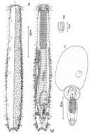
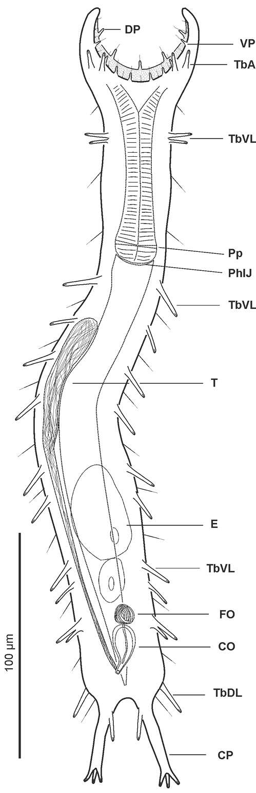
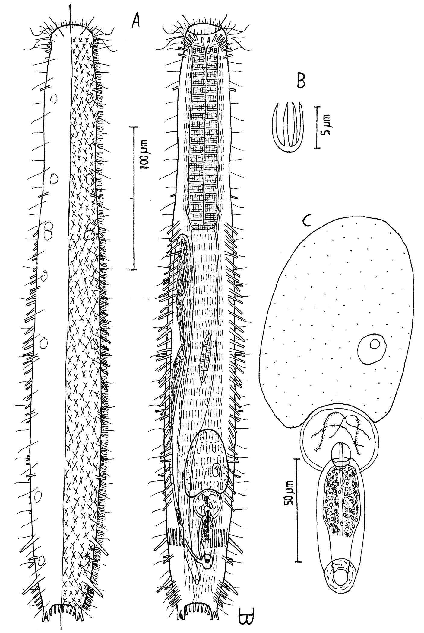
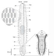
Figure 2.Pseudostomella dolichopoda sp. n. schematic drawing. Habitus as seen from the ventral side. CO caudal organ CP caudal pedicle DP dorsal papillae E egg FO frontal organ PhIJ pharyngeo-intestinal junction Pp pharyngeal pores T testicle TbA anterior adhesive tubes TbDL dorsolateral adhesive tubes TbL lateral adhesive tubes TbVL ventrolateral adhesive tubes VP ventral papillae.
-
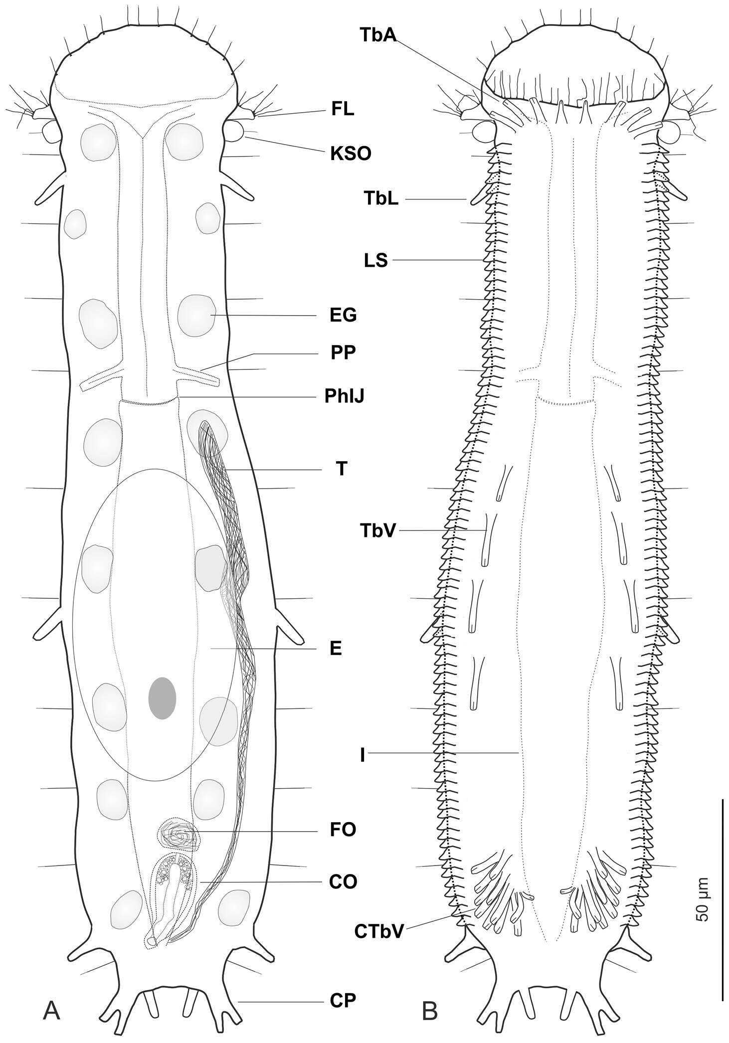
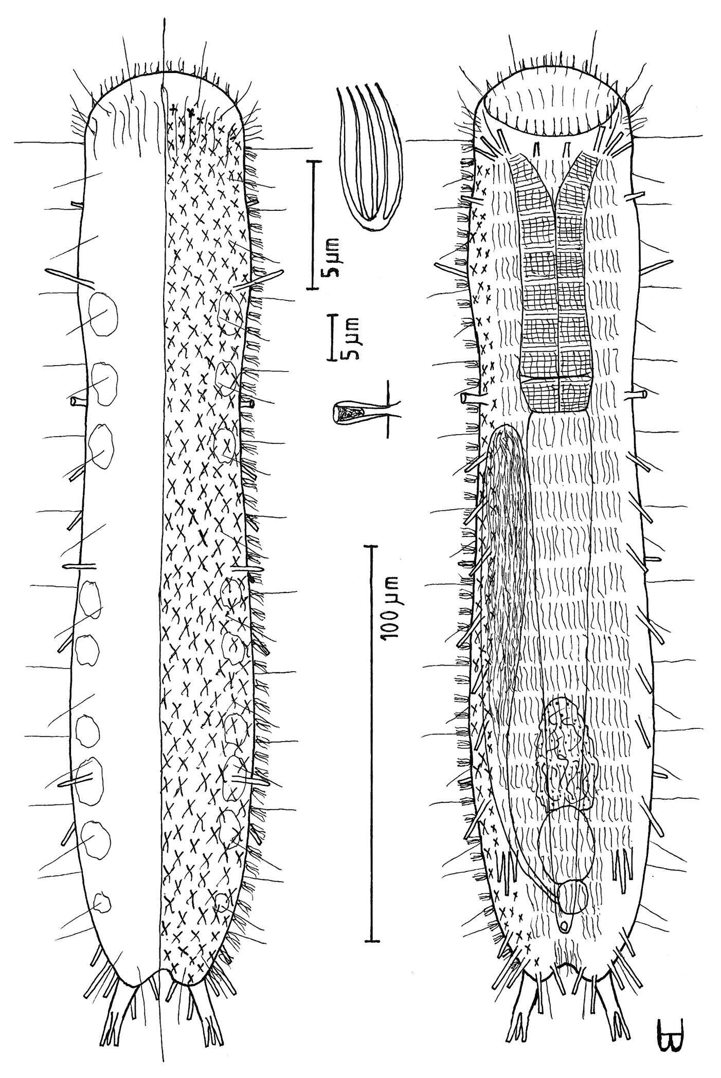
Figure 1.Ptychostomella lamelliphora sp. n. schematic drawings. A Habitus as seen from the dorsal side showing the internal anatomy B Habitus as seen from the ventral side. CO caudal organ CP caudal pedicle CTbV cluster of ventral adhesive tubes E egg EG epidermal gland FL fleshy lobe FO frontal organ I intestine KSO Knob-like sensory organ LS lamellate scales PhIJ pharyngeo-intestinal junction Pp pharyngeal pores T testicle TbA anterior adhesive tubes TbL lateral adhesive tubes TbV ventral adhesive tubes.
-
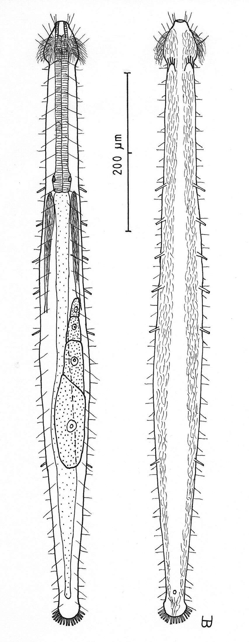
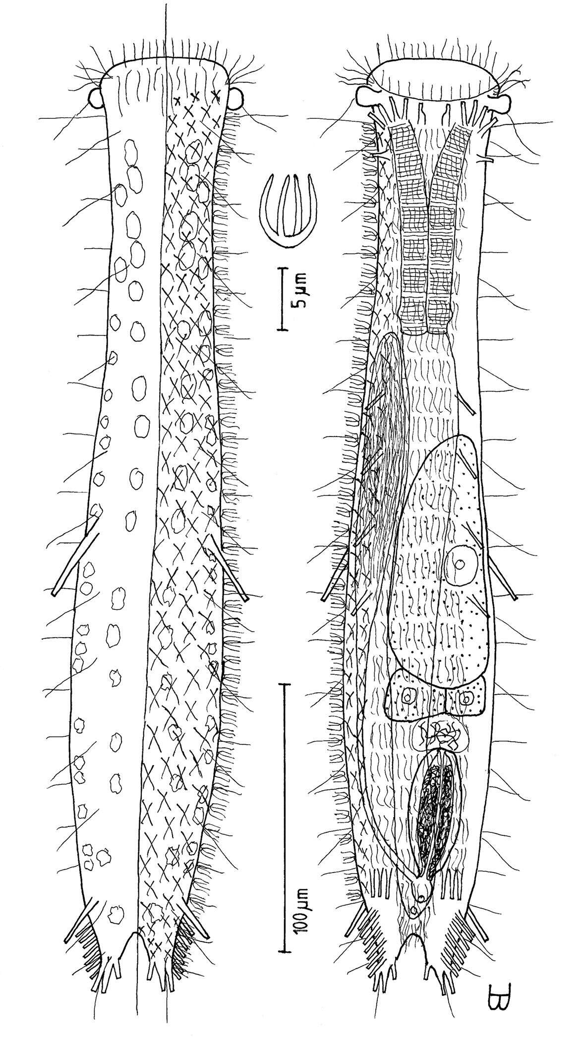
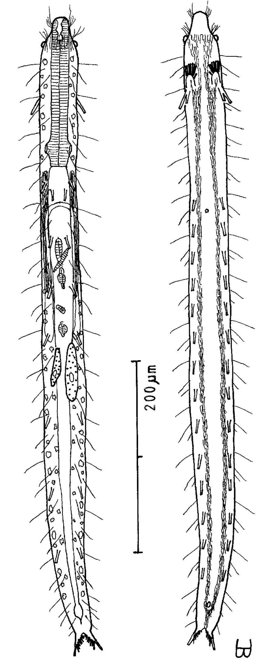
Figure 2.Cephalodasys dolichosomus sp. n. dorsal and ventral views of a mature adult (Lt=772, LPh=215 µm) from Main Beach, Ras Mohamed National Park, S. Sinai, Egypt; dorsal with dorsal and lateral body cilia, digestive and reproductive tracts; ventral with adhesive tubes and locomotor ciliary bands.
-
All Biocode files are based on field identifications to the best of the researcher’s ability at the time.
-
All Biocode files are based on field identifications to the best of the researcher’s ability at the time.
-
All Biocode files are based on field identifications to the best of the researcher’s ability at the time.
-
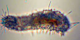
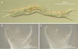
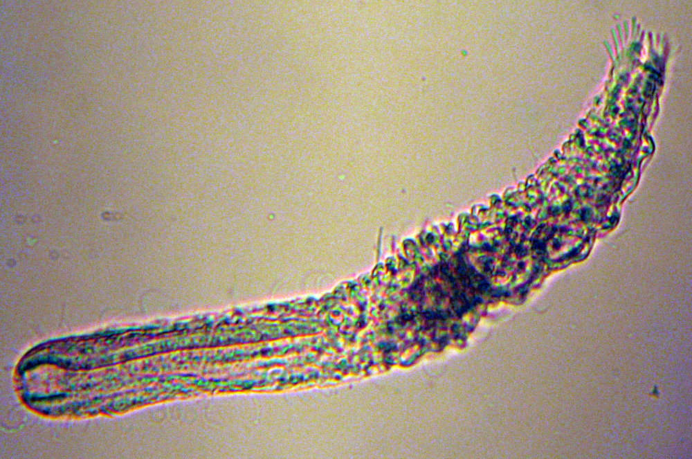
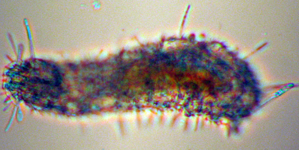
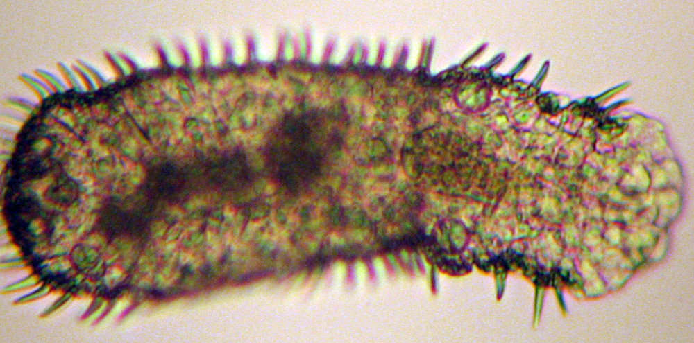
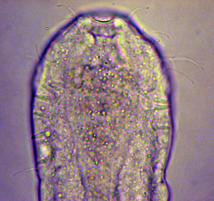
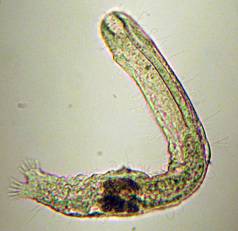
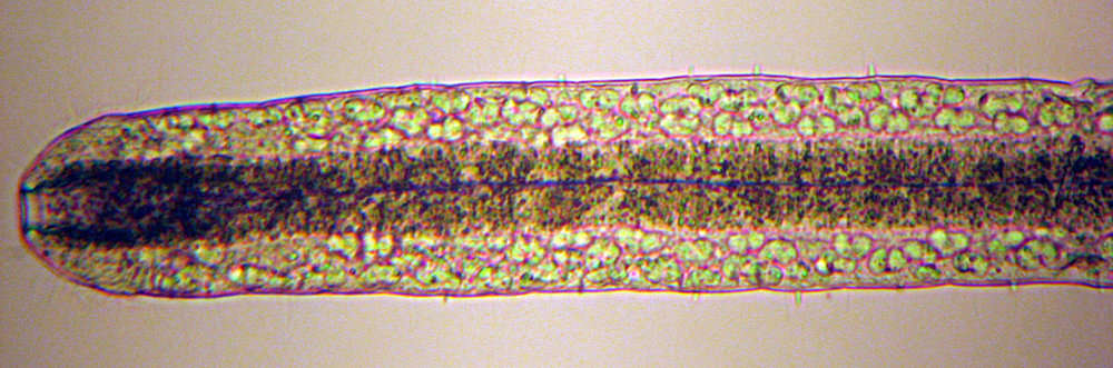
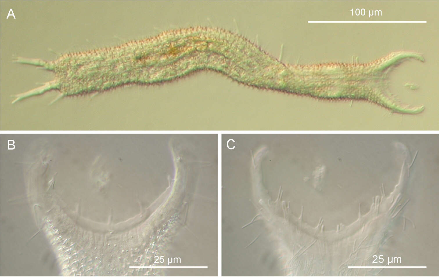
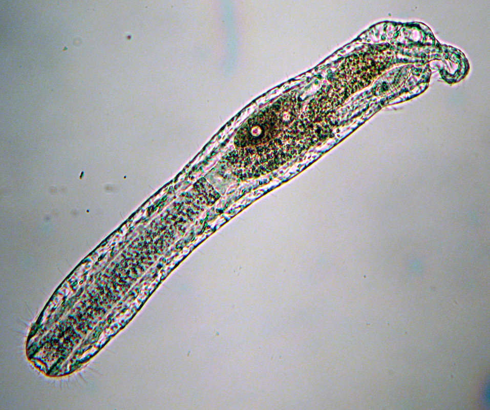
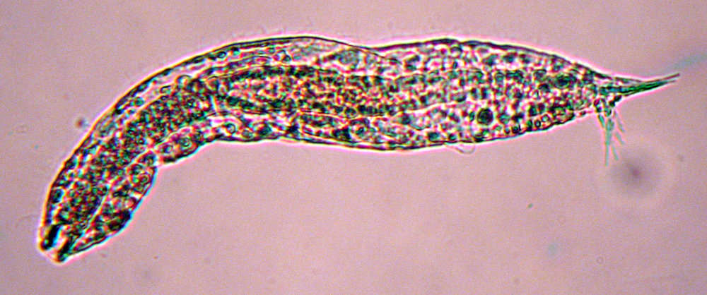
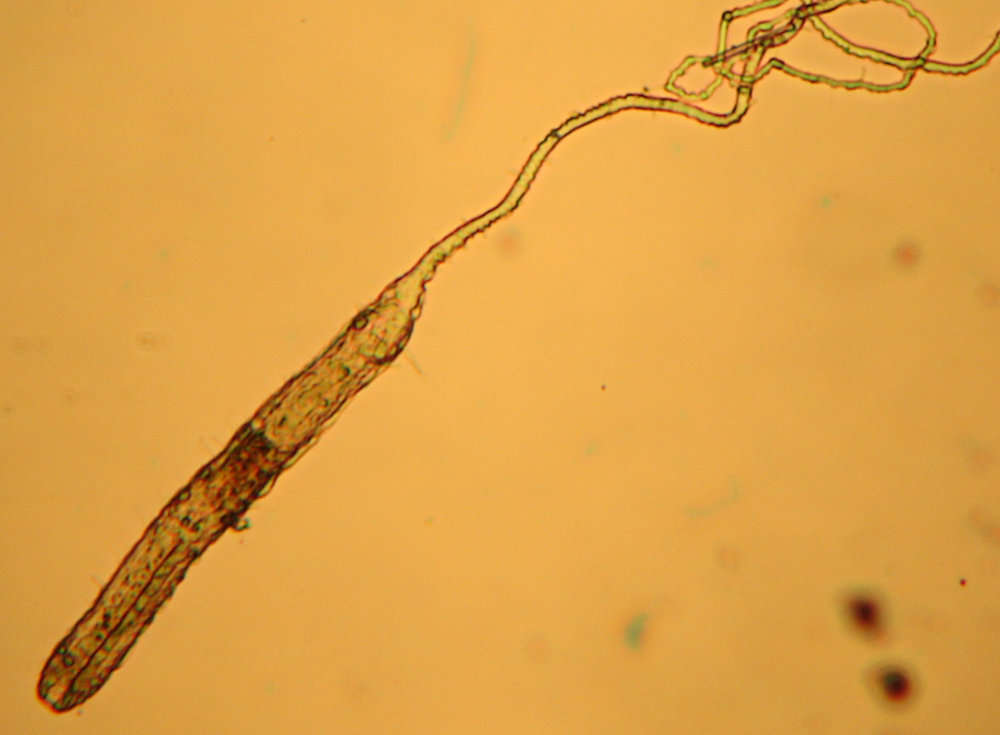
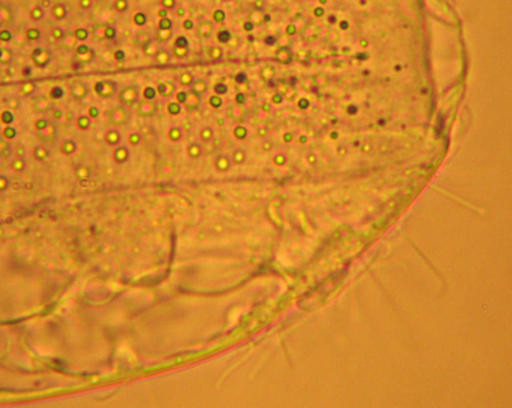
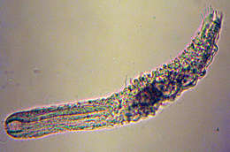
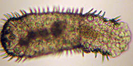
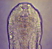
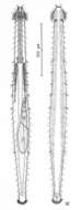
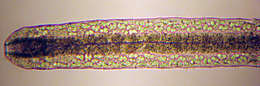
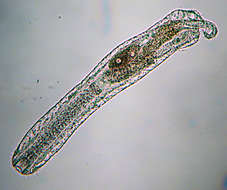
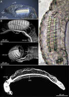
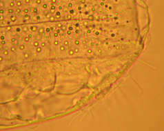
Figure 3.Pseudostomella dolichopoda sp. n. DIC photomicrographs. A habitus B close-up of the anterior region, dorsal view C Close-up of the anterior region, ventral view.
-
Rick Hochberg, Sarah Atherton, Vladimir Gross
Zookeys
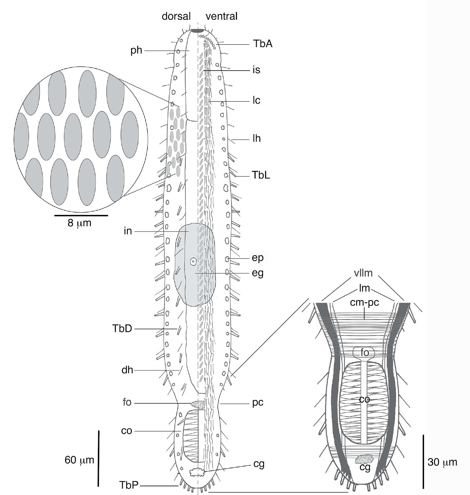
Figure 1.Schematic of Lepidodasys ligni sp. n. showing the crossed-helical scale pattern and a closeup of the musculature in the caudal region. Abbreviations: cg caudal gland; cm-pc circular musculature of the posterior constriction; co caudal organ; dh dorsal sensory hairs; eg mature egg; ep epidermal gland; fo frontal organ; in intestine; is interciliary scales; lc locomotory cilia; lh lateral sensory hairs; lm longitudinal muscle; pc posterior constriction; ph pharynx; TbA anterior adhesive tubes; TbD dorsal adhesive tubes; TbL lateral adhesive tubes; TbP posterior adhesive tubes; vllm ventrolateral longitudinal muscle.
-
Figure 14.Tetranchyroderma rhopalotum sp. n. A dorsal and ventral views of a mature adult (Lt=344, LPh=102 µm) from the Giftun Village Spit Outside, near Hurghada, Egypt; dorsal with tetrancrous surface (over half of the body), dorsal and lateral body cilia, and dorsal adhesive tubes; ventral with digestive and reproductive tracts, other adhesive tubes, and the locomotor ciliary band B dorsal tetrancre, with a separate scale bar.
-
All Biocode files are based on field identifications to the best of the researcher’s ability at the time.
-
All Biocode files are based on field identifications to the best of the researcher’s ability at the time.
-
All Biocode files are based on field identifications to the best of the researcher’s ability at the time.
-
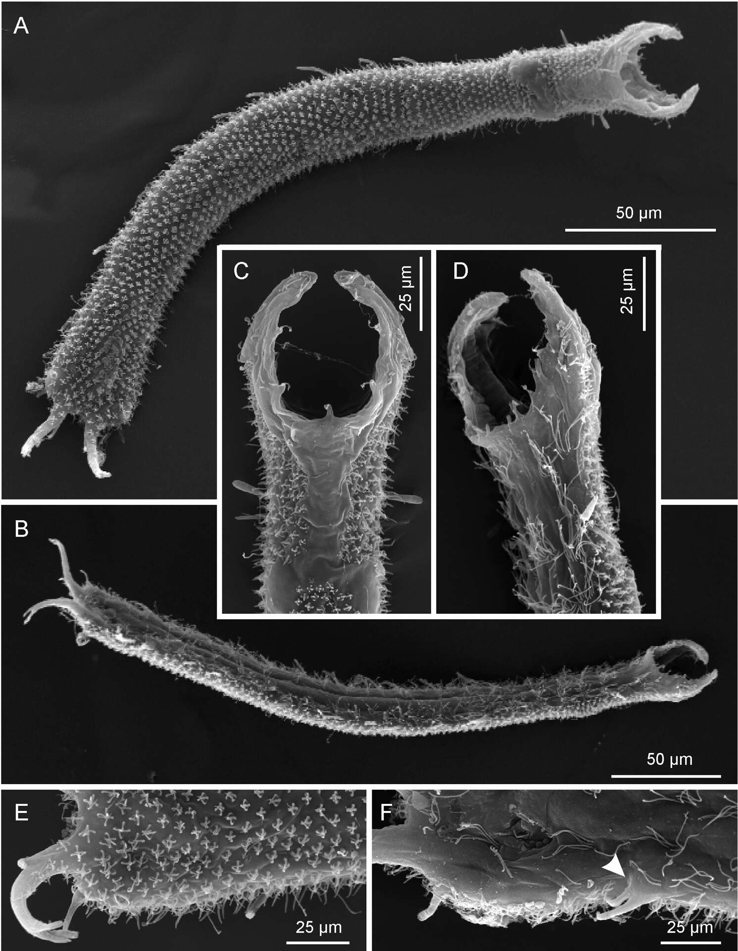
Figure 4.Pseudostomella dolichopoda sp. n. SEM photomicrographs. A habitus, dorsal view B habitus, ventral view; C close-up of the anterior region, dorsal view D close-up of the anterior region, ventrolateral view E close-up of the posterior region, dorsal view F close-up of the posterior region, ventral view, arrow shows the two ventrolateral adhesive tubes borne from a common base.
-
Rick Hochberg, Sarah Atherton, Vladimir Gross
Zookeys
Figure 3.The reproductive and muscular systems of Lepidodasys ligni sp. n. A Differential interference contrast photograph of the posterior end showing the accessory reproductive organs. B, C Confocal images (47 × 0.35 µm optical sections) of the musculature of the posterior end in lateral (B) and dorsal (C) views D Closeup of the caudal organ with DIC microscopy E Lateral view of an entire specimen revealing the muscular system (73 × 0.4 µm optical sections). Abbreviations: cg caudal gland; cm-co circular muscles of the caudal organ; cm-pc circular muscles of the posterior constriction; co caudal organ; coc caudal organ canal; dlm dorsal longitudinal muscle; dllm dorsal lateral longitudinal muscle; fo frontal organ; hm helicoidal muscle (end position on midgut); lh lateral sensory hair; ph pharynx; TbP posterior adhesive tube; scm somatic circular muscles (thoughout trunk); vllm ventrolateral longitudinal muscle.
-
Figure 15.Tetranchyroderma sinaiensis sp. n. A dorsal and ventral views of a mature adult (Lt=423, LPh=146 µm) from the Na’ama Bay, S. Sinai, Egypt; dorsal with tetrancrous surface (over half of the body), dorsal and lateral body cilia, and dorsolateral adhesive tubes; ventral with digestive and reproductive tracts, oyher adhesive tubes, and the locomotor ciliary band B dorsal tetrancre C caudal organ, frontal organ and ovum; B. and C. with separate scale bars.
-
All Biocode files are based on field identifications to the best of the researcher’s ability at the time.
-
All Biocode files are based on field identifications to the best of the researcher’s ability at the time.
-
Figure 16.Tetranchyroderma xenodactylum sp. n. A dorsal and ventral views of a mature adult (Lt=246, LPh=87 µm) from the Nabq, S. Sinai, Egypt; dorsal with pentancrous surface (over half of the body), dorsal and lateral body cilia, and TbDL; ventral with digestive and reproductive tracts, adhesive tubes, and the locomotor ciliary band B dorsal pentancre C the strange finger-like structure that protrudes laterally at the PhJIn; B. and C. with separate scale bars.
-
Figure 17.Paraturbanella levantia sp. n. dorsal and ventral views of a mature adult (Lt=657, LPh=163 µm) from Bir Mesud, Alexandria, Egypt; dorsal with pestle organs, pattern of glands, dorsal and lateral body cilia, digestive and reproductive tracts; ventral with adhesive tubes and locomotor ciliary bands.