-
Jaime Gonzalez-Cueto, Sigmer Quiroga, Jon Norenburg
Zookeys
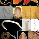
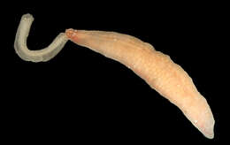
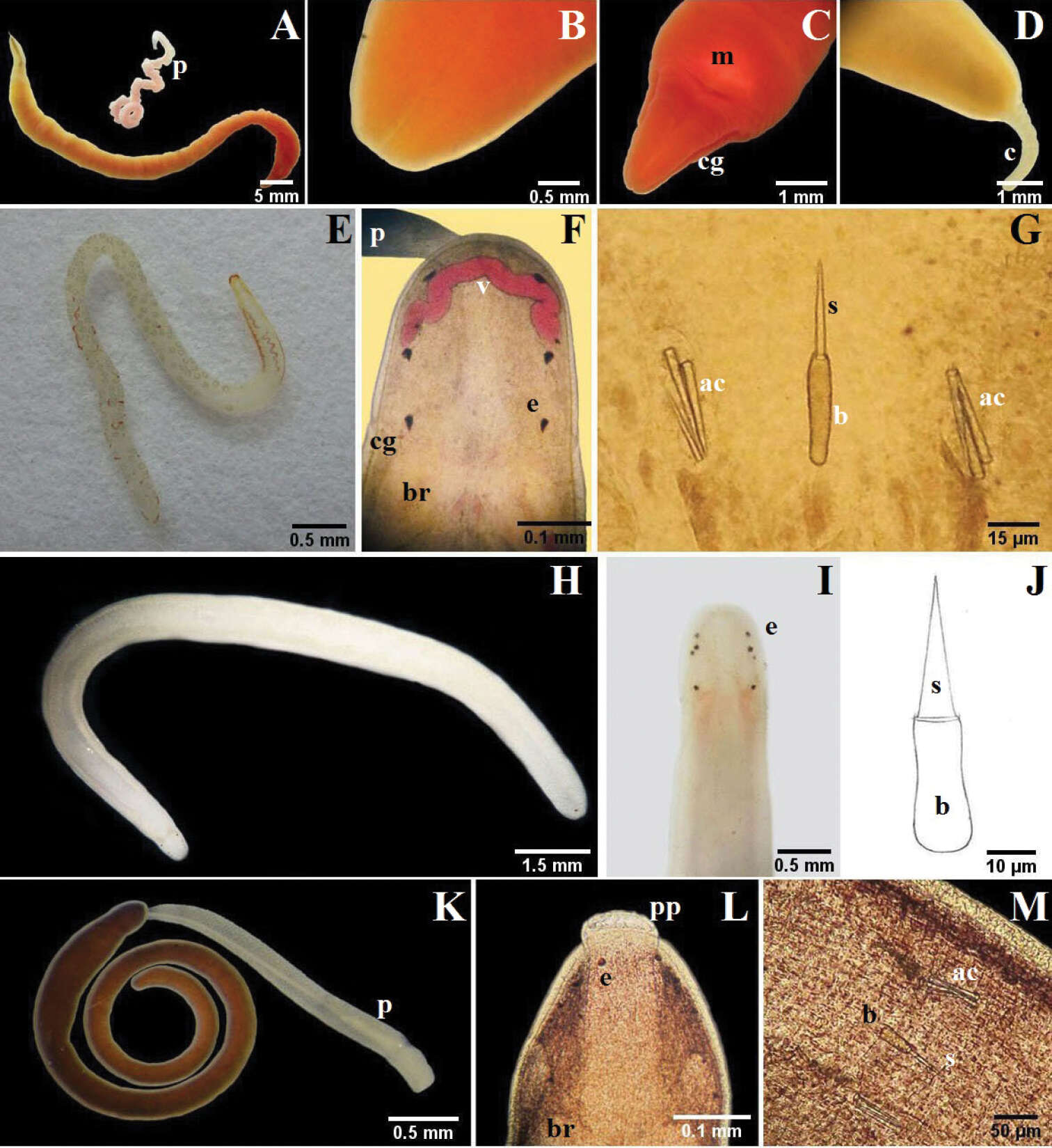
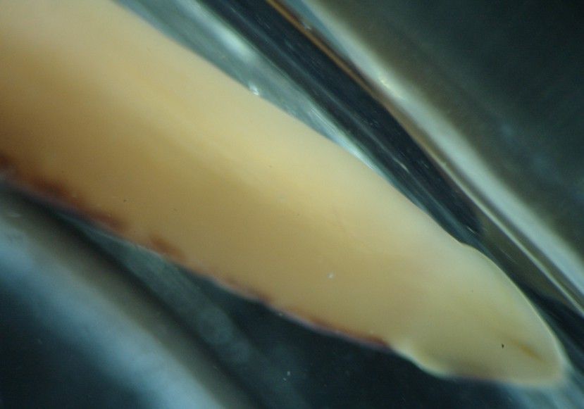
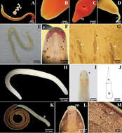
Figure 3.A–D Micrura ignea: A entire specimen, the worm has expulsed the proboscis B dorsal detail of the head C ventral detail of the head D detail of the tail E–G Amphiporus cruentatus: F dorsal detail of the head G detail of the stylets H–J Amphiporus cf. ochraceus: I dorsal detail of the head J drawing of the stylet K–M Amphiporus texanus: K entire worm L dorsal detail of the head M detail of the stylets. ac accessory stylet, b base of the stylet, br brain, c cirrus, cg cephalic grooves, e eyes, m mouth, p proboscis, pp proboscis pore, s sylet, v blood vessel.
-
Often has this obvious reddish "brain" visible through the body wall. In Sweden they are often very pale, but in the U.K they can be considerably darker.
-
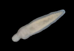
App 3 cm long. Characteristic furrows and ridge on head.
-
-
Santa Pola, Valencia, Spain
-
Touro, Galicia, Spain
-
Jaime Gonzalez-Cueto, Sigmer Quiroga, Jon Norenburg
Zookeys
Figure 3.A–D Micrura ignea: A entire specimen, the worm has expulsed the proboscis B dorsal detail of the head C ventral detail of the head D detail of the tail E–G Amphiporus cruentatus: F dorsal detail of the head G detail of the stylets H–J Amphiporus cf. ochraceus: I dorsal detail of the head J drawing of the stylet K–M Amphiporus texanus: K entire worm L dorsal detail of the head M detail of the stylets. ac accessory stylet, b base of the stylet, br brain, c cirrus, cg cephalic grooves, e eyes, m mouth, p proboscis, pp proboscis pore, s sylet, v blood vessel.
-
-
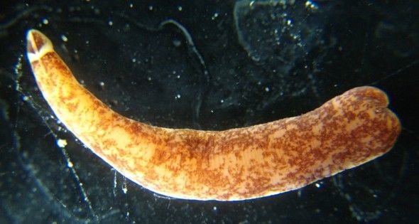
The animal has a pair of raised longitudinal middorsal ridges (colored white on its head). The ridges, separated by a darker pigmented area, continue back along the body for several times the length of the head then fade out. This individual is hard to focus on because it is crawling rapidly. Photo by Dave Cowles, July 2012
-
-
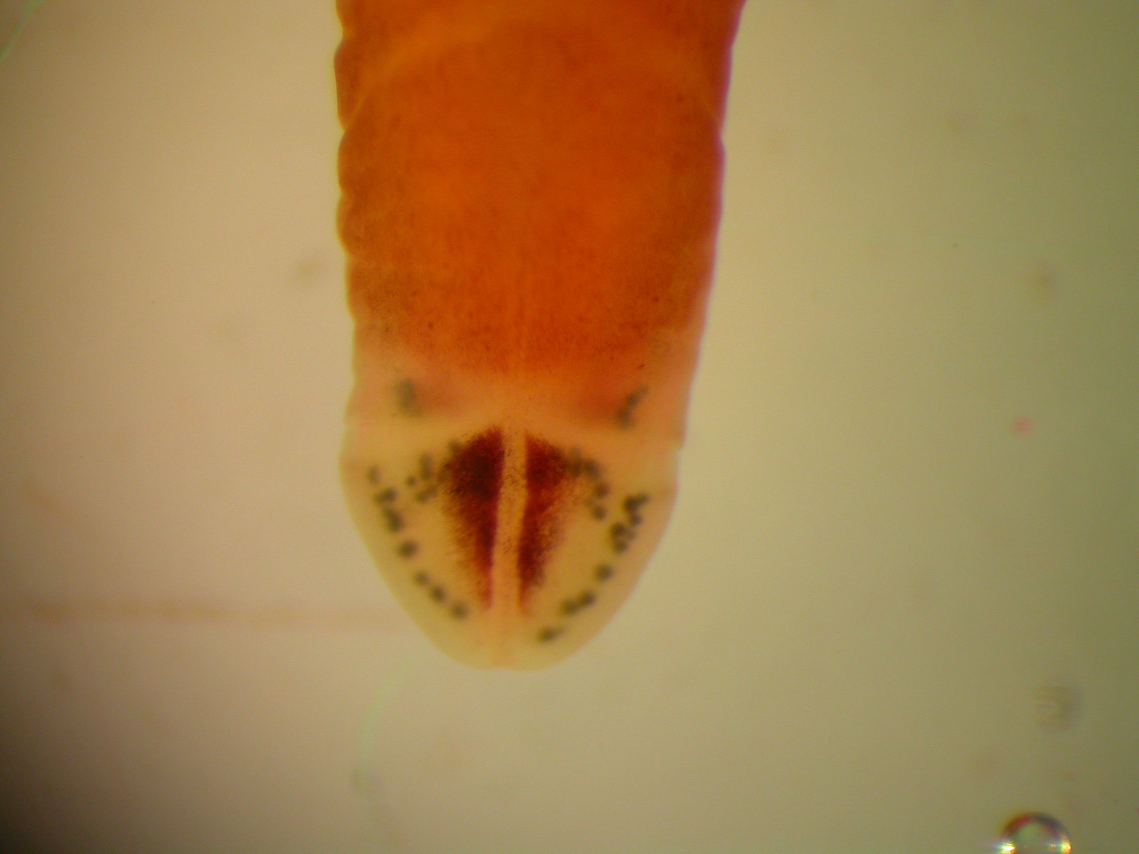
This view of the side of the head shows rows of numerous black eyes. Photo by Dave Cowles, July 2012
-
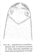
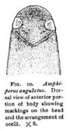
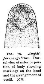
Dorsal view of anterior portion of body showing markings on the head and the arrangement of ocelli.Coe, W. R. (1901). The Nemerteans of the Expedition. Proceedings of the Washington Academy of Sciences, Vol. 3, 1-110.
-
This view of the underside of the head (the worm is crawling on the water's surface film) shows the anterior but ventral mouth. Photo by Dave Cowles, July 2012
-
-
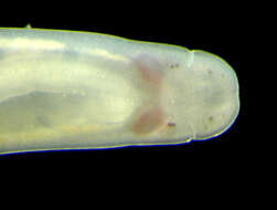
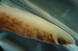
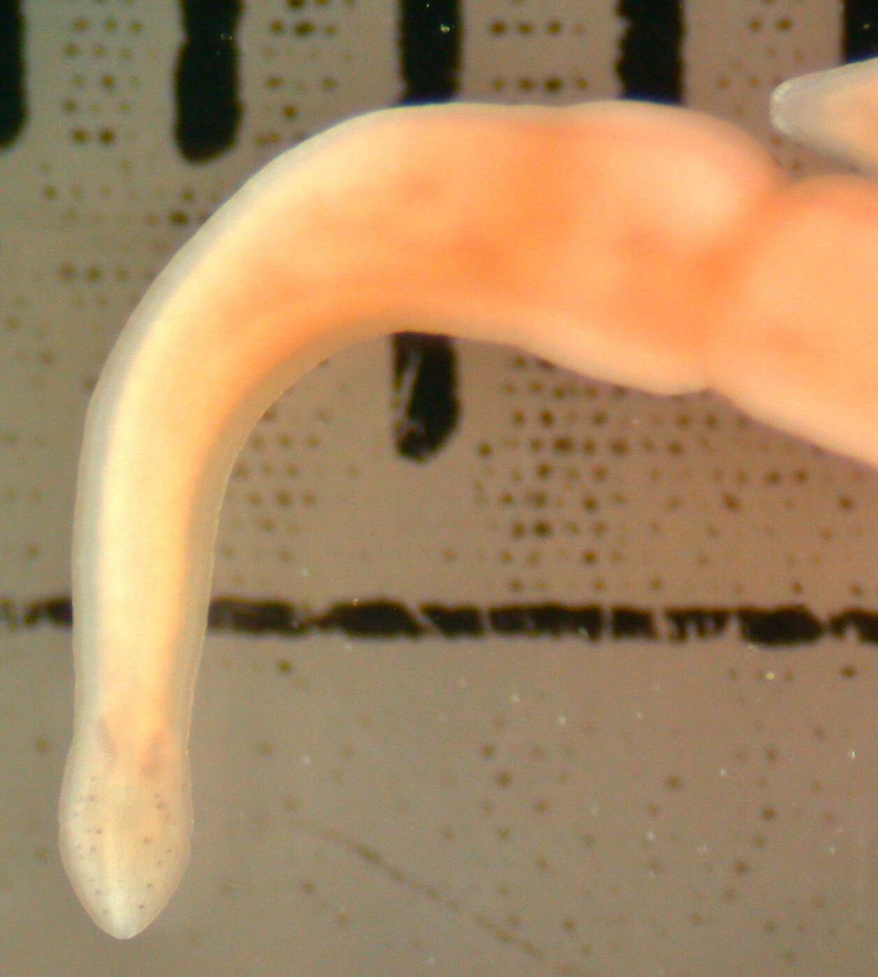
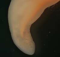
Amphiporus bimaculatus, probably an anterior fragment (Photo by: Dave Cowles, July 2012)
-
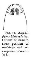
Outline of head to show position of markings and arrangement of ocelli.Coe, W. R. (1901). The Nemerteans of the Expedition. Proceedings of the Washington Academy of Sciences, Vol. 3, 1-110.
-
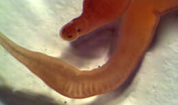
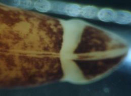
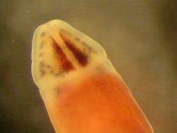
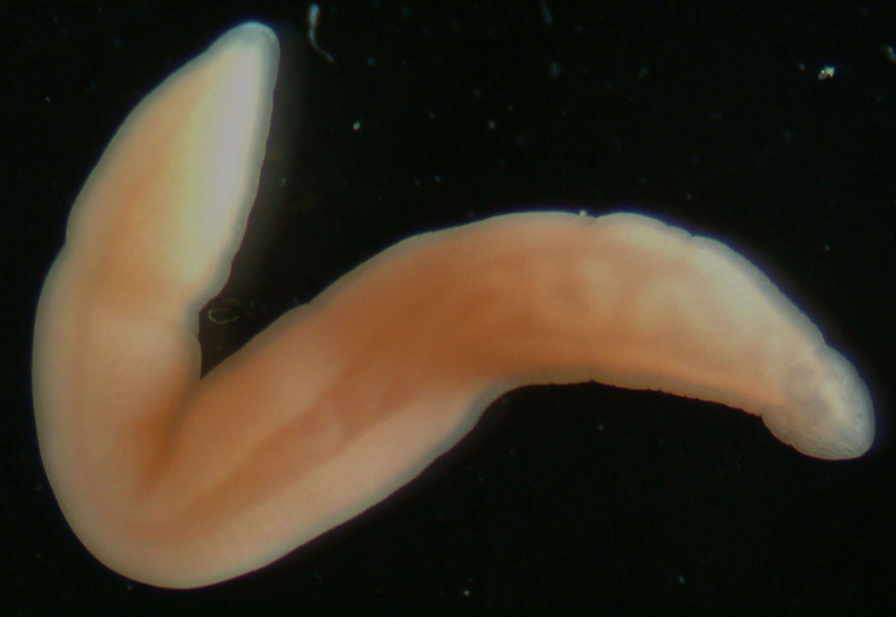
Amphiporus bimaculatus: head
-
Amphiporus bimaculatus: head
-
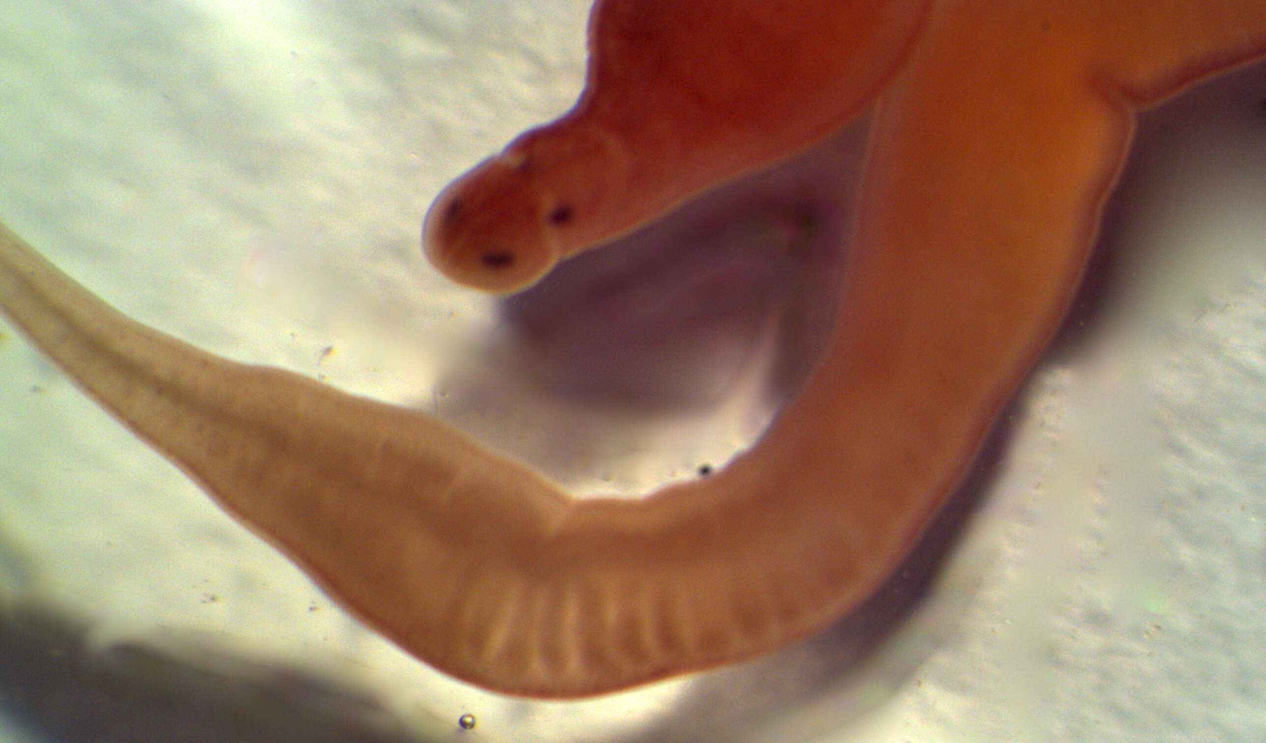
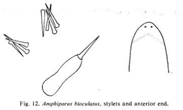
Stylets and anterior endBrunberg, L. On the Nemertean Fauna of Danish Waters. Ophelia, 1(1), 77-111.
-
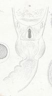
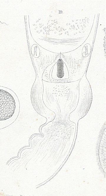
Plate13.19 Stylet-region of Amphiporus bioculatus
-
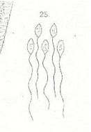
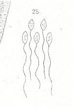
Plate7.25 Spermatozoa of Amphiporus bioculatus
-
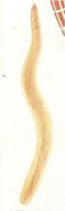
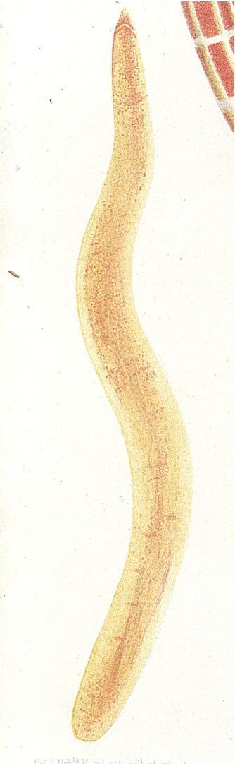
Plate8.3 Amphiporus bioculatus n. s.