Automatic identification method for seroperitoneum B-mode ultrasound image

A technology for ascites and automatic identification, applied in the field of biomedicine, can solve the problems of insufficient mastery of ultrasound examination technology, unable to achieve unified and standardized operation, difficult to make accurate judgments, etc., and achieve the effect of reducing subjective limitations.

Pending Publication Date: 2021-06-25
SHANGHAI CHANGZHENG HOSPITAL +1
View PDF7 Cites 0 Cited by
  • Summary
  • Abstract
  • Description
  • Claims
  • Application Information

AI Technical Summary

Problems solved by technology

It has become an important indicator for judging whether there is abdominal organ injury in the assessment of the injury condition of critically ill patients. However, due to the lack of mastery of ultrasound examination technology by clinical front-line d

Method used

View more

Image

Smart Image Click on the blue labels to locate them in the text.
Viewing Examples
Smart Image
  • Automatic identification method for seroperitoneum B-mode ultrasound image
  • Automatic identification method for seroperitoneum B-mode ultrasound image
  • Automatic identification method for seroperitoneum B-mode ultrasound image

Examples

Experimental program
Comparison scheme
Effect test

Embodiment 1

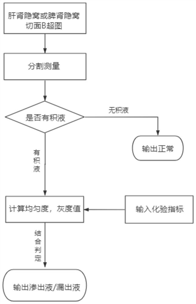
[0052] refer to figure 1 , the present embodiment provides a method for automatic identification of peritoneal effusion B-ultrasound images, comprising the steps of:

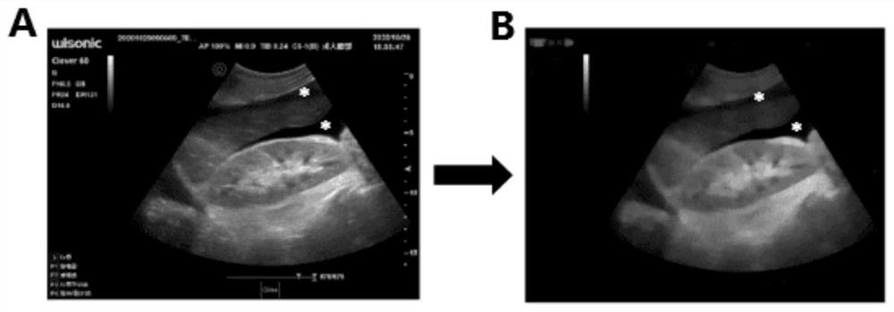
[0053] Step 1, establish a data set: do a transverse scan at the 9th or 10th intercostal space on the posterior axillary line of the patient, move the probe up and down to obtain B-ultrasound images of the hepatic and renal recesses; place the probe on the 10th or 11th rib of the left hypochondriac At the same time, B-ultrasound images of the left intercostal spleen and kidney oblique section were obtained; at the same time, ascites samples were collected by paracentesis, and basic information such as age, height, and weight of the patients were recorded, and data sets were established respectively. The data in this example come from B-ultrasound images of volunteers from Shanghai Changzheng Hospital, including different peritoneal effusions.

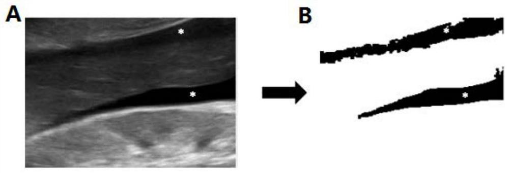
[0054] Step 2, preprocessing the image and segmenting the peritonea...

Login to view more

PUM

No PUM Login to view more

Abstract

The invention provides an automatic identification method for a seroperitoneum B-mode ultrasound image, and the method comprises the following steps: 1, respectively carrying out ultrasonic scanning on a standard section of a liver and kidney crypt or a spleen and kidney crypt to obtain a B-mode ultrasound image marked with seroperitoneum, and establishing a seroperitoneum data set; 2, performing image processing on the acquired B-mode ultrasound image and segmenting a seroperitoneum area in the image; 3, carrying out pixel statistics on the segmented seroperitoneum area, carrying out quantitative analysis on seroperitoneum conditions, and judging whether seroperitoneum exists or not; 4, carrying out characteristic value calculation on the segmented seroperitoneum region, carrying out correlation analysis on the characteristic value and a laboratory test and examination result, and judging the type of the seroperitoneum. According to the method provided by the invention, the seroperitoneum area in the B ultrasonic image can be automatically segmented, quantitative and qualitative analysis is performed on the seroperitoneum area, subjective limitation of manual judgment is reduced, result judgment is quickly and accurately performed on the seroperitoneum condition, and the method is suitable for large-scale popularization.

Description

technical field [0001] The invention relates to the technical field of biomedicine, in particular to an automatic B-ultrasonic image recognition method for peritoneal effusion. Background technique [0002] Trauma is a common problem facing the world today. Patients with severe trauma are critically ill and require timely medical assistance. The key is that clinicians can quickly and accurately assess the patient's condition. Ultrasound has become an important means of bedside evaluation because of its simplicity, convenience, safety, effectiveness, and ease of repeatability. Internal hemorrhage caused by blunt injury of abdominal viscera or ascites secondary to other serious diseases can easily accumulate in the lowest part of the abdominal cavity. Ultrasound can sensitively identify the ascites in the lowest hepatic and renal recesses in the supine position. It has become an important indicator for judging whether there is abdominal organ injury in the assessment of the i...

Claims

Login to view more

Application Information

Patent Timeline
no application Login to view more
IPC IPC(8): A61B8/00
CPCA61B8/5215
Inventor 赵佳琦周洁文李晓兵周潇忱马小琳聂生东
Owner SHANGHAI CHANGZHENG HOSPITAL
Who we serve
  • R&D Engineer
  • R&D Manager
  • IP Professional
Why Eureka
  • Industry Leading Data Capabilities
  • Powerful AI technology
  • Patent DNA Extraction
Social media
Try Eureka
PatSnap group products