


What are Pinguecula (yellowish patch or bump on the white of the eye) and Pterygium (surfer’s eye)?
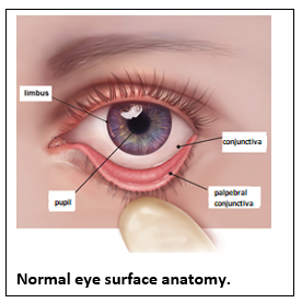
Pinguecula and Pterygium; The membrane structure that covers the white part of our eye is called the conjunctiva. The clear glass-like tissue in the front of the eye is the cornea.
The border of the cornea and conjunctiva is formed by the limbus. Pinguecula and pterygium are abnormal tissue growths on the membrane that covers the white of the eye (conjunctiva).
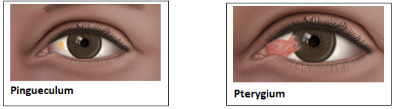
Pinguecula is a yellow-white, raised tissue growth on the conjunctiva. It does not cross the limbus border. It usually develops as a result of calcium, cholesterol and protein accumulation on the white area of the eye on the nose side. It is usually seen in middle-aged and elderly people.
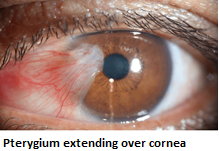
Pterygium is a vascular, triangular tissue growth on the inner edge of the eye that extends from the limbus to the cornea. When it covers the cornea, it can cause vision loss. It is also known as a fleshy growth on the eye due to its appearance.
Why do pinguecula and pterygium develop? In whom are these diseases more common?
It is thought that intense exposure to ultraviolet light from the sun and being in a dusty and windy environment play a role in the development of both pinguecula and pterygium. Therefore, it is more common in occupations that require long-term work in the open air, such as sailors and farmers. It is very rare in children. These diseases are most common between the ages of 30-50. It is more common in light-skinned and colored-eyed people than in dark-colored people.
What can be done to prevent the development of pinguecula and pterygium?
Measures to be taken to prevent the development of pinguecula and pterygium or to slow their growth:
- Use of sunglasses to protect from UV rays
- Use of protective glasses if working in a dusty environment
- Use of artificial tear drops if the eyes are dry
What are the symptoms of Pinguecula and Pterygium?
 Complaints in pinguecula and pterygium vary by the size of the lesion. In the early period, most patients do not have any complaints and do not require treatment. In general, both diseases show redness and swelling on the conjunctiva.
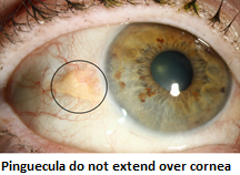
The pinguecula is more yellowish in color and does not extend to the cornea. Due to the swelling in the conjunctiva, the distribution of tears is disturbed and patients experience burning, stinging and foreign body sensation. It may be necessary to use artificial tear drops at this stage.
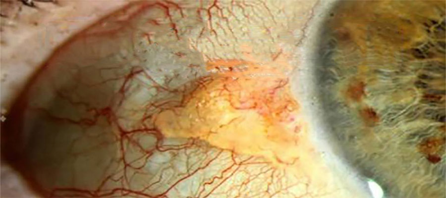
In the pterygium, a vascular tissue extending over the cornea is seen. In hot environments (after a bath, etc.) the veins expand, so the redness increases and the lesion appears to have enlarged. If the pterygium extends centrally on the cornea, it may obscure the pupil or cause astigmatism. In this case, the patient has a decrease in vision.
Pinguecula and Pterygium treatment
Most of the time, treatment is not necessary for pinguecula and pterygium. However, if the lesions cause discomfort in the eyes or loss of vision or if the appearance is aesthetically disturbing, treatment becomes necessary.
Pinguecula:
- Artificial tear drops reduce discomfort and foreign body sensation in the eyes.
- Cortisone eye drops can be used temporarily in cases such as excessive redness and swelling.
Pterygium:
Treatment of pterygium depends on the size of the lesion, its growth rate, and the complaints it causes. If the pterygium is small, cortisone drops and artificial tear drops are sufficient for redness and stinging complaints. However, if the pterygium extends towards the pupil, enlarges during follow-up and reduces vision, the only treatment option is surgery. Additionally, if the appearance of the pterygium bothers the patient, surgery can be performed for aesthetic purposes.
Pterygium surgery:
There are many different techniques in pterygium surgery. Clinical studies and research have shown that the best method is the conjunctival autograft (stem cell transplant) technique. In this surgery, the pterygium tissue is cut and removed.
Conjunctival tissue taken from the intact area is transplanted to the place where the removed tissue is. Along with the new tissue, healthy stem cells are also transplanted. With this method, both the recurrence of the disease is less and less scars remain in the recovering area.
Aesthetically, the best image is provided. The surgery takes about 30 minutes. The surgery is performed under local anesthesia. Drops are used for a few weeks after surgery.