
Microcytic Hypochromic Anemia: Causes and Diagnosis
let us explore the basics of microcytic hypochromic anemia.
What is Microcytic Hypochromic Anemia?
Microcytic hypochromic anemia is a specific type of anemia characterized by red blood cells that are smaller than usual and have reduced hemoglobin content. These characteristics give them their names – “microcytic” for small size and “hypochromic” for reduced hemoglobin.
Common Causes of Microcytic Hypochromic Anemia
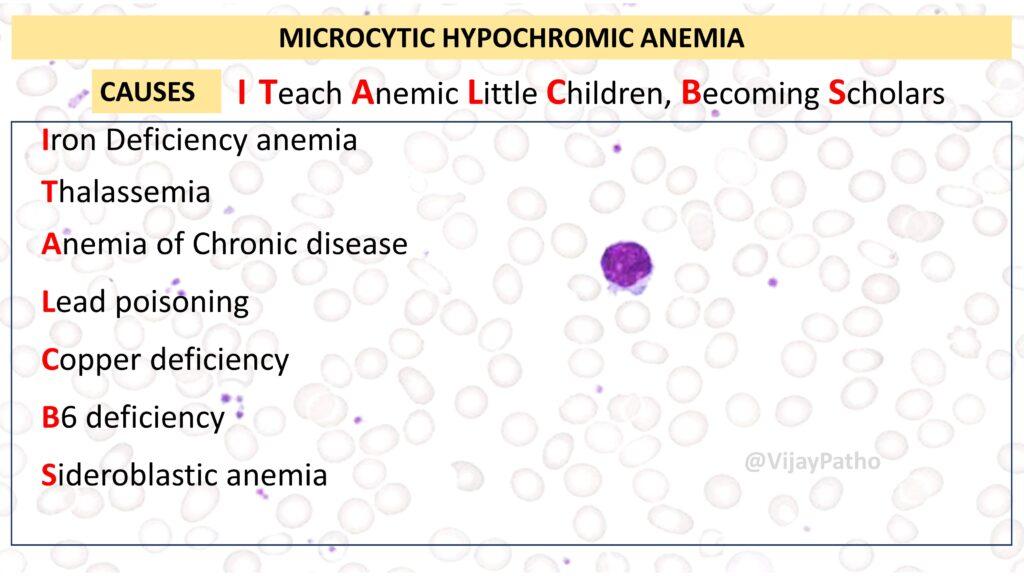
To remember the common causes of microcytic hypochromic anemia, you can use the mnemonic: “I Teach Anemic Little Children Becoming Scholars.” Let’s break it down:
I for Iron Deficiency Anemia: Inadequate iron supply hampers hemoglobin production.
T for Thalassemia: Genetic conditions disrupt globin chain synthesis, leading to reduced hemoglobin production.
A for Anemia of Chronic Disease: Inflammatory signals hinder iron availability for red blood cell formation.
L for Lead Poisoning: Lead inhibits enzymes critical for hemoglobin synthesis.
C for Copper Deficiency: Reduced copper levels affect iron release, leading to inadequate hemoglobin production.
B for B6 Deficiency: Vitamin B6 is essential for heme synthesis, and its deficiency can result in microcytic anemia.
S for Sideroblastic Anemia: This condition involves the disruption of heme synthesis, often due to genetic factors.
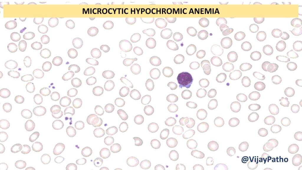
 Morphology of Microcytic Hypochromic Anemia
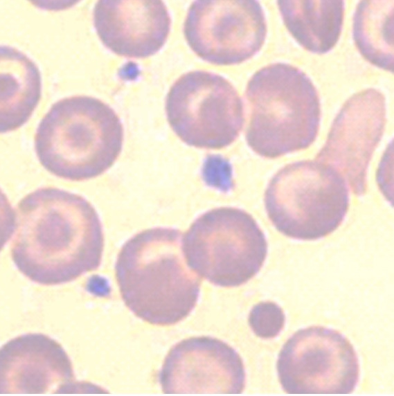
Microcytic hypochromic anemia is characterized by small-sized red blood cells with reduced hemoglobin content. Peripheral blood smears may also show variable sizes and shapes of red blood cells, known as anisocytosis.
Diagnostic Approach
When diagnosing microcytic hypochromic anemia, clinicians typically start with a serum iron profile, including:
Serum iron estimation
Serum ferritin levels
Total iron binding capacity (TIBC)
Transferrin levels
Based on the results, different scenarios may arise:
Decreased serum iron and ferritin with increased TIBC: Suggests iron deficiency anemia.
Decreased serum iron, variable ferritin, and decreased TIBC: Indicates anemia of chronic disease.
Increased serum iron and ferritin with normal TIBC and dimorphic blood smear: May point to sideroblastic anemia.
Increased serum iron and ferritin with decreased TIBC and basophilic stippling of RBCs: Suggests lead poisoning.
All parameters are normal: Perform hemoglobin electrophoresis to check for thalassemia.
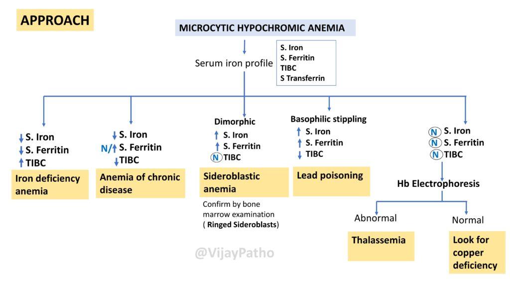
Remember that this approach is based on serum iron profile results, and there are additional, less common causes of microcytic hypochromic anemia that may require further investigation.
Thank you for reading, and if you found this information helpful, stay tuned for more informative content.
Watch the video here


Recent Comments