
Introduction
Oral ulceration is a painful, common clinical finding, affecting up to 25% of the population, with many seeking advice and treatment from their general dental practitioner (GDP). The aetiology of oral ulceration can range from trauma to the oral mucosa, to an underlying systemic disease, a side effect to medications, or recurrent aphthous stomatitis (RAS). Rarely, an ulcer may be the presenting finding of an intraoral malignancy. Therefore, it is essential to understand the aetiology of the ulceration as this underpins its management.
Ulceration may be managed within the primary dental care setting by removing any causative factors, such as trauma sources if present, and managing the patient’s symptoms with topical treatments such as local anaesthetic sprays and topical corticosteroids. In some cases, liaising with the patient’s general medical practitioner (GMP) may be necessary, in order to investigate any underlying deficiency states (iron, B12, folate) or systemic diseases. However, in cases involving ulceration resistant to topical treatments, severe ulceration or suspected malignancy, referral to a local oral medicine or oral and maxillofacial unit is required.
Aetiology of oral ulceration
Traumatic ulceration
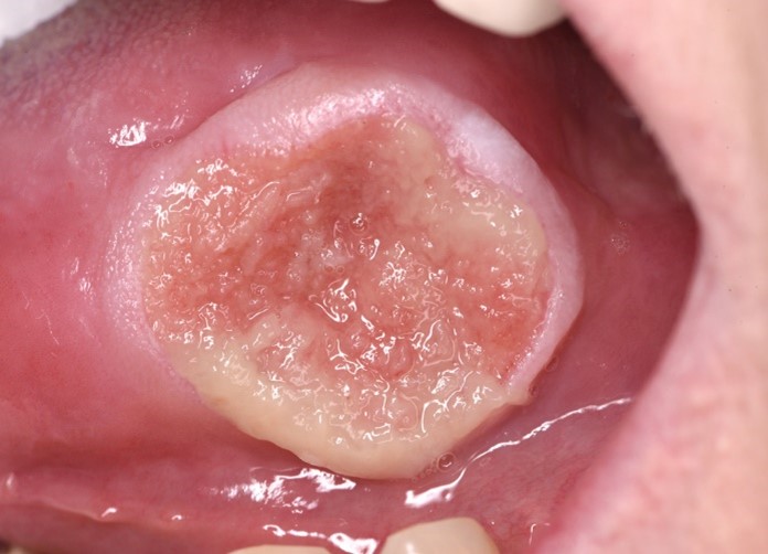
Trauma to the oral mucosa from a fractured tooth, a sharp cusp or a denture clasp may result in the formation of an ulcer (Table 1). In these cases, ulcers are variable in size and appearance (Figure 1). They are often painful with raised white borders with a yellow base, and tend to affect the buccal mucosa, tongue, and lower lip.

Recurrent aphthous stomatitis
RAS ulceration usually affects the non-keratinised mucosa. Its aetiology is unclear, but it is thought to be driven by a dysfunctional cell-mediated immune response (Table 2).1 Predisposing factors include: stress; trauma; deficiency states (iron, B12 or folate); smoking cessation; menstruation; or, the use of sodium lauryl sulfate- (SLS) containing toothpaste. There are three distinct forms of aphthous ulceration, which are characterised by:
-
the size of the ulcer;
-
the number of ulcers present at one time;
-
the length of time to resolve; and,
-
the presence of any scarring after healing.
Types of RAS
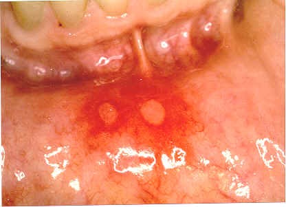
Minor aphthae: painful small ulcers (usually <1cm), have a fibrinous base surrounded by a red inflammatory halo and occur on the non-keratinised mucosa. Ulcers appear in crops of one to six that heal within 10 days in the absence of scarring (Figure 2).
Major aphthae: differ from minor aphthae in their size (>1cm), pain and healing time, which will usually be several weeks. When major aphthae heal there can be scarring present.
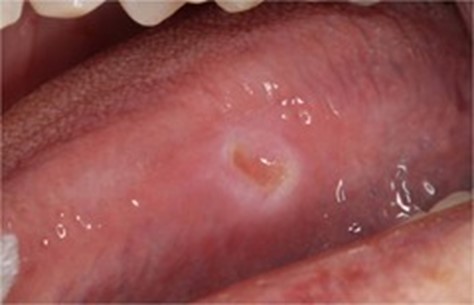
Herpetiform ulceration: the least common form of RAS. These are multiple pinpoint ulcers ranging in size from 1-3mm that are markedly painful and coalesce into larger ulcers after a few days. They can take approximately 14 days to heal but do not scar helping to differentiate them from major aphthae. They tend to affect the ventral tongue and upper lip (Figure 3).


Systemic causes
Systemic disease can result in ulceration, which has the same clinical features as ulceration in RAS. However, the aetiology differs from RAS as the ulceration is secondary to the systemic disease rather than due to immune dysregulation in RAS.
Gastrointestinal diseases such as coeliac disease or Crohn’s disease may cause oral ulceration. It can be helpful to ask any patient presenting with ulceration whether they have had any gastrointestinal symptoms (Tables 3 and 4). An onward referral to the patient’s GMP would be prudent for further investigation. This may initially include blood investigations to detect any deficiency states resulting from blood loss or malabsorption, and a faecal calprotectin test, which may be elevated in patients with active gastrointestinal inflammation. The patient’s ulceration can be managed symptomatically in the same way as with RAS. However, appropriate management of their gastrointestinal disease may lead to the resolution of the ulceration.
Multisystem inflammatory disorders such as Beçhet’s disease and MAGIC syndrome (a rare syndrome characterised by features of both Beçhet’s disease and the recurrent inflammation of cartilage as seen in relapsing polychondritis) can also cause oral ulceration. These patients may also suffer from genital ulceration and ocular manifestations.2 Therefore, when taking an ulcer history, it is important to enquire if patients experience ulceration on other body sites.
Various autoimmune conditions can cause extensive oral ulceration. Pemphigus vulgaris affects the oral mucosa (as well as other mucous membranes) causing vesicles, which quickly burst to leave superficial erosions with ragged edges before the onset of skin vesicles or bullae, which also break down to give painful ragged erosions. Patients presenting with these symptoms should be urgently referred to an oral medicine consultant or dermatologist for assessment and management, which involves immunosuppressive treatment.
More commonly seen in patients presenting to their GDP is lichen planus. In its erosive form, there are shallow areas of ulceration with a yellow fibrin plaque covering the surface of the erosion. Sometimes these patients will have striae at the periphery of the ulcerated area and may also have skin involvement with <5mm itchy purplish papules with a shiny surface, and striae often on the forearms and wrists.
Drug-induced oral ulceration
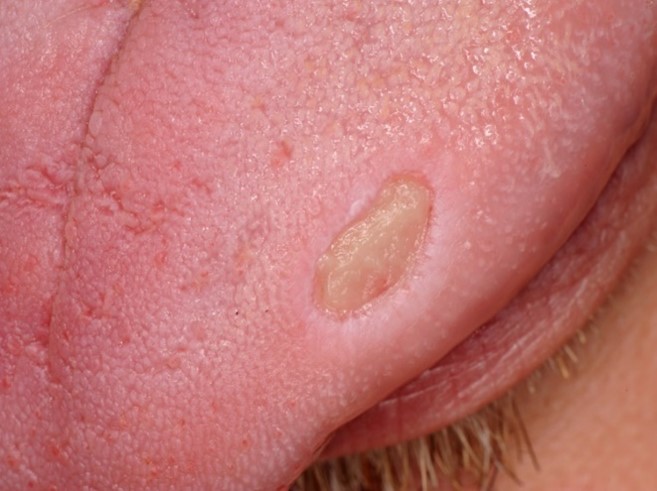
Oral ulcers may be induced by systemic medications and can present as single or multiple areas of ulceration.3 Drug-induced ulcers are typically flat with a whitish base and a raised clear margin (Figure 4). These ulcers tend to be resistant to topical treatments. Below is a list of the classes of medications, which are often linked to drug-related ulceration:
-
antihypertensives (e.g., bisoprolol);
-
bisphosphonates (e.g., alendronic acid);
-
immunosuppressants (e.g., methotrexate);
-
potassium channel activators (e.g., nicorandil); and,
-
antimalarials (e.g., chloroquine).

Should drug-induced ulceration be suspected, you should liaise with the patient’s GMP or the consultant who prescribed the medication to enquire if the dosage could be reduced or temporarily stopped to allow healing of the ulcer (Table 5).
Patients undergoing chemotherapy (and those receiving radiation to the head and neck region) may develop oral mucositis. This is a very painful condition causing inflammation and widespread ulceration of the oral mucosa, which usually develops four to seven days after starting a chemotherapy regimen. The ulcers tend to be deep with an irregular outline occurring on the non-keratinised mucosa and have a fibrinopurulent exudate. These patients will usually be closely reviewed by their oncologist, who will often manage the oral mucositis. Typical management involves chlorhexidine digluconate 0.2% mouthwash to prevent secondary infection, as well as local analgesics and mucosal coating agents such as Gelclair. The patient will also often receive systemic analgesia and their dose of chemotherapy may be adjusted to enable the resolution of the mucositis, which usually occurs in two to four weeks.
Malignancy
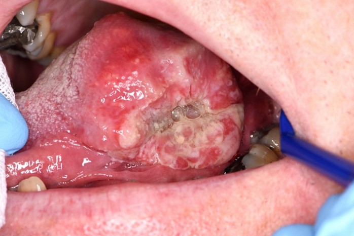
The most common intra-oral malignant neoplasm is oral squamous cell carcinoma (OSCC), which clinically can present as a non-healing ulcer (Figure 5). While an OSCC can develop at any site within the mouth, the most common sites are the floor of the mouth, the postero-lateral tongue border, and the gingivae.

A clinician should be suspicious of OSCC in any patient who is presenting with a solitary unexplained ulcer, which is persistent (>14 days), particularly if they have any risk factors for the development of an OSCC including tobacco or betel nut use, heavy alcohol consumption, or past or family history of oral cancer (Table 6). Other worrying symptoms include unexplained paraesthesia of the lower lip or tongue, and an unexplained persistent lump in the neck.
The classical clinical features of an ulcerated OSCC involve an ulcer with raised rolled margins and which is indurated on palpation. These ulcers rarely cause pain, but patients may report discomfort in the later stages, as well as a tendency of the ulcer to bleed to mild trauma or spontaneously.
After taking a thorough history from the patient and examination, if a practitioner suspects the ulcerated area represents oral cancer the patient should be referred urgently to their local oral medicine/oral surgery or maxillofacial department for assessment, confirmation of diagnosis and onward management.
Case report
A 56-year-old female was referred to the Oral Medicine Department at Belfast Royal School of Dentistry for a non-healing ulcer on the left lateral border of her tongue. She reported that this ulcer was painful on contact with foods and had been present for six weeks. The patient had no recollection of any preceding trauma or cause.
Her past medical history included eosinophilic granulomatosis with polyangiitis that was being managed with rituximab. She was otherwise fit and well. She was a non-smoker and did not consume alcohol regularly.
Extraoral examination did not display any cervical lymphadenopathy or facial asymmetry. However, the patient did have notable pallor of the face. Intraoral examination revealed a large 3x3cm raised ulcerated lesion on the lateral border of the tongue, with rolled margins and a speckled surface. The ulcer was indurated and painful to palpate (Figure 6) and lay adjacent to a lingually positioned LL5 with a fractured restoration. The differential diagnoses considered were:
-
traumatic ulcer;
-
squamous cell carcinoma; and,
-
granulomatosis disease.

An incisional biopsy was taken alongside routine blood investigations to aid in diagnosis. The biopsy revealed that the ulcer was consistent with a traumatic aetiology. The blood investigations found that the patient had a microcytic anaemia due to low iron. Therefore, a working diagnosis was made of a traumatic ulcer secondary to the fractured LL5 with delayed healing associated with low iron.
The patient’s LL5 was temporised and Difflam Spray was prescribed for symptomatic relief. She was also referred to her GMP to correct her iron levels. At review after six weeks, the ulcer was significantly reduced in size and had resolved at a further two-month follow-up.
Referral
The UK’s National Institute for Health and Care Excellence (NICE) produced guidelines in 2015 for head and neck cancers, which recommend referral on a suspected cancer pathway for people with unexplained ulceration in the oral cavity lasting for greater than three weeks, or a persistent and unexplained lump in the neck. Furthermore, referral to an oral medicine or oral maxillofacial unit may be made for ulceration with the following features4:
-
an ulcer that is indurated with a raised rolled margin;
-
ulceration that cannot be adequately managed with topical treatments;
-
investigations that cannot be undertaken in the primary dental care setting; and,
-
recurrent ulceration of unknown aetiology.
Summary
Oral ulceration can occur due to a variety of reasons. Investigations should be undertaken to identify any predisposing cause but most often ulceration will be due to trauma or RAS. Management is aimed at symptomatic relief through local analgesic preparations or, if unsuccessful, topical corticosteroid treatments. Patients who are resistant to treatment or who have red-flag symptoms should be urgently referred to oral medicine or oral and maxillofacial surgery units for further investigation.