Microbiology Case Study: A Friend or Foe with No Cell Wall

A 28 year old woman underwent an elective myomectomy for menorrhagia caused by fibroids. Postoperatively, she developed fevers. A CT scan of the abdomen and pelvis showed a complex pelvic fluid collection measuring 5.4 by 4.4 by 7.0 cm. Drainage was attempted by Interventional Radiology without success. She then underwent an exploratory laparotomy with drainage of the collection, evacuation of a hematoma, and removal of an IUD. The abscess fluid was sent to the Microbiology lab for culture.

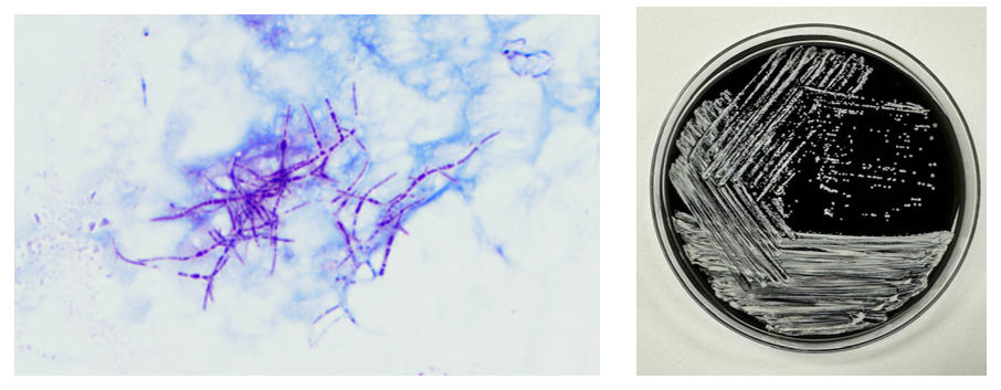
The Gram stain of the fluid revealed 1+PMN with no organisms. After 48 hours of incubation, there was few to moderate growth of pinpoint clear colonies on blood agar, with characteristic miniscule appearance of a central area of dense growth and peripherally less dense (Figure 1). Upon closeup reviewing of the colonies revealed a “fried egg” appearance (Fig 2).

There was no growth on MacConkey plate. Final identification by MALDI-TOF demonstrated Mycoplasma hominis.

Figure 1. Blood agar plate growing clear small colonies of M. hominis.
Figure 2. Characteristic colonies of organism with peripheral growth and denser central growth with fried-egg appearance.

Discussion

Mycoplasma hominis is a facultative anaerobe of the Mycoplasma genus, which is among the smallest free-living organisms known. They are fastidious and differentiated from other bacteria by their small size and absence of cell walls. Infections with M. hominis predominately involve the urogenital tract of females, causing pelvic inflammatory disease, bacterial vaginosis, and postpartum fever.1 Extragenital manifestations of M. hominis infections are rare but include extragenital abscesses, CNS infections in neonates, and septic arthritis.2 Reports of Mycoplasma hominis infections vary based on demographics (country, age, and number of sexual partners) but have been isolated in 4 to 30% of urogenital infections in females. 1,3

The lack of cell walls confers both diagnostic and clinical challenges. Mycoplasmas is not seen on the routine Gram stain smears. Instead of a cell wall, they have a triple-layered membrane composed of sterol.3 When able to grow in culture, colonies are small and have a ‘fried egg’ appearance on agar.4 Of the Mycoplasma species, M. hominis is the least fastidious and the most common Mycoplasma isolated on conventional culture agar media. 4-6 Non-traditional culture-based diagnosis is often made via molecular testing. 7 It is often detected in coinfections with Trichomonas vaginalis and thought to be a symbiotic relationship. 8-9

Since M. hominis is also found to be colonized in the genital tract of normal healthy individuals, it can be challenging to establish the clinical and diagnostic significance of M. hominis. With evolving molecular diagnostic assays targeting STI agents, it has been a controversial topic for assay manufacturers or labs that develop their own in-house assays whether there is a clinical utility for M. hominis PCR as part of STI multiplex PCR panels.

In contrast to Mycoplasma pneumoniae, M. hominis has intrinsic resistance to macrolides. 10 Preferred treatment regimens for M hominis infections include tetracyclines, clindamycin and fluoroquinolones. 11 However, resistance to members of these antibiotic classes has been reported and differs based on country of origin. 11 The absence of a cell wall explains the inherent resistance of Mycoplasma species to the beta-lactam antibiotic class. Its isolation in coinfections, particularly Trichomonas has been controversial in its contribution to emerging Trichomonas metronidazole resistance. 12

References

  1. Verteramo, R., Pastella, A., Calzolari, E., et al. An epidemiological survey of Mycoplasma hominis and Ureaplasma urealyiticum in gynaecological outpatients, Rome, Italy. Epidemiology & Infection,2013, 141(12), 2650-2657
  2. Zheng X, Olson DA, Tully JG, Watson HL, Cassell Gh, Gustafson DR, Svien KA, Smith TF: Isolation of Mycoplasma hominis from a brain abscess. J Clin Microbiol. 1997, 35: 992-994.
  3. Thomas Prescott Atkinson, Mitchell F. Balish, Ken B. Waites, Epidemiology, clinical manifestations, pathogenesis and laboratory detection of Mycoplasma pneumoniae infections, FEMS Microbiology Reviews, Volume 32, Issue 6, November 2008, Pages 956–973
  4. Razin S. Mycoplasmas. In: Baron S, editor. Medical Microbiology. 4th edition. Galveston (TX): University of Texas Medical Branch at Galveston; 1996. Chapter 37.
  5. Meloni GA, Bertoloni G, Busolo F, Conventi L. Colony morphology, ultrastructure and morphogenesis in Mycoplasma hominis, Acholeplasma laidlawii and Ureaplasma urealyticum. J Gen Microbiol. 1980;116(2):435-443.
  6. Christiansen G, Jensen LT, Boesen T, Emmersen J, Ladefoged SA, Schiotz LK, Birkelund S: Molecular biology of Mycoplasma. Wien Klin Wochenschr. 1997, 109: 557-561
  7. Baczynska, A., Svenstrup, H.F., Fedder, J. et al. Development of real-time PCR for detection of Mycoplasma hominisBMC Microbiol 4, 35 (2004).
  8. Tine RC, Dia L, Sylla K, Sow D, Lelo S, Ndour CT. Trichomonas vaginalis and Mycoplasma infections among women with vaginal discharge at Fann teaching hospital in Senegal. Trop Parasitol. 2019 Jan-Jun;9(1):45-53.
  9. Vancini, R.G., Benchimol, M. Entry and intracellular location of Mycoplasma hominis in Trichomonas vaginalis . Arch Microbiol 189, 7–18 (2008).
  10. Pereyre S, Gonzalez P, De Barbeyrac B, Darnige A, Renaudin H, Charron A, Raherison S, Bébéar C, Bébéar CM. Mutations in 23S rRNA account for intrinsic resistance to macrolides in Mycoplasma hominis and Mycoplasma fermentans and for acquired resistance to macrolides in M. hominis. Antimicrob Agents Chemother. 2002 Oct;46(10):3142-50.
  11. Krausse R, Schubert S. In-vitro activities of tetracyclines, macrolides, fluoroquinolones and clindamycin against Mycoplasma hominis and Ureaplasma ssp. isolated in Germany over 20 years. Clin Microbiol Infect. 2010;16(11):1649-1655.
  12. Dessì, D., Margarita, V., Cocco, A., Marongiu, A., Fiori, P., & Rappelli, P. (2019). Trichomonas vaginalis and Mycoplasma hominis: New tales of two old friends. Parasitology, 146(9), 1150-1155. doi:10.1017/S0031182018002135

-Dr. Alex Shaffer is a first-year ID fellow (2023-2025) at Montefiore Einstein. Alex is interested in the diagnostic stewardship projects and has been actively involved in various activities with ID and micro lab faculty.

-Phyu Thwe, Ph.D, D(ABMM), MLS(ASCP)CM is Associate Director of Infectious Disease Testing Laboratory at Montefiore Medical Center, Bronx, NY. She completed her medical and public health microbiology fellowship in University of Texas Medical Branch (UTMB), Galveston, TX. Her interests includes appropriate test utilization, diagnostic stewardship, development of molecular infectious disease testing, and extrapulmonary tuberculosis.

Microbiology Case Study: A 53 Year Old Male with Malaise, Bilateral Flank Pain and Decreased Urine Output

Case History

A 53 year old male presented to the emergency department with a one-day history of malaise, bilateral flank pain and decreased urine output. His past medical history was notable for decompensated cirrhosis due to alcohol use disorder complicated by esophageal varices and gastric ulcers, peritoneal ascites, several recent episodes of upper GI bleeding, monoclonal gammopathy of unknown significance, and remote prostate cancer status post prostatectomy. Oral history indicated the patient was actively drinking shortly before presentation and had recently consumed oysters on the half shell, broiled shrimp, and crab meat. While being seen in the emergency department, the patient quickly progressed to septic shock.     

Admission laboratory studies demonstrated severe metabolic acidosis, pancytopenia, and acute renal failure. Initial CT abdomen/pelvis demonstrated cirrhotic liver changes, varices, distal esophageal wall thickening, bilateral perinephric fat stranding extending into the pelvis and perivesical fat. The patient was admitted to the MICU for intubation/mechanical ventilation and administration of multiple pressors, as well as empiric meropenem, vancomycin and micafungin given concern for septic shock.Initial blood cultures drawn in the ED flagged positive with gram negative rods (both aerobic and anaerobic bottles) less than 24 hours after collection.

Laboratory Identification

Blood cultures were processed for culture workup and molecular identification. No identification was able to be established using a commercial multiplex PCR-based molecular panel on the positive blood culture broth. Subsequent growth of the organism on MacConkey agar 18 hours later revealed non-lactose fermenting colonies (Image 1A). The organism was oxidase-negative, positive for catalase and indole (Image 1B) and exhibited hydrogen sulfide production when inoculated on triple sugar iron (TSI) media. The organism was definitively identified as Edwardsiella tarda by MALDI-TOF MS and was broadly susceptible to all antibiotic tested including beta-lactams and fluoroquinolones.

Discussion

Edwardsiella tarda is an infrequently isolated member of the Enterobacterales which is most often associated with gastroenteritis. Reports of additional presentations include peritonitis, intra-abdominal abscess, and wound infections are increasing.1 Bacteremia is rare, and can lead to cholangitis, cholecystitis, and liver abscess via hematogenous spread.     Patients of advanced age (>65 years) and those with hepatobiliary diseases including liver cirrhosis and alcohol abuse and iron storage disorders are at increased risk for extraintestinal disease..2 Colonization of the gastrointestinal tract is believed to be a precipitating even leading to bacteremia. Cases of gastroenteritis are usually self-limiting, although ciprofloxacin or trimethoprim/sulfamethoxazole can be utilized in cases of prolonged duration.5 Prompt and accurate diagnosis of E. tarda bacteremia is important as this presentation is associated with an elevated mortality rate,4 particularly among those with liver disease.     

As members of the Enterobacterales, Edwardsiella sp. share common biochemical features of other members of the order including being catalase-positive and oxidase-negative. Oxidase negativity aids in the distinction between E. tarda and Aeromonas/Plesiomonas, both species which are also associated with aquatic environments and cause gastrointestinal infections.5 E. tarda is non-lactose fermenting and reduces sulfur containing amino acids to hydrogen sulfide. This can result in confusion with members of the genus Salmonella4 as colonies exhibit similar appearances on medias including Hektoen Enteric agar and Xylose-lysine deoxycholate agar formulated for recovery and presumptive identification of Salmonella from gastrointestinal specimens. Importantly, E. tarda is also indole-positive which will biochemically differentiate these two genera.

E. tarda is associated with freshwater or brackish aquatic environments. This organism is a well-known pathogen in aquaculture industries causing serious infections in fish and significant economic loss. As such, vaccines and prophylactic antibiotics are utilized to prevent E. tarda infection among aquatic animals, and diagnostic approaches including specialized multiplex PCRs are available. Human infection is often associated with consumption of contaminated fish and seafood, and diagnosis is almost exclusively dependent on microbiological culture by contrast. Clinical multiplex molecular panels for both gastrointestinal and bloodstream infections lack the ability to detect E. tarda. In this patient’s case, recent consumption of seafood serves as the most likely event leading to E. tarda gastrointestinal colonization and is consistent with previously reported cases of E. tarda bacteremia.3 The patient was treated with meropenem eventually narrowed to piperacillin/tazobactam and blood cultures cleared. He was weaned off pressors and extubated.     Unfortunately, the patient decompensated over the course of three weeks due to worsening shock and acidosis. He was moved to comfort care and expired soon thereafter.

References

  1. Hirai et. al. 2015.  Edwardsiella tarda bacteremia. A rare but fatal water- and foodborne infection: Review of the literature and clinical cases from a single centre. Can. J. Infect. Dis. Med. Microbiol. 26(6): 313-318.
  2. Hasegawa M., and Sanmoto, Y.  2024. Recurrent cholangitis and bacteremia due to Edwardsiella tarda: a case report.     Oxford Med. Case Rep. Volume 2024, Issue 1, January 2024, omad148, https://doi.org/10.1093/omcr/omad148
  3. An et. al.     2023.     Case Report: Disseminated Edwardsiella tarda infection in an immunocompromised patient.     Front. Cell. Infect. Microbiol. 20 November 2023.
  4. Janda, J.M, and Abbott, S.L. 1993. Infections Associated with the genus Edwardsiella: the Role of Edwardsiella tarda in human disease. Clin. Infect. Dis. Oct;17(4):742-8.
  5. Janda, J.M and Abbott, S.L. 1999. Unusual Food-Borne Pathogens: Listeria monocytogenes, Aeromonas, Plesiomonas, and Edwardsiella species. Clin. Lab. Med. 19(3):553-582.

-Andrew Clark, PhD, D(ABMM) is an Assistant Professor at UT Southwestern Medical Center in the Department of Pathology, and Associate Director of the Clements University Hospital microbiology laboratory. He completed a CPEP-accredited postdoctoral fellowship in Medical and Public Health Microbiology at National Institutes of Health, and is interested in antimicrobial susceptibility and anaerobe pathophysiology.

-Francesca Lee, MD, is an associate professor in the Departments of Pathology and Internal Medicine (Infectious Diseases) at UT Southwestern Medical Center

Microbiology Case Study: A 72 Year Old Male with Postoperative Fever

Case description

A 72 year-old male with severe aortic stenosis resulting in heart failure underwent aortic valve replacement and became febrile with mild shortness of breath on post-operative day one.  Additional pertinent medical history includes end-stage renal disease secondary to diabetic nephropathy with kidney transplantation seven months prior which was complicated by delayed graft function requiring hemodialysis and immunosuppression. The patient’s presentation was suspicious for either a post-operative fever or a complication of hemodialysis. Physical examination did not suggest infection at either the surgical site or his arteriovenous fistula. A chest CT revealed a moderate, loculated left pleural effusion with left lower lobe atelectasis and nodular consolidations in the right lung, raising concern for pneumonia. A bronchial biopsy was obtained, and histopathological evaluation was consistent with an abscess, but no organisms were visualized. Culture from a left bronchial brushing was also obtained at the same time. Two days later, the patient developed a progressively enlarging forehead lesion (Image 1).

Image 1. Progressively enlarging forehead lesion. Microscopic evaluation following biopsy of the lesion revealed organisms morphologically consistent with Nocardia  species.

Laboratory Identification

Microscopic evaluation of the biopsy of the lesion for culture revealed branching, beaded gram positive filamentous bacteria which stained positive using a modified acid-fast stain, suggestive of Nocardia species. This was corroborated by the bronchial culture, where similar modified acid-fast organisms were visualized and grew chalky white colonies identified as Nocardia sp. by MALDI-TOF MS (Image 2). The isolate was referred to a reference laboratory for definitive speciation and antimicrobial susceptibility testing, and the patient was started empirically on ceftriaxone and trimethoprim/sulfamethoxazole.  Speciation and susceptibility testing subsequently identified the organism as Nocardia brasiliensis with susceptibility to both ceftriaxone and trimethoprim/sulfamethoxazole, and treatment was thereafter continued for 40 days.

Image 2. Modified acid-fast stain of a bronchial culture from the patient showed a branching, modified acid-fast rods consistent with Nocardia.  Cultures of both the lesion and bronchial brushing revealed white, chalky colonies on buffered charcoal yeast extract agar consistent with Nocardia sp.

Discussion

Nocardia is a saprophytic bacterium that is commonly found in soil worldwide. Gram staining of Nocardia sp. usually reveals delicate, weakly or erratically staining, beaded, branching Gram-positive rods. There are currently 130 known species of Nocardia,1 of which more than 50 have been reported to have clinical relevance.2 Disease due to Nocardia is known to occur due to inhalation of the organism from the environment or traumatic inoculation. It is thought to be an opportunistic pathogen most commonly affecting immunocompromised patients3 like the patient in this case.

Manifestations of nocardiosis vary widely depending on the species responsible, with most common presentations being lymphocutaneous, pulmonary, or disseminated disease. Here, we present a patient who displayed both lymphocutaneous and pulmonary forms of Nocardia infection. While prognosis is poorer in immunocompromised patients, with appropriate treatment, recovery rate is high in both lymphocutaneous and pulmonary forms, unlike in disseminated disease, where studies have found mortality rates between 44% to 85%.4

Diagnosis of Nocardia infections is mainly performed through either direct visualization of the organism or through culture. The modified acid-fast stain is commonly performed but has limited sensitivity and should generally be performed in conjunction with the Gram stain.5 Recovery in culture is most reliable from respiratory and/or tissue samples, while blood culture, by contrast, has poorer yield. While some strains, as in this patient, appear within several days in culture, some strains may take up to 2 to 3 weeks to detect5 necessitating extended incubation and consultation with the clinical laboratory.

Treatment requires prolonged courses of antibiotics. Susceptibilities vary by species, making it important to obtain species identification to identify appropriate therapy to guide empiric therapy. Susceptibility testing is performed to allow further tailoring of antibiotic regimens. In this case, the patient was treated with a combination of trimethoprim/sulfamethoxazole and ceftriaxone, both of which cover the majority of clinically relevant Nocardia species, until susceptibility testing revealed that the empiric treatment provided adequate coverage for the Nocardia brasiliensis identified, supporting continuation of the chosen empiric regimen.

References

  1. Genus Nocardia. List of Prokaryotic names with Standing in Nomenclature. https://www.bacterio.net/genus/nocardia (2023).
  2. Hamdi AM, Fida M, Deml SM, Abu Saleh OM, Wengenack NL. Retrospective Analysis of Antimicrobial Susceptibility Profiles of Nocardia Species from a Tertiary Hospital and Reference Laboratory, 2011 to 2017. Antimicrob Agents Chemother. 2020 Feb 21;64(3):e01868-19. doi:10.1128/AAC.01868-19.
  3. McNeil MM, Brown JM. The medically important aerobic actinomycetes: epidemiology and microbiology. Clin Microbiol Rev. 1994 Jul;7(3):357-417. doi:10.1128/CMR.7.3.357.
  4. Traxler RM, Bell ME, Lasker B, Headd B, Shieh WJ, McQuiston JR. Updated Review on Nocardia Species: 2006-2021. Clin Microbiol Rev. 2022 Dec 21;35(4):e0002721. doi: 10.1128/cmr.00027-21.
  5. Saubolle MA, Sussland D. Nocardiosis: review of clinical and laboratory experience. J Clin Microbiol. 2003 Oct;41(10):4497-501. doi: 10.1128/JCM.41.10.4497-4501.2003.

-Albert Budhipramono, MD, PhD, is currently a PGY1 Clinical Pathology Resident at the University of Texas Southwestern Medical Center in Dallas, Texas.


-Clare McCormick-Baw, MD, PhD is an Assistant Professor of Clinical Microbiology at UT Southwestern in Dallas, Texas. She has a passion for teaching about laboratory medicine in general and the best uses of the microbiology lab in particular.

-Andrew Clark, PhD, D(ABMM) is an Assistant Professor at UT Southwestern Medical Center in the Department of Pathology, and Associate Director of the Clements University Hospital microbiology laboratory. He completed a CPEP-accredited postdoctoral fellowship in Medical and Public Health Microbiology at National Institutes of Health, and is interested in antimicrobial susceptibility and anaerobe pathophysiology.

Hematology Case Study: Brrrr, It’s Cold Outside!

A CBC was received on a 70-year-old surgical inpatient at our facility and was run in automated mode on our Sysmex XN analyzer. On the first run, the analyzer gave flags for RBC agglutination and MCHC >37.5. These flags require evaluation of the high MCHC with investigation of a cold agglutinin, lipemia or icterus. A smear review for RBC agglutination was also indicated. The sample was incubated at 37°C for 30 minutes and the CBC was repeated. Results are shown below in Table 1.

Table 1. CBC results before warming at 37°C and after 15 minutes incubation.

On Run 2, the MCHC is now below 37.5, so there was no operator alert to incubate further or to investigate the high MCHC. This result was validated with the comment “37°C results, possible cold agglutinin.” However, there was still an RBC agglutination flag, a high MCV, and the hemoglobin and hematocrit still don’t look great, i.e. they don’t follow ‘the rules of 3’. We know that these rules are only valid for normal samples, but if we look at the previous results from 2 days ago, the Hgb 9.3 and Hct 28.4 did follow the ‘rules of 3’ and additionally, today’s results look like the patient’s hemoglobin has not changed but the hematocrit has dropped. The RBC did go up on the second run, but it is considerably lower than 2 days ago. The indicies are also inconsistent with the previous sample. “Hmmm…What could cause this?” Instead of validating, this is what you should be asking yourself.

Cold agglutinins are known for causing pre-analytical and analytical spurious results with CBCs. With cold agglutinins, IgM antibodies bind to the RBCs after exposure to cold, causing RBC agglutination which leads to a classic pattern of false results. There is an increased MCV because the RBCs are clumped and sticking to one another, making the analyzer think these larger clumps are individual RBCs. This, in turn, will make the RBC count appear deceased because large clumps of RBCs going through the RBC aperture are counted as one RBC. The hematocrit is lowered because the volume of a clump is less than those cells individually. Hemoglobin concentration, on the other hand, is not affected by cold agglutinins, but because the hematocrit and RBC are falsely lowered, this makes the MCH and MCHC spuriously markedly increased. Cold agglutinins may be subtle, like this one, but some have extremely high MCHC and MCV, and extremely low RBC and Hct. Clues that you have a cold agglutinin are not only the high MCHC, but also flags from your analyzer such as “RBC agglutination”. The hematocrit will likely seem too low for the hemoglobin, and you may even see a hemoglobin that is higher than the hematocrit (yes it happens!) and RBC values so low as to be incompatible with life.

The first thing is to do if you get these spurious results is to compare the parameters and instrument flags and decide if the results are consistent with a cold agglutinin. Other factors that may cause a high MCHC include lipemia, icterus, low sodium, abnormal proteins, hemolyzed samples and samples from patients with hemoglobinopathies. The patterns of result with these samples will show a high MCHC, but with a normal or low MCV, and you won’t usually see an RBC agglutination flag. A sample with cold agglutinins may also appear grainy or clumpy to the naked eye. With a suspected cold agglutinin, warming the sample for 15-30 minutes will allow the RBCs to disperse and improve the results. However, these results must be reviewed, and if there are still instrument flags and/or if there is still clumping, the results may not yet be corrected or ‘correct’. Be sure to review a smear from the tube before warming and after. The first smear confirms the presence of the cold agglutinins and clumping, and the second smear should confirm the resolution of the clumping after warming.

This sample did have the characteristic grainy appearance of a cold agglutinin, and the post warming results did look a bit better, but the RBC agglutination flag was still present, and a review of the smear showed that the sample still had RBC clumping. After warming for another 30 min, the sample was quickly mixed and placed back on the analyzer. The results of this 3rd run are shown below, in Table 2.

Figure 1. EDTA tube with RBC agglutination
Table 2. CBC results on 3 runs. Run 3, after 60 min of incubation.

Notice that hemoglobin has not changed as it is not affected by the cold agglutinins, but after warming for 60 minutes, the RBC, hematocrit and indicies all now look consistent with the previous sample drawn 2 days ago, and a review of the smear showed no RBC agglutination. These results from the 3rd run are ready to validate.

Cold agglutinins are IgM autoantibodies that react best at 4°C but may also react at room temperature. They are generally not clinically significant and may be found in many healthy individuals. These natural cold autoantibodies occur at low titers, less than 1:64, and have no activity at higher temperatures. However, because they react at room temperature, they are notorious as a pre-analytical and analytical factor that causes spurious CBC results. They can also cause difficulties in Blood Banking during ABO/Rh typing and antibody detection.

Cold agglutinins have various clinical manifestations. Benign cold agglutinins generally do not cause hemolytic anemia and need no treatment. Most benign cold autoantibodies have anti-I specificity, are polyclonal, low titer, and do not react above 30°C. Cold agglutinins associated with Mycoplasma pneumoniae, and infectious mononucleosis are usually clinically insignificant. In cases where they do cause hemolytic anemia, the antibodies are polyclonal IgM with normal κ and λ light chains. The anemia is acute and generally spontaneously resolves in several weeks without treatment.

Though most cold agglutinins are benign and do not cause RBC destruction, when they do, they can cause hemolytic anemia that varies in severity from mild to life-threatening. This chronic cold agglutinin disease (CAD) is now known to be a form of autoimmune hemolytic anemia caused by a bone marrow lymphoproliferative disorder. Chronic CAD is a cold-autoantibody autoimmune hemolytic anemia (cAIHA) that is caused by an autoantibody produced by the clonal B cell lymphocytes. This antibody is usually monoclonal IgM with κ light chains and “I” or “i” specificity. These pathological cold agglutinins are high titer and usually react at 28°C to 32°C, and even up to 37°C. The highest temperature at which the antibodies continue to be activated is called the thermal amplitude. Because these can act at higher thermal amplitude, they may lead to CAD. In CAD the IgM autoantibodies bind to red cell antigens at 30-32°C, typically in the cooler extremities. IgM’s structure, a large immunoglobin pentamer, makes it an effective activator of the classical complement system. As the blood circulates to the central parts of the body, the RBCs warm up and the IgM antibodies dissociate from the RBC membranes, but the complement activation will continue, leading to RBC hemolysis and a cAIHA.

Chronic CAD occurs most often in adults over 50, is more common in women, and produces anemia with varying severity. Patients may be seasonally affected. In the winter, the temperature of blood may fall below 30°C in the extremities, activating the cold agglutinins. Patients may experience acrocyanosis of the hands, feet, ears, and nose with exposure to cold. They may also experience other cold related symptoms such as numbness and Raynaud’s. Patients with chronic CAD and mild anemia are therefore monitored with a ‘wait and see’ plan and advised to avoid cold temperatures. In patients with more severe anemia, it is found that targeting the underlying lymphoproliferative disorder provides the best treatment. Rituximab has been used to achieve partial remission. Therapeutic plasma exchange is also used in severe cases to rapidly remove cold agglutinins.

I have been thinking about cold agglutinins recently because of the number I have seen come into our lab this winter. As I am writing this, watching the temperature outside drop in anticipation of more snow coming in tonight, cold agglutinins came to my mind again. I used to live in the cold northern Northeast, but after moving further south, we see fewer cold agglutinins in hematology than I used to see. This winter we have had some cold spells, and, interestingly, I’ve seen more cold agglutinins. That led me to ask myself if cold agglutinin disease is really more common when patients are exposed to cold temperatures. I remember learning myself, and telling my students that that the treatment for mild to moderate CAD was to advise patients to move to someplace warmer. It has been assumed for many years that CAD worsens in colder climates or seasons. Interestingly, there have been a number of studies done since the 1950’s that examined the relationship of cold temperatures and CAD. The studies used hemoglobin, bilirubin and LDH for monitoring. Early case reports had findings that supported the theory of more anemia and higher LDH in the winter. (Dacie, Lyckholm)One recent article in 2022 found a 4-fold difference in the incidence of CAD between cold (Norway) and warm (Italy) climates. (Berentsen). However, atthe same time another study found that there was no statistically significant seasonal variation in hemoglobin, but that LDH levels were higher in winter. It concluded that these conditions should be monitored through all seasons because of the risk of hemolysis and thrombotic episodes. It was also demonstrated that though there may not be obvious statistical difference in CAD between cold and warm months, that there is a large variability of disease severity across patients and even with an individual patient. (Roth).

In conclusion, when working in any department of the laboratory, quality results are important. Results on the patient chart that vary considerably from day to day because sometimes a cold agglutinin has been effectively resolved in lab testing and other days results after 15-30 minutes of warming are just reported without a good review of the smear and the parameters, are confusing, and could affect patient care. If one tech reports the results after 30 min incubation with values that are still spurious, and the next tech resolves the agglutination with further warming, the lab will be reporting out inconsistent results. A patient who actually has stable CBC results may have deltas and what appear to be erratic results. Cold agglutinins do take time to resolve, but with over 80% of samples autoverifying with the use of auto verification, we have time to work on these problem samples. If something doesn’t look or feel right about a sample, look at all the parameters, check the instrument flags and operator alerts, check the previous results and investigate any changes. It is important to review results carefully, because we want to report out the best results possible.

References

S Berentsen, W Barcellini, S D’Sa, U Randen, THA Tvedt, B Fattizzo, E Haukås, M Kell…

Blood, The Journal of the American Society of Hematology, 2020•ashpublications.org

Climent F, Cid J, Sureda A. Cold Agglutinin Disease: A Distinct Clonal B-Cell Lymphoproliferative Disorder of the Bone Marrow. Hemato. 2022; 3(1):163-173. https://doi.org/10.3390/hemato3010014

Nikousefat Z, Javdani M, Hashemnia M, Haratyan A, Jalili A. Cold Agglutinin Disease; A Laboratory Challenge. Iran Red Crescent Med J. 2015 Oct 17;17(10):e18954. doi: 10.5812/ircmj.18954. PMID: 26566452; PMCID: PMC4636857.

Patriquin, C.J. and Pavenski, K. (2022), O, wind, if winter comes … will symptoms be far behind?. Transfusion, 62: 2-10. https://doi.org/10.1111/trf.16765

Rodak, Bernadette F., et al. Hematology: Clinical Principles and Applications. 5th ed. St. Louis, Mo., Elsevier Saunders, 2016

Röth A, Fryzek J, Jiang X, Reichert H, Patel P, Su J, et al. Complement-mediated hemolysis persists year round in patients with cold agglutinin disease. Transfusion. 2022; 62: 51–59. https://doi.org/10.1111/trf.16745

Socha-small

-Becky Socha, MS, MLS(ASCP)CMBBCM graduated from Merrimack College in N. Andover, Massachusetts with a BS in Medical Technology and completed her MS in Clinical Laboratory Sciences at the University of Massachusetts, Lowell. She has worked as a Medical Technologist for over 40 years and has taught as an adjunct faculty member at Merrimack College, UMass Lowell and Stevenson University for over 20 years.  She has worked in all areas of the clinical laboratory, but has a special interest in Hematology and Blood Banking. She currently works at Mercy Medical Center in Baltimore, Md. When she’s not busy being a mad scientist, she can be found outside riding her bicycle.

Shish Kebabs, Pears and Grapes: All “Clue(s)” to Diagnosing Microbial Infections on Pap Smears 

What is the test?

In the 1920s, the Greek physician Georgios Papanikolaou developed a method of cervical cancer screening, now reliably used and colloquially known as a “Pap smear”. During a Pap smear, a healthcare provider swabs cells from the cervix for further analysis by the lab​1​, including cytopathologic examination. Regular utility of Pap smears in women aged 21 to 65 has decreased the incidence and mortality of cervical cancer by at least 80% in the United States alone​2​. 95% of cervical cancer cases worldwide are caused by persistent human papilloma virus (HPV) infection of the cervix​3​. However, other sexually transmitted infections (STIs) and common non-STIs with associated morphological changes can also be identified on Pap smears. 

Bacteria

Bacterial vaginosis (BV) is the most common cause of abnormal discharge in young women, characteristically with a “fishy” ammonia-like odor. Although not strictly considered an STI, this infection is associated with severe rare complications including pelvic inflammatory disease (PID), infertility and premature labor. The vagina typically have an abundance of Lactobacillus spp., which is a gram positive bacilli (Figure 1). However, if the normal vaginal flora is disrupted with high abundance of gram negative bacteria such as Gardnerella vaginalis or Mobiluncus, then BV may occur. On pap smears, ‘clue cells’ which are squamous epithelial cells with coccobacilli, appear as dark purple staining and cloudy appearance suggest BV. In cervicovaginal samples typically found in women using intrauterine device usage, clumps of filamentous bacteria suggestive of Actinomyces species may also be visible. For cases pertaining to bacterial etiologies, staining of the rods or filaments usually warrant suspicion of infection. Other the other hand although Chlamydia trachomatis is the most common bacterial STI in the United States, with up to 4 million new cases every year, diagnosis on pap smears is not the definitive diagnosis since the cytopathology is non-specific. The interpretations include visualization of inflammatory exudates and inflammatory cells. Oftentimes, further testing is needed. 

Figure 1. Lactobacilli spp. bacilli seen as part of normal flora (red arrow, top left and top middle). Example of a clue cell (red arrow): intermediate squamous cell coated with grape-like short coccobacilli (Gardnerella vaginalis) with a shift in normal flora (top right). What appears to be a clue cell (red arrow) at first glance is actually an intermediate squamous cell covered with normal flora upon closer inspection (bottom left). Follicular cervicitis due to Chlamydia infection (bottom right). Note lymphohistiocytic aggregates with polymorphous lymphocytes and histiocytes (red arrow). Tangible body macrophages may also be identified (ThinPrep, 40X).

Parasite

Trichomonas vaginalis is the most prevalent non-viral STI in the United States, strongly associated with an increased risk of HIV infection among women​4​. Majority of the patients may exhibit symptoms such as burning, itching and vaginal discharge. T. vaginalis is a parasitic flagellated protozoan although in some cases, a flagella may be found. Typically, it is of a pear-shape and staining of a nucleus may be visualized (Figure 2).

Figure 2. Pear-shaped round-oval Trichomonas spp. (red arrows) ranging in size from 15-30 µm present singly and in groups, known as “Trich parties”. Note pale vesicular eccentrically located nucleus and eosinophilic cytoplasmic granules. Reactive cytological changes (black arrow) such as nuclear enlargement and perinuclear halos are also identified. (ThinPrep, 40X. Case courtesy of Dr. Edina Paal, VA Medical Center, Washington DC).

Fungus

Yeast infections (vulvovaginal candidiasis) due to Candida infection occur in 75% of women at some point in their lives. Classic clinical presentation includes itching, erythema, and thick white “cottage-cheese” discharge. There is also an increased association with HIV infection, diabetes, and any cause of immunosuppression (e.g. transplant, chemotherapy, steroids). Morphologically, budding yeast forms (conidia, small and oval measuring 3-6 µm) and pseudohyphae (long filamentous spores) are found. The combination of pseudohyphae and yeast forms are referred to as “sticks and stones” and frequently, squamous cells lined up along the pseudohyphae are found referred to as having “shish kebab” appearance (Figure 3)5. Note that no true septation is found.

On pap smears, sometimes there could be superficial mucosal infections that may appear associated with enlarged hyperchromatic nuclei with halos, which can be confused with low grade squamous intraepithelial lesions.

Figure 3. Shish kebab: entangled streaming intermediate and superficial squamous cells along Candida spp. pseudohyphae (red arrow, top). Partially treated Candida infection. Note poorly formed granular pseudohyphae (red arrow) with significant treatment effect (red arrow, bottom) (ThinPrep, 40X).

Viruses

HPV with its associated risk of cervical cancer remains the most crucial microorganism to detect on Pap smears. Given HPV’s association to cancer development, it is crucial to examine the samples for cervical lesions and their associated pathologies. HPVs in the low-risk category typically is associated with low-grade squamous intraepithelial lesions and HPVs in the high-risk category is associated with high-grade squamous intraepithelial lesions and invasive squamous cell carcinoma.  Other viral etiologies seen in PAP smears include Herpes simplex virus (HSV) and cytomegalovirus (CMV). Infections with HSV can be asymptomatic but the most common symptom is the development of vesiculopustular or small ulcerative lesions on the genitalia. The classic cytopathological findings of HSV infection are the 3 “Ms”: multinucleation, nuclear molding, and margination of chromatin. CMV infections are rare, usually asymptomatic and can be transient. On the pap smears, the infected cells may appear with intranuclear inclusions surrounded by a halo.

Conclusions

While Pap smears are routinely performed on women and can provide a presumptive diagnosis, current developments in molecular technologies (e.g. Nucleic acid amplification tests (NAATs)) is transforming the field. There are several FDA-approved molecular platforms available for clinical diagnostic labs to test for HPV and can even genotype the strain to determine risk levels. Crucially, the remaining sample from liquid-based Pap tests​6​ can also be submitted for both NAAT testing and HPV-DNA testing, facilitating a quicker turnaround time and obviating the need for additional patient sampling.  Most recently, a PCR-based test is now on the market (Cepheid, Sunnyvale, CA) that can diagnose all three etiologies, BV, Candidiasis, and Trichomoniasis, within 60 minutes from a single specimen.  In summary, accurate examination of the Pap smear often incidentally provides the first step in the diagnosis and further work-up of all these infectious diseases.

References 

​​1. Pap Smear: MedlinePlus Medical Test [Internet]. [cited 2024 Feb 4]. Available from: https://medlineplus.gov/lab-tests/pap-smear/ 

​2. PDQ® Screening and Prevention Editorial Board. Cervical Cancer Screening (PDQ®): Health Professional Version. National Cancer Institute [Internet]. 2022 [cited 2024 Feb 4];1–26. Available from: https://www.cancer.gov/types/cervical/hp/cervical-screening-pdq 

​3. Lei J, Ploner A, Elfström KM, Wang J, Roth A, Fang F, et al. HPV Vaccination and the Risk of Invasive Cervical Cancer. New England Journal of Medicine. 2020 Oct 1;383(14):1340–8.  

​4. Davis A, Dasgupta A, Goddard-Eckrich D, El-Bassel N. Trichomonas vaginalis and Human Immunodeficiency Virus Coinfection Among Women Under Community Supervision: A Call for Expanded T. vaginalis Screening. Sex Transm Dis [Internet]. 2016 Sep 15 [cited 2024 Feb 5];43(10):617–22. Available from: https://pubmed.ncbi.nlm.nih.gov/27631355/ 

​5. Kamal Meherbano M.  The Pap smear in inflammation and repair. Cytojournal [Internet]. 2022 Apr 30; 19:29. Available from: doi: 10.25259/CMAS_03_08_2021

​6. Hawthorne CM, Farber PJ, Bibbo M. Chlamydia/gonorrhea combo and HR HPV DNA testing in liquid-based pap. Diagn Cytopathol [Internet]. 2005 Sep [cited 2024 Feb 5];33(3):177–80. Available from: https://pubmed.ncbi.nlm.nih.gov/16078250/ 

-Zoon Tariq is a pathology resident at George Washington University. Her interests include surgical pathology and cytopathology.

-Rebecca Yee, PhD, D(ABMM), M(ASCP)CM is the Chief of Microbiology, Director of Clinical Microbiology and Molecular Microbiology Laboratory at the George Washington University Hospital. Her interests include bacteriology, antimicrobial resistance, and development of infectious disease diagnostics.

Microbiology Case Study: Traveler’s Diarrhea in a 59 Year Old Patient

Case History

A 59-year-old man presented to the Emergency Room with bright red blood per rectum, associated with nausea, vomiting, abdominal cramping, and persistent watery diarrhea. Several days earlier, he had returned from a three-week trip to the Dominican Republic. On physical examination, he was afebrile. His abdomen was soft and not tender or distended.

A stool sample was sent to the Microbiology Lab for PCR testing, and both Vibrio and Vibrio cholera targets were detected. The stool was then plated for culture confirmation. Hemolytic colonies grew on the blood agar plate (Figure 1), and yellow (original medium color green) colonies grew on thiosulfate citrate bile sucrose selective agar due to sucrose fermentation (Figure 2). Gram stain from these colonies showed gram negative, curved, comma-shaped rods (Figure 3) and MALDI-ToF identification revealed Vibrio albensis. The specimen was sent to the Department of Public Health for confirmation, which reported Vibrio cholerae O1 serovar Ogawa with O1 antigen typing. 

Figure 1. Growth on blood agar
Figure 2. TCBS-sucrose fermentation
Figure 3. Gram negative, curved, comma-shaped rods

Discussion

Vibrio albensis is a gram negative, halophilic bacterium belonging to the Vibrionaceae family. V. albensis is a recently identified species within the Vibrio genus and is believed to be a member of the Vibrio cholera complex. Although primarily considered non-pathogenic, V. albensis has been associated with rare cases of human infections, particularly in individuals with compromised immune systems.1 While previously reported studies indicated V. albensis as a non-O1, non-O130 serogroup of V. cholerae,2the Department of Public Health confirmed our patient’s isolate as O1 serovar Ogawa by O1 antigen typing. V. albensis is an emerging pathogen with limited information regarding its clinical significance and optimal management. The infections are predominantly associated with contaminated seawater or seafood exposure. The primary transmission mode is through open wounds or ingesting raw or undercooked seafood.3

Clinical presentation of this organism can be similar to V. cholerae, as diffuse watery diarrhea, the hallmark of cholera, or asymptomatic. Other case reports presented bacteremia, septicemia, and urinary tract infections.4,5

Laboratory diagnosis of Vibrio cholerae albensis infection involves isolating and identifying the bacterium from stool samples. This can be achieved using selective culture media, such as thiosulfate-citrate-bile salts-sucrose (TCBS) agar, which allows for the growth of V. cholerae and its variants.6

Antimicrobial resistance is a growing concern in the management of cholera. Studies have reported varying resistance levels to commonly used antibiotics in V. cholerae, including V. cholerae albensis. It is essential to monitor antimicrobial susceptibility patterns to guide appropriate treatment strategies.7 Further research is needed to better understand the epidemiology, clinical manifestations, and optimal treatment strategies for V. albensis infections as members of V. cholerae complex are being identified/recognized with more advanced diagnostic tools.

References

  1. Baker-Austin C, et al. (2016). Vibrio albensis sp isolated from a mesophilic bacterial culture, abalone (Haliotis spp.), and seawater. International Journal of Systematic and Evolutionary Microbiology, 66(1), 187-192.
  2. Ahmed AOE, Ali GA, Hassen SS, Goravey W. Vibrio albensis bacteremia: A case report and systematic review. IDCases. 2022 Jun 30;29:e01551. doi: 10.1016/j.idcr.2022.e01551. PMID: 35845827; PMCID: PMC9283503.

3. Sharma P, et al. (2018). Vibrio albensis: An Emerging Pathogen Causing Necrotizing Fasciitis. Journal of Clinical Microbiology, 56(3), e01454-17.

4. Araj GF, Taleb R, El Beayni NK, Goksu E. Vibrio albensis: An unusual urinary tract infection in a healthy male. J Infect Public Health. 2019 Sep-Oct;12(5):712-713. doi: 10.1016/j.jiph.2019.03.018. Epub 2019 Apr 10. PMID: 30981654.

5. Sack RB, et al. (2004). Cholera. The Lancet, 363(9404), 223–233.

6. Centers for Disease Control and Prevention (CDC). (2021). Laboratory Methods for the Diagnosis of Vibrio cholerae. Retrieved from https://www.cdc.gov/cholera/laboratory.html

7. Ceccarelli, M., et al. “Editorial–Differences and similarities between Severe Acute Respiratory Syndrome (SARS)-CoronaVirus (CoV) and SARS-CoV-2. Would a rose by another name smell as sweet.” European review for medical and pharmacological sciences 24.5 (2020): 2781-2783.

-Eros Qama, MD, is a 2nd year AP/CP pathology resident in the Department of Pathology at Montefiore Medical Center in Bronx, NY

-Phyu Thwe, Ph.D, D(ABMM), MLS(ASCP)CM is Associate Director of Infectious Disease Testing Laboratory at Montefiore Medical Center, Bronx, NY. She completed her medical and public health microbiology fellowship in University of Texas Medical Branch (UTMB), Galveston, TX. Her interests includes appropriate test utilization, diagnostic stewardship, development of molecular infectious disease testing, and extrapulmonary tuberculosis.

Microbiology Case Study: The Importance of Process Workflows and Stains in Positive Blood Culture Bottles with No Organisms Seen

Case History

An 83 year old female with history of type 2 diabetes presented to the emergency room (ER) with two month history of dysuria and diarrhea. Upon admission, Clostridiodes difficile (C. difficile ) GDH/TOX with reflex to nucleic acid amplification test (NAAT), urine culture, and 2 sets of blood culture specimens were collected for testing. C. difficile test was positive for GDH and Toxin A/B production. Urine culture was positive with 50,000 – 99,000 colony forming units (CFU)/mL of Klebsiella pneumoniae. Patient was administered ceftriaxone, metronidazole and vancomycin and discharged three days post admission. On day of discharge, blood cultures were documented as “no growth at 3 days”. The patient visited her primary care physician the day after discharge. Two days later, the primary care office was notified of a corrected report. The patient’s blood culture collected on day of admission was being corrected from no growth to a positive with growth of Fusobacterium nucleatum. The patient was contacted to check on her status and medication compliance was verified. Patient continued to demonstrate symptom improvement during follow up at physician’s office 10 days post discharge.

What happened here?

The blood culture was collected at an acute care hospital. The system microbiology laboratory policy for a first blood culture positive that is read as no organisms seen (NOS) from an acute care hospital laboratory, is to inoculate appropriate media and prepare three smears. Gram stain is to be performed on one of the smears and if organisms are not seen, a stained and unstained smear, along with inoculated media, are to be sent to the core microbiology laboratory (core lab). The bottle is then to be reloaded on the blood culture instrument.

The policy for a second positive alert from the same NOS bottle is to inoculate appropriate media and prepare 3 cytospin smears. Gram stain is to be performed on one of the cyto-smears and if organisms are still not seen, a stained and unstained smear are to be sent to the core lab along with the positive bottle and inoculated media. The core lab is to use the unstained cytospin smear to make and read an acridine orange (AO) stain.

In this patient’s case, the anaerobic blood culture bottle from one set flagged as positive on day 4 of incubation. The gram stain was read as NOS and inoculated media and smears were sent to the core lab. The bottle was reloaded on the instrument and the bottle alerted as positive a second time that day. The cytospin gram stain was read as NOS and inoculated media, smears, and blood culture bottle were sent to the core lab. At the core lab, a new unspun smear was made from the bottle. AO was performed on this smear and read as NOS.

In our laboratory, if a gram stain result is not entered for a blood culture, it will auto verify as a negative blood culture every 24 hours with a final negative result at 5 days. Both blood culture sets on this patient resulted as no growth at 5 days.

At the 48 hour (6 days post-collection) culture read, BAP, CHOC, and MAC demonstrated no growth, but the CDC ANA had growth. The isolate was setup for MALDI identification and resulted as 99.9% Fusobacterium nucleatum.

Per protocol, a review of smears was performed on this discrepant smear-culture. The unstained cyto-smear sent by the acute care laboratory had not been stained with AO and read per protocol. Rather, as was mentioned above a Gram stain was made on a new unspun smear and read as NOS. Reviewing this AO stain during the investigation revealed long thin fluorescing rods with tapered ends (Fig 1)

The cyto-smear that had been sent after the second positive alert, but did not get stained and read was also stained with AO and long thin fluorescing rods with tapered ends were observed (Fig 2).

At this point in the investigation, the unspun gram stain from the 1st positive alert that was sent over from the acute care hospital laboratory was also reviewed. Knowing that the bottle was positive for F. nucleatum the technologist shared that she eventually saw rare long thin gram-negative rods with pointed ends that matched what was seen in the AO (Fig 3). As part of the investigation, safranin was added for an additional 10 minutes revealing the organism more clearly (Fig 4).

Figure 1. Acridine orange of unspun slide.
Figure 2. Acridine orange of cytosmear
Figure 3. Gram stain of unspun smear.
Figure 4. Gram stain of unspun smear with 10 minutes of safranin

Discussion

Fusobacterium is an obligate anaerobic gram-negative rod that gram stains as a light staining thin rod with pointed ends. Fusobacterium are found in oropharyngeal flora and are commonly seen in oral biofilms. It is a primary pathogen seen in peri-implantitis, root canal infections, dentoalveolar abscesses and spreading odontogenic infections. It can also be a pathogen seen in abscesses in various parts of the body and seen in the blood stream.

Due to the staining characteristics of Fusobacterium species, they often blend into the background of gram stains from positive blood cultures. As a result, the miss in gram stain and delay in culture growth combined with the late detection of Fusobacterium on blood culture instrumentation for these fastidious organisms can result in a false negative report that is only caught after the organism grows from the original NOS bottle.

Safranin is a secondary stain or counterstain, utilized in the Gram stain, that will stain the colorless gram negative bacteria pink or red. Legionella, Brucella melitensis, Legionella and Campylobacter species are all reported to be enhanced with safranin left on for 2 minutes and it is recommended if anaerobes are suspected and not seen to leave on for 3-5 minutes or use basic fuchsin in order to enhance the morphology of these organisms. For this reason, some laboratories routinely use basic fuchsin as the counterstain.

Acridine orange (AO) is a fluorochromatic dye which binds to nucleic acids of microorganisms and human cells. Acridine orange is a fluorochrome stain that binds to the nucleic acid of cells and bacteria.  RNA and single‐stranded DNA will appear orange and double‐stranded DNA found in human cells, with the exception of red blood cells, will appear yellow or yellow‐green, when visualized under UV light. Therefore, bacteria and fungi stain bright orange while epithelial, white blood cells, and background debris will appear pale green to yellow. To name a few, some common applications for AO include routine screening of normally sterile body fluids and rule‐out of Gram‐positive microorganisms versus crystal violet stain precipitate. Of important note, while not able to differentiate the actual organism, it does work for detecting Mycoplasma and Ureaplasma.

Conclusion

Blind subculturing of NOS gram stains from positive blood cultures, longer staining with safranin, and AO stains are beneficial to be added to the micro lab armamentarium. They are especially beneficial when added into the protocol for processing sterile body site specimens in which organisms, that stain lightly and blend in the background, may be suspected. For further review, Special Media or Stains for Fastidious and Infrequently Encountered Organisms can be found in the Clinical Microbiology Procedures Handbook 5th edition.

References

Kononen E, Nagy E, Conrads G. 2023. Bacteroides, Porphyromonas, Prevotella, Fusobacterium, and Other Anaerobic Gram-Negative Rods. Manual of Clinical Microbiology 13th edition. ASM Press, Washington, DC.

Tille PM.  Bailey & Scott Diagnostics Microbiology, 14th ed., St. Louise, Mosby, Inc., 2017.

Dallas SD and Harrington A. 2023. 3.2 Staining Procedures. Clinical Microbiology Procedures Handbook 5th edition. ASM Press, Washington, DC.

-Jennifer Tedrick MLS(ASCP), Technical Specialist

-Maureen Bythrow, M(ASCP), Microbiology Manager

-Frances Valencia-Shelton, PhD, D(ABMM), SM(ASCP)CM is the Clinical Infectious Diagnostics Director for the Baptist Health System in Jacksonville, FL. She is actively engaged in the Jacksonville Area Microbiology Society and the American Society for Microbiology. Her interests include defining and utilizing clinical best-practice for testing and reporting. She is equally interested in learning with and educating others in the field of clinical microbiology.

Microbiology Case Study: A 62 Year Old Lung Transplant Recipient with Shortness of Breath

Presenting History 

A 62 year old male, former smoker, with status post double lung transplant three months prior, presented to the lung transplant clinic for a follow-up appointment in July complaining of shortness of breath, which had worsened over the past 3 weeks and prompted the need for O2 again with minimal daily activities. He denies any chest pain, fevers, headaches, dizziness, N/V/D. He was admitted for further management of possible organ rejection and worsening respiratory function tests.  

The patient was started on IV solumedrol followed by a prednisone taper. A chest CT (non-contrast) showed patent bronchial anastomoses and stable bilateral small right greater than left loculated pleural effusions. Respiratory pathogen panel results were negative, and Cryptococcus Antigen and titer were negative. He then underwent bronchoscopy and biopsy, showing no signs of rejection. BAL was sent to microbiology for cultures. Fungal culture grew 3 days after incubation (Fig 1), and the Lactophenol cotton blue (LCB) prep shows septate hyphae with long and short conidiophores in small groups, which was identified as Scedosporium spp.

Figure 1: (left to right) Sabouraud agar, Blood agar, Chocolate Agar
Figure 2: Lactophenol cotton blue (left) low magnification and (right) high magnification

Discussion

Scedosporium apiospermum is an environmental mold increasingly reported as an opportunist organism due to the increasing use of corticosteroids, immunosuppressants, antineoplastics, and indiscriminate use of broad-spectrum antibiotics.1 The organism can cause various diseases, including colonization in cystic fibrosis, neurological infection associated with near-drowning incidents, and disseminated disease in immunocompromised individuals.2,3

Laboratory diagnosis of a Scedosporium infection is primarily based on the histopathologic exam from a direct specimen or microscopic examination of lactophenol cotton blue prep of fungal culture growth combined with the clinical or radiographic findings suggesting infection. Since the microconidia of Scedosporium could resemble Blastomyces spp, care should be taken to rule out the dimorphic mold. Scedosporium grows well and faster than Blastomyces on routine mycological media such as Sabouraud’s glucose agar, blood agar, and chocolate agar. Patient’s travel/demographic history is particularly important since Blastomyces is commonly found in Ohio and Mississippi River Valley regions and endemic in Southcentral and Southeastern US whereas Scedosporium is ubiquitous.4 

Scedosporium growth is also observed on the media with a high concentration of cycloheximide5 which is inhibitory for clinical Aspergillus species. A competing fungal flora of rapidly growing Aspergillus and Candida species is frequently present. Isolation using benomyl agar6 or cycloheximide-containing agar is then recommended. Culture of sputum or bronchoalveolar lavage (BAL) or secretions from the trachea or external ears, particularly in CF patients, may be hampered by their mucoid consistency. 

Typically, fungal identification is achieved primarily via microscopic examination in clinical microbiology laboratories. At the same time, more laboratories have adopted matrix-assisted laser-desorption-ionization Time-of-Flight (MALDI-ToF) for more accurate and rapid identification. Microscopic examination from a fungal culture requires a significantly longer time for mold sporulation. With MALDI-ToF, identification can be achieved rapidly as soon as sufficient growth for protein extraction. Nucleic-acid-based identification methods, such as DNA polymerase chain reaction (PCR) combined with ITS (Internal transcribed spacer) or 28s rRNA, can also be used for identification directly from clinical samples or the mold grown on culture.7 Histopathologic examination is helpful for determining the presence of invasive mold infection, but it is not possible to establish definitive identification without culture because various hyaline molds have a similar appearance. For this reason, culture is still an essential part of the diagnostic evaluation. Culture is also vital for testing in vitro susceptibility since Scedosporium spp can be resistant to multiple antifungal agents.7

References

[1] Khan A, El-Charabaty E, El-Sayegh S. Fungal infections in renal transplant patients. J Clin Med Res. 2015;7:371–8.

[2]  K.J. Cortez, E. Roilides, F. Quiroz-Telles, J. Meletiadis, C. Antachopoulos, T. Knudsen, et al. Infections caused by Scedosporium spp Clin Microbiol Rev, 21 (1) (2008), pp. 157-197

 [3] W.J. Steinbach, J.R. Perfect Scedosporium species infections and treatments J Chemother, 15 (2003), pp. 16-27

[4] Kim MK, Smedberg JR, Boyce RM, Miller MB. The Brief Case: “Great Pretender”-Disseminated Blastomycosis in Western North Carolina. J Clin Microbiol. 2021 Nov 18;59(12):e0304920. doi: 10.1128/JCM.03049-20. Epub 2021 Nov 18. PMID: 34792387; PMCID: PMC8601235.

[5] Rippon JW. , Medical Mycology. The Pathogenic Fungi and the Pathogenic Actinomycetes3rd edn, 1998PhiladelphiaSaunders

[6] Summerbell RC. The benomyl test as a fundamental diagnostic method for medical mycology, J Clin Microbiol, 1993, vol. 31 (pg. 572-577)

[7] De Pauw B, Walsh TJ, Donnelly JP, Stevens DA, Edwards JE, Calandra T, Pappas, et al. European Organization for Research and Treatment of Cancer/Invasive Fungal Infections Cooperative Group; National Institute of Allergy and Infectious Diseases Mycoses Study Group (EORTC/MSG) Consensus Group. Revised definitions of invasive fungal disease from the European Organization for Research and Treatment of Cancer/Invasive Fungal Infections Cooperative Group and the National Institute of Allergy and Infectious Diseases Mycoses Study Group (EORTC/MSG) Consensus Group. Clin Infect Dis. 2008 Jun 15;46(12):1813-21. doi: 10.1086/588660. PMID: 18462102; PMCID: PMC2671227.

-Abdon Lopez Torres, M.D., is a second year AP/CP resident of Pathology Department at Montefiore Medical Center, Bronx, NY. He completed his medical degree in Saint George’s University in Grenada. He’s interested in pursuing a surgical pathology fellowship after completing his residency.

-Phyu Thwe, Ph.D, D(ABMM), MLS(ASCP)CM is Associate Director of Infectious Disease Testing Laboratory at Montefiore Medical Center, Bronx, NY. She completed her medical and public health microbiology fellowship in University of Texas Medical Branch (UTMB), Galveston, TX. Her interests includes appropriate test utilization, diagnostic stewardship, development of molecular infectious disease testing, and extrapulmonary tuberculosis.

Microbiology Case Study: Disseminated Disease Confirmed by Bone Marrow Biopsy in a Patient with HIV

Case History

A 35 year old female patient with a past medical history of uncontrolled HIV, retinitis caused by cytomegalovirus and recurrent colitis presented to the Emergency Department with body pain, fever, severe neutropenia, and diarrhea. CT scan revealed worsening sigmoid/rectal wall thickening. Patient also presented with esophageal candidiasis. Blood workup revealed that the patient had sickle cell disease (HBSC), anemia (Hgb 5.6 gm/dl) that required multiple transfusions, and elevated white blood cell count (up to 17,000). The patient also had leukopenia (neutropenia and lymphopenia), which, in addition to the anemia without hemolysis or bone marrow compensation and CD4 count <50, led to strong suspicious of disseminated mycobacteria infection.  A bone marrow biopsy was performed and AFB staining revealed loose granulomas and numerous acid-fast bacilli seen. Culture of the bone marrow grew out acid-fast bacilli further identified as Mycobacterium avium complex (MAC).

Discussion

Mycobacterium avium complex (MAC) is made up of several nontuberculosis mycobacterial (NTM) species that require genetic testing to be speciated.1 MAC is predominantly made of the slow-growers mycobacteria (SGM) such as M. avium, M. intracellulare, M. chimaera, and M. colombiense.2,3 Most species of nontuberculosis mycobacteria are found in environmental sources. The MAC organisms are found throughout the environment, particularly in the soil and water, mainly in the Southeast of the United States.1 Human diseases are most likely from exposure to environmental sources either through direct inhalation, implantation or indirect consumption or contamination food or water. MAC is considered the most commonly encountered group of slow growers.

The MAC cause pulmonary disease that is clinically similar to tuberculosis, mostly in immunocompromised patients with CD4 cell counts less than 200/μL, such as those with HIV/AIDS. They are the most frequent bacterial cause of illness in patients with HIV/AIDS and immunosuppression.1,4  MAC is also the most common nontuberculosis mycobacterial species responsible for cervical lymphadenitis in children. Additionally, hypersensitivity pneumonitis-like symptoms can occur which were initially thought to be an allergic reaction only, but current studies suggests infection and inflammation. Traditionally, MAC cause chronic respiratory disease, populations such as middle-aged male smokers and postmenopausal females with bronchiectasis (also known as Lady Windermere syndrome). 

Diagnostic testing for pulmonary infection caused by MAC includes acid-fast bacillus (AFB) staining and culturing of the appropriate specimens. Respiratory specimens are the most commonly tested specimen type. If disseminated MAC (DMAC) infection is suspected, culture specimens should include blood and urine. Blood cultures are typically used to confirm the diagnosis of DMAC in an immunocompromised patient with clinical signs and symptoms 5. MAC can also be isolated from bodily fluids and other tissues, such as lymph nodes and bone marrow. If diarrhea is present, stool cultures can be collected. Skin lesions should be cultured if clinically warranted. To determine pulmonary involvement, imaging studies of the chest should be performed. Lymph node biopsy or complete lymph node excision is usually used to diagnose MAC lymphadenitis in children. Skin testing (MAC tuberculin test) has little value in establishing a diagnosis.6 Routine screening for MAC in respiratory or GI specimens is not recommended.

Organisms part of the MAC are not stained well by the dyes used in Gram stain, but instead are acid-fast positive. The ability of an organism to hold onto the carbol-fuchsin stain after being treated with a mixture of ethanol and hydrochloric acid is referred to as “acid-fast.” The high lipid content (around 60%) in mycobacteria’s cell wall makes them acid-fast. SGM require more than 7 days of incubation.  Growth of M. avium species can be visualized in both LJ and 7H11 media 5. Colony morphology can be smooth or rough. Biochemical reactions to both niacin and nitrate reduction are negative. Upon growth, colonies can be identified using the MALDI-TOF mass spectrometry 6. However, depending on the database and technology used, reports from the MALDI-TOF may report MAC as M. avium complex or into the individual subspecies. Molecular techniques such as polymerase chain reaction or whole genome sequencing, as well as high-performance liquid chromatography, are required for species identification. Direct detection of nucleic acid in clinical specimens by PCR methods have been reported, although most tests are laboratory-developed and FDA-approved. Molecular technologies typically target the 16S rRNA gene, the 16S-23S internal transcribed spacer (ITS) region or the heat shock protein 65 (hsp65) gene. Prior to PCR, the AccuProbe test was the first commercial molecular assay for identification of mycobacteria by targeting 16S RNA 7. In Japan, an enzyme immunoassay (EIA) kit was used to detect serum IgA antibodies to MAC-specific glycopeptidolipid core antigen. This could be useful for serodiagnosis of pulmonary infections caused by the MAC. This EIA kit’s sensitivity and specificity have been reported to be 54-92% and 72-99%, respectively 8. Other serologic tests are also being investigated. 

While this may not aid in the direct detection of MAC infection, a complete blood count (CBC) in DMAC patients frequently shows anemia and, on rare occasions, pancytopenia due to bone marrow suppression caused by the infection, though either leukocytosis or leukopenia may be present. Hypogammaglobulinemia may be another possibility 9. Patients with DMAC typically have elevated transaminase and alkaline phosphatase levels on liver function tests. An HIV test should be performed if pulmonary or disseminated MAC infection is suspected.

            MAC is extremely resistant to antituberculosis medications, and a combination of up to six medications is often needed for effective treatment. The preferred medications at the moment are ciprofloxacin, rifabutin, ethambutol, or azithromycin combined with one or more of these other medications 4. For patients with HIV, azithromycin is currently advised as a preventative measure. Of note, preventive treatment of MAC colonization in asymptomatic patients is also not advised. The Clinical and Laboratory Standards Institute (CLSI) recommends performing antimicrobial susceptibility testing using broth microbroth dilution technique. Breakpoints for clarithromycin, amikacin, moxifloxacin, and linezolid are reported 10. Although ethambutol, rifampin, and rifbutin are useful, no official breakpoints are available as there are no strong correlation studies showing the relationship between minimal inhibitory concentrations (MIC) and clinical outcomes.

Figure 1. Acid-fast staining of the bone marrow aspirate revealed many acid-fast bacilli (left, 100X; right, 50X).

References

1.            Akram SM, Attia FN. Mycobacterium avium Complex. StatPearls. Treasure Island (FL) ineligible companies. Disclosure: Fibi Attia declares no relevant financial relationships with ineligible companies.: StatPearls Publishing Copyright © 2023, StatPearls Publishing LLC.; 2023.

2.            Miskoff JA, Chaudhri M. Mycobacterium Chimaera: A Rare Presentation. Cureus. 2018;10(6):e2750.

3.            Murcia MI, Tortoli E, Menendez MC, Palenque E, Garcia MJ. Mycobacterium colombiense sp. nov., a novel member of the Mycobacterium avium complex and description of MAC-X as a new ITS genetic variant. International journal of systematic and evolutionary microbiology. 2006;56(Pt 9):2049-2054.

4.            Kwon YS, Koh WJ, Daley CL. Treatment of Mycobacterium avium Complex Pulmonary Disease. Tuberculosis and respiratory diseases. 2019;82(1):15-26.

5.            Hamed KA, Tillotson G. A narrative review of nontuberculous mycobacterial pulmonary disease: microbiology, epidemiology, diagnosis, and management challenges. Expert review of respiratory medicine. 2023:1-16.

6.            Body BA, Beard MA, Slechta ES, et al. Evaluation of the Vitek MS v3.0 Matrix-Assisted Laser Desorption Ionization-Time of Flight Mass Spectrometry System for Identification of Mycobacterium and Nocardia Species. Journal of clinical microbiology. 2018;56(6).

7.            Ichiyama S, Iinuma Y, Yamori S, Hasegawa Y, Shimokata K, Nakashima N. Mycobacterium growth indicator tube testing in conjunction with the AccuProbe or the AMPLICOR-PCR assay for detecting and identifying mycobacteria from sputum samples. Journal of clinical microbiology. 1997;35(8):2022-2025.

8.            Hernandez AG, Brunton AE, Ato M, et al. Use of Anti-Glycopeptidolipid-Core Antibodies Serology for Diagnosis and Monitoring of Mycobacterium avium Complex Pulmonary Disease in the United States. Open forum infectious diseases. 2022;9(11):ofac528.

9.            Gordin FM, Cohn DL, Sullam PM, Schoenfelder JR, Wynne BA, Horsburgh CR, Jr. Early manifestations of disseminated Mycobacterium avium complex disease: a prospective evaluation. The Journal of infectious diseases. 1997;176(1):126-132.

10.         CLSI. [Performance Standards for Susceptibility Testing of Mycobacteria, Nocardia spp., and Other Aerobic Actinomycetes, 1st ed. CLSI M62. Clinical and Laboratory Standards Institute; 2018

-Dr. Abdelrahman Dabash is currently a PGY-2 pathology resident at George Washington University. He was born in Dakahlia, Egypt, and was raised in Al-Khobar, KSA. He attended the Faculty of Medicine at Cairo University, where he received his doctorate degree. He worked as an NGS analyst for 2 years prior to coming to GWU. His academic interests include Gastrointestinal pathology, hematopathology, and molecular pathology. In his spare time, he enjoys playing soccer, swimming, engaging in outdoor activities, and writing Arabic calligraphy. Dr. Dabash is pursuing AP/CP training.

-Rebecca Yee, PhD, D(ABMM), M(ASCP)CM is the Chief of Microbiology, Director of Clinical Microbiology and Molecular Microbiology Laboratory at the George Washington University Hospital. Her interests include bacteriology, antimicrobial resistance, and development of infectious disease diagnostics.

Microbiology Case Study: A Case of Alpha-Gal Syndrome and Information on the Lone Star Tick

A 72 year old male presented to UVMMC in July, after being found unconscious and not breathing in his home. The patient presented with swelling of the throat and tongue, which had obstructed his airway. In addition to the swelling, the patient also presented with a hive-like rash along his upper torso and arms along with low blood pressure. The patient was successfully treated by an injection of epinephrine and asked about food allergies, as his clinical presentation was indicative of anaphylaxis. Having declared no food allergies, the patient was asked what he had eaten before the episode, noting that he had a beef burger for dinner hours earlier, which was not unusual for his diet. The attending physician noted the time between the man’s last meal and symptoms of anaphylaxis, which seemingly ruled out a food allergy. The patient was eventually discharged home, with recommendations to monitor his diet and return if symptoms resumed.

Two days later, the patient returned to UVMMC with coughing, shortness of breath, swelling of his tongue and throat, and heartburn. Once again, the patient was treated with injectable epinephrine, which alleviated his symptoms. When asked again about his diet, the man mentioned that hours earlier at dinner he had pork chops, which was also not unusual for his diet. Upon closer examination, a circular rash was observed on the patient’s right shoulder and the patient was tested for Lyme Disease. While awaiting the results of the test, the patient was asked about any exposures to ticks. Upon the mention of tick exposure, the man recalled seeing one a week prior crawling on his arm while he was watering his garden. Insisting that he did not feel a bite and quickly brushed the tick off of his arm, the man described the tick as being brown with a singular white dot on the center of its body. When the Lyme Disease test returned negative, the attending physician ordered a blood test, looking for specific antibodies to alpha-gal. The test returned positive, and the man was diagnosed with Alpha-Gal Syndrome (AGS) from exposure to a Lone Star Tick (Amblyomma americanum) bite. The patient was then referred to an allergist for symptom management.

Figure 1. Image of the rash discovered on the patient’s right shoulder

Lone star ticks (Amblyomma americanum) are aggressive human-biting ticks that actively seek out potential hosts through the use of CO2 trails and vibrational movements.4 This strategy is a distinct behavior when compared to other tick species that commonly employ the ‘ambush strategy’ involving lying in wait for a potential host to pass by.4,5 A complete life cycle for a lone star tick involves three distinct stages, including a larval, nymph and adult stage.3 While the bite of a larval tick is considered less dangerous due to it feeding for the first time and being less likely to have exposure to infected hosts, there is a risk that certain pathogens can be passed from the mother tick to the larvae.4 All three stages of the Lone Star tick’s life cycle require a blood meal from three different hosts, and all stages will feed on humans along with other vertebrate animals.3 These ticks live primarily in areas of woodlands where there is plenty of undergrowth and tall grasses.5

Due to changes in the climate, such as shorter, milder winters and an increased abundance of preferred hosts, the Lone Star tick has increased in both abundance and distribution over the last several decades.3 Despite these concerning trends, these ticks are commonly found throughout the eastern, southeastern, and south-central regions of the United States.3 Because Lyme Disease places such a huge burden on public health populations, the Lone Star tick is often overshadowed in public health messaging by black-legged ticks such as “deer” ticks (Ixodes scapularis) due to their Lyme-carrying abilities.4 In contrast, the Lone Star tick is incapable of transmitting the spirochete that causes Lyme Disease (Borrelia burgdorferi)3, which is a reason why the patient’s blood test was negative for the pathogen in the current case.

Despite being incapable of carrying Lyme Disease, symptoms associated with a Lone Star tick bite may present similarly to that of Lyme Disease including the presence of a rash on the skin.3,4 While similar, this rash is considered distinct from the rash observed in Lyme Disease and has been termed Southern Tick-Associated Rash Illness (STARI).3 While the specific etiologic agent has not yet been identified, the rash is often accompanied by fatigue, headache, fever, and muscle pains and will usually present within seven days of a tick bite.3 While no diagnostic test is available to distinguish STARI from Lyme disease, diagnosis is usually based on symptoms, geographic location, possibility of a tick bite, and the presentation of the rash which is typically a red circle expanding to around 8cm in diameter.3

Lone Star ticks can transmit a variety of bacterial and viral pathogens, but they are most commonly associated with Alpha-Gal Syndrome (AGS).2,3,4,5 Alpha-Gal refers to the sugar molecule galactose-alpha 1,3-galactose, which is commonly found in most mammals except people, fish, reptiles, and birds.2 The sugar molecule is found in meats (pork, beef, rabbit, lamb, venison, etc.), as well as in mammalian products such as gelatin, cow’s milk, or milk products.2 Lone Star ticks transmit this sugar to humans by feeding on hosts and maintaining trace amounts of alpha-gal within their salivary glands, which is then injected into the next host.2,4 In humans, the immune system reacts to alpha-gal in the bloodstream similarly to a foreign invader, initiating an IgE-mediated allergic response.4 Symptoms will often vary between each individual but can include hives, nausea, vomiting, heartburn, dizziness or fainting, and anaphylaxis, among many other symptoms.2,4

It is estimated that between 2010 and 2022, more than 110,000 people were suspected of having AGS, and diagnosis is usually confirmed by blood tests which look for specific antibodies to the sugar.2 Interestingly, not every exposure to alpha-gal will result in an allergic reaction, and unlike food allergies where exposure can result in immediate reaction symptoms, it could take up to several hours after ingestion of an animal product containing alpha-gal for symptoms to appear in AGS patients.4 Unfortunately, there is no treatment for AGS, but patients are typically managed by an allergist with recommendations of carrying an injectable epinephrine device, avoiding foods containing alpha-gal, taking antihistamines as needed, and monitoring or adjusting other medications which may be manufactured using gelatin capsules.2,4

References

1 [Figure 1 Image]: ACP Internist. (n.d.). MKSAP Quiz: Evaluation for a Skin Eruption [website]. Accessed online on December 5th 2023 from, https://acpinternist.org/archives/2016/10/mksap.htm

2 CDC. (2023). Alpha-gal Syndrome [website]. Accessed online on November 17th 2023, from https://www.cdc.gov/ticks/alpha-gal/index.html

3 CDC. (2018). Lone star tick a concern, but not for Lyme disease [website]. Accessed online on November 17th, 2023 from https://www.cdc.gov/stari/disease/index.html

4 Kennedy, A. C., BCE1, & Marshall, E. (2021). Lone Star Ticks (Amblyomma americanum):: An Emerging Threat in Delaware. Delaware journal of public health, 7(1), 66–71. https://doi.org/10.32481/djph.2021.01.013

5 Vermont Department of Health. (2023). Information on Ticks in Vermont [website]. Accessed online on November 17th, 2023 from https://www.healthvermont.gov/disease-control/tickborne-diseases/information-ticks-vermont

-Maggie King is a Masters student in the Department of Pathology and Laboratory Medicine at the University of Vermont.

-Christi Wojewoda, MD, is the Director of Clinical Microbiology at the University of Vermont Medical Center and an Associate Professor at the University of Vermont.