
- Home
- Search
- Images
- Species Checklists
- US States: O-Z >
- US National Parks
- Central America
- South America
- US National Parks
- Southern Subpolar Region
|
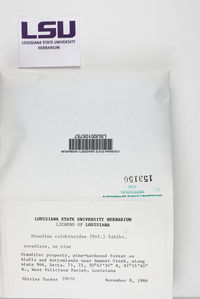
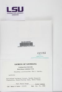
Rinodina colobinoides (Nyl.) Zahlbr.
 |
|
|
Family: Physciaceae
[Lecanora colobinoides Nyl., moreLecanora erysiphaea Nyl., Rinodina erysiphaea (Nyl.) Zahlbr., Rinodina guianensis Aptroot, Rinodina sipmanii Aptroot, Rinodina sorediata H. Magn.]  John Sheard |
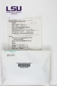
MB#404267 Basionym. Lecanora colobinoides Nyl., Acta Soc. Sci. Fenn. 7: 444 (1863). Rinodina erysiphaea (Nyl.) A. Zahlbr., Cat. Lich. Univ. 7: 514 (1931). Rinodina sorediata H. Magn., Acta Horti Gotob. 17: 292 (1947). Rinodina guianensis Aptroot, Proc. Kon. Ned. Akad. Wetensch. C90: 240 (1987). Rinodina sipmanii Aptroot, Proc. Kon. Ned. Akad. Wetensch. C90: 241 (1987). Exsiccatae. Sipman Lich. Latino. 42 (U as R. sipmanii). Description. Thallus thin, dark grey, sometimes brownish, comprised of dispersed areoles, to 0.40-1.60 mm wide; surface plane and shining; margin indeterminate; prothallus absent; vegetative propagules present; blastidia arising from margins of larger areoles, frequently spreading to cover surface with granular blastidia, 35-60 µm wide, sometimes branched, some blastidia becoming sorediate; soredia 25-50 µm in diam. Apothecia narrowly attached, usually frequent but not contiguous, to 0.50‑0.90 mm in diam., or sometimes absent; disc brown, more rarely black, becoming convex or half‑globose; thalline margin concolourous with thallus, entire or crenulate, or incomplete, 0.05‑0.10 mm wide, often becoming excluded by loss of algae, often with orange pigment in apothecial base and thallus medulla; excipular ring present, raised prior to exclusion of thalline margin. Apothecial Anatomy. Thalline exciple 50‑100 µm wide laterally; cortex 5-10 µm wide; epinecral layer 5-10 µm wide; crystals absent from cortex and medulla; cortical cells pigmented or not, to 4.5‑6.0 µm wide algal cells to 8.0‑11.5 µm long; thalline exciple not expanded below; proper exciple hyaline, 10‑15 µm wide laterally, expanding to 15‑50 µm wide above, or pigmented when thalline margin absent, then 20-25 µm wide laterally and 40-45 µm wide above, cells then becoming ovoid, 5.0-6.5 x 10.0-11.0 µm; hypothecium hyaline or yellowish, 35‑90(-120) µm deep; hymenium 70‑120 µm high, not inspersed; paraphyses; 1.5-3.0 µm wide, not conglutinate, with apices to 3.5‑6.0 µm in diam., lightly pigmented, immersed in dispersed pigment to form a red-brown, or sometimes reddish orange epihymenium, then K+ purple-rose; asci 50‑75 x 16‑20 µm. Ascospores 8/ascus, Type A or B development, Pachysporaria‑type II, (14.0-)17.0-18.0(-20.5) x (7.0-)8.5-9.0(-10.5) µm, average l/b ratio 1.9-2.1, some young spores with acute apices and curved; lumina irregularly spherical, often becoming elongate, lacrimiform; torus lacking or narrow; walls not or lightly ornamented (x1250) in oldest spores. Pycnidia abundant in some specimens, narrowly attached, becoming irregularly globose, sometimes multiple by proliferation from base, multilocular, lacking distinct ostiole, ca. 0.30 mm in diam.; conidiophores type-VIII; conidia bacilliform, 4.0-6.0 x ca. 1.0 µm. Chemistry. Spot tests all negative, or K+ purple-rose below apothecia and in medulla; secondary metabolites not detected (Giralt et al. 1995). Substrate and Ecology. Corticolous, recorded on Magnolia fraseri, Prosopsis glandulosa, Prunus, Quercus and Sabal palmetto. Most collections are at or close to sea level with the exception of the Great Smoky Mountain National Park record and the types of R. colobinoides and R. erysiphaea from Bogota which are at 2 600 m. Distribution. First reported from North America by Tucker (1979) where it is primarily distributed around the Coastal Plain of the Gulf of Mexico. The species is also reported from tropical South America, Caribbean, coastal Portugal and India (Giralt et al. 1995). The Great Smoky Mountain National Park record is an addition to the tropical lichen species present in the southern Appalachians, a region considered to be a Pleistocene refugium for vascular plants and bryophytes with present day tropical or subtropical distributions (Billings and Anderson 1966, Sheard et al. 2008). Notes. The species is characterized by its Pachysporaria-type spores, blastidiate condition and the frequent orange pigment (K+ purple-rose) in tissues of the apothecia base and sometimes in the thallus. Rinodina guianensis is comparable but the presence of Type B spore development in this species was the primary reason that Giralt et al. (1995) did not include this species in R. colobinoides. However, this character has subsequently been observed in R. colobinoides and R. sipmanii. Rinodina guianensis cannot therefore be maintained as separate species, particularly as this type of spore development is now known to occur in other species with Pachysporaria-type II spores. The epihymenium of the South American R. colorans Vain. contains the same or a very similar pigment and despite its larger (19.0-25.0 x 8.5-12.5 µm), Pachysporaria-type spores and lack of vegetative propagules may therefore be related to R. colobinoides. Rinodina colobinoides is discussed in greater detail by Giralt et al. (1995) and has previously been labeled as R. isidiosa Merrill in some North American herbaria but the name was never published. Specimens examined. BEQUIA ISLAND. Admiralty Bay, H.A. Imshaug 30945 (MSC). BERMUDA. Paget Par., Elbow Beach, Franz Berger 20167 (personal herb.). DOMINICA. Parish St Peter, Pointe Round, H.A. Imshaug 33484 (MSC). FRENCH GUYANA. Cayenne, A. Aptroot 15082 (U). GRAND CAYMAN. 'Castle', H.A. Imshaug 24561 (MSC). GUYANA. E Demerara Dist., Mahaica River, H. Sipman 19505; West Berbice Dist., Onverwacht, H. Sipman 19629; SURINAM. Paramaribo, A. Aptroot 14082 (all U). U.S.A. FLORIDA. Miami‑Dade Co., S tip Key Biscayne, S.C. Tucker 25300 (SBBG); Polk Co., Tieger Creek Preserve, 1989, E.M. Wheeler (NY); Seminole Co., Sanford, 1916, S. Rapp (FH); S. Rapp 302 (MICH). LOUISIANA. East Baton Rouge Par., Miller Farm, S.C. Tucker 8847 (LSU). TENNESSEE. Sevier Co., Great Smoky Mountains Nat. Park, Twin Creeks Trail, T. Tønsberg 37441 (BG). TEXAS. Harris Co., Houston, 1869, H.W. Ravenel (BM, FH, US); Hidalgo Co., S of McAllen, T.H. Nash 8617 (ASU). Selected References. Magnusson (1947a as R. sorediata H. Magn.), Giralt et al. (1995 Fig. 1C), Giralt (2001 Plate XIV: C). |

")
")
")
")
")

")
")
")

")
")

")

")
![]()
![]()

![]()
![]()
![]()
![]()
![]()