fracture
[frak´chur] 1. the breaking of a part, especially a bone.
2. a break in continuity of bone; it may be caused by trauma, twisting due to muscle spasm or indirect loss of leverage, or by disease that results in
osteopenia. See illustration.
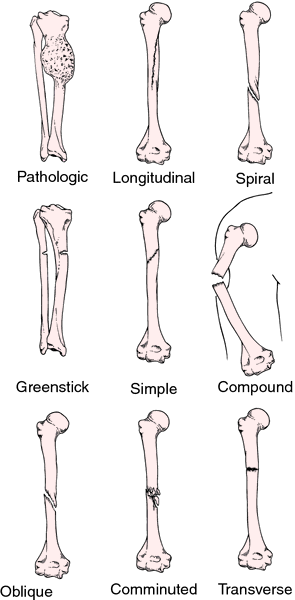
Types of fractures.
Treatment. Immediate first aid consists of
splinting the bone with no attempt to reduce the fracture; it should be splinted “as it lies,” which means supporting it in such a way that the injured part will remain steady and will resist jarring if the victim is moved. Later it will be treated by
reduction, which means that the broken ends are pulled into alignment and the continuity of the bone is established so that healing can take place. Fracture healing is truly a process of regeneration. Fractures heal with normal bone, not with scar tissue. Closed reduction is performed by manual manipulation of the fractured bone so that the fragments are brought into proper alignment; no surgical incision is made. Open fractures are highly contaminated and must be débrided and copiously irrigated in the operating room. A fracture may also require internal fixation with pins, nails, metal plates, or screws to stabilize the alignment. Once closed reduction is accomplished, the bone is immobilized by application of a
cast or by an apparatus exerting
traction on the distal end of the bone.
avulsion fracture separation of a small fragment of bone cortex at the site of attachment of a ligament or tendon.
Barton's fracture fracture of the distal end of the radius into the wrist joint.
Bennett's fracture fracture of the base of the first metacarpal bone, running into the carpometacarpal joint, complicated by subluxation.
blow-out fracture fracture of the orbital floor caused by a sudden increase of intraorbital pressure due to traumatic force; the orbital contents herniate into the maxillary sinus so that the inferior rectus or inferior oblique muscle may become incarcerated in the fracture site, producing diplopia on looking up.
Colles' fracture fracture of the lower end of the radius, the distal fragment being displaced backward.
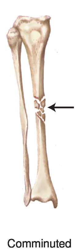
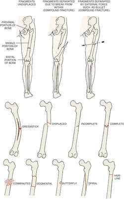
comminuted fracture one in which the bone is splintered or crushed, with three or more fragments. See illustration.
complete fracture one involving the entire cross section of the bone.
compression fracture one produced by compression.
depressed fracture (depressed skull fracture) fracture of the skull in which a fragment is depressed.
direct fracture one at the site of injury.
dislocation fracture fracture of a bone near an articulation with concomitant dislocation of that joint.
double fracture fracture of a bone in two places.
Duverney's fracture fracture of the ilium just below the anterior inferior spine.
fissure fracture a crack extending from a surface into, but not through, a long bone.
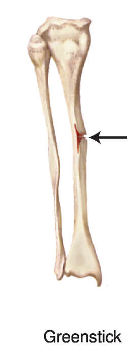
greenstick fracture one in which one side of a bone is broken and the other is bent, most commonly seen in children. See illustration.
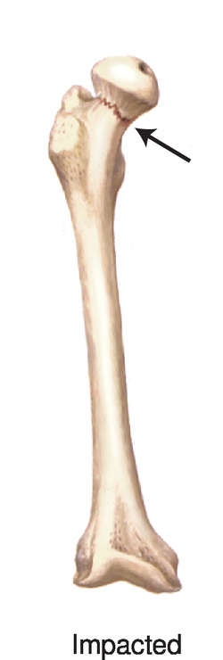
impacted fracture fracture in which one fragment is firmly driven into the other.
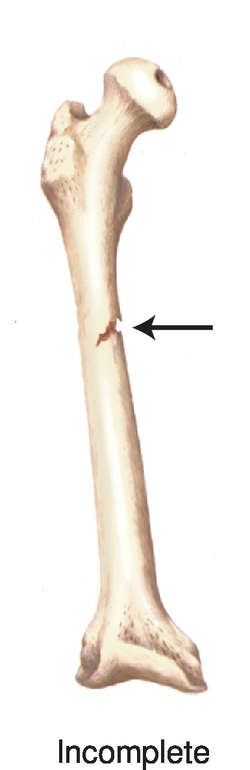
incomplete fracture one that does not involve the complete cross section of the bone.
indirect fracture one distant from the site of injury.
intrauterine fracture fracture of a fetal bone incurred in utero.
Jefferson's fracture fracture of the atlas (first cervical vertebra).
lead pipe fracture one in which the bone cortex is slightly compressed and bulged on one side with a slight crack on the other side of the bone.
Le Fort fracture bilateral horizontal fracture of the maxilla. Le Fort fractures are classified as follows: Le Fort I fracture, a horizontal segmented fracture of the alveolar process of the maxilla, in which the teeth are usually contained in the detached portion of the bone. Le Fort II fracture, unilateral or bilateral fracture of the maxilla, in which the body of the maxilla is separated from the facial skeleton and the separated portion is pyramidal in shape; the fracture may extend through the body of the maxilla down the midline of the hard palate, through the floor of the orbit, and into the nasal cavity. Le Fort III fracture, a fracture in which the entire maxilla and one or more facial bones are completely separated from the craniofacial skeleton; such fractures are almost always accompanied by multiple fractures of the facial bones.
longitudinal fracture one extending along the length of the bone. See illustration.
Monteggia's fracture one in the proximal half of the shaft of the ulna, with dislocation of the head of the radius.
oblique fracture one in which the break extends in an oblique direction. See illustration.
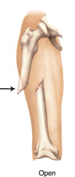
open fracture one in which a wound through the adjacent or overlying soft tissue communicates with the outside of the body; this must be considered a surgical emergency. The compounding may come from within (by a bone protruding through the skin) or from without (e.g., by a bullet wound communicating with the bone). See illustration. Called also
compound fracture.
pathologic fracture one due to weakening of the bone structure by pathologic processes such as neoplasia or osteomalacia; see illustration. Called also
spontaneous fracture.
pertrochanteric fracture fracture of the femur passing through the greater trochanter.
ping-pong fracture an indented fracture of the skull, resembling the indentation that can be produced with the finger in a ping-pong ball; when elevated it resumes and retains its normal position.
Pott's fracture fracture of lower part of the fibula with serious injury of the lower tibial articulation.
Smith's fracture reversed Colles' fracture.
spiral fracture one in which the bone has been twisted and the fracture line resembles a spiral. See illustration.
sprain fracture the separation of a tendon from its insertion, taking with it a piece of bone.
stellate fracture one with a central point of injury, from which radiate numerous fissures.
Stieda's fracture a fracture of the internal condyle of the femur.
transcervical fracture one through the neck of the femur.
transverse fracture one at right angles to the axis of the bone. See illustration.
trophic fracture one due to a nutritional (trophic) disturbance.
Miller-Keane Encyclopedia and Dictionary of Medicine, Nursing, and Allied Health, Seventh Edition. © 2003 by Saunders, an imprint of Elsevier, Inc. All rights reserved.
fracture
(frak'chur) [L. fractura, a break] 1. An injury that upon assessment is painful, swollen, and deformed.

TYPES OF FRACTURES
TYPES OF FRACTURES
TYPES OF FRACTURES
TYPES OF FRACTURES
TYPES OF FRACTURES
TYPES OF FRACTURES
TYPES OF FRACTURES

TYPES OF FRACTURES

TRACTION APPLIED TO A FRACTURE OF THE LOWER EXTREMITY
2. A break of a bone. See:
illustrationCauses
Fractures may be due to pathology, direct violence, indirect violence, or muscular contraction. In a pathological fracture, bones break, spontaneously and without trauma, due to certain diseases and conditions like cancer, osteomalacia, syphilis, and osteomyelitis, In a fracture due to direct violence, the bone breaks at the spot where the force was applied, as in fracture of a crushed tibia. In a fracture due to indirect violence, the bone is fractured by a force applied at a distance from the site of fracture and transmitted to the fractured bone, as a fracture of the clavicle by a fall on an outstretched hand. In a fracture due to muscular contraction, the bone breaks from a sudden, violent contraction of the muscles.
Signs
Signs include loss of the power of movement, pain with acute tenderness over the site of fracture, swelling and bruising, deformity and possible shortening, unnatural mobility, and crepitus or grating heard when the ends of the bone rub together.
Treatment
Immediate first aid includes splinting of the fracture site and joints above and below it to limit further movement and displacement. Applying a cold pack to the fracture site and elevating it above the level of the heart may limit pain and swelling. Radiography should be used to identify the fracture and the exact position of the bone fragments.
The physician reduces the fracture. The bone is kept in position by a cast or splint until union has taken place. Afterwards the limb is restored to complete function by physical therapy and exercise.
In open or compound fractures, bleeding must be arrested before the fracture is treated. Initially, the open fracture should be covered with a clean or sterile dressing and the fracture site immobilized. Open reduction may be required. The wound is then washed and cleaned with sterile saline. If the area is grossly contaminated, mild soap solution may be used provided it is thoroughly washed away with generous amounts of sterile saline. When the wound is clean, a sterile dressing is secured by a bandage. The bone may then be immobilized by external fixation until the wound heals.
Skeletal traction may be used instead of a cast or external fixator for certain fractures, such as femoral shaft fractures. Pins are placed in the bone, and the bone ends are held in place by pulleys and weights until union occurs.
If the bone does not heal, a weak electric current applied to the bone ends (bone stimulation) may promote healing. Hip fractures require gentle handling and immobilization to prevent displacement of the fracture, aggravation of bleeding, or disruption of a pelvic hematoma. Open reduction with internal fixation may be required and is performed when the patient is judged to be hemodynamically stable.
CAUTION!
First aid for fractures of the spine requires extreme care in moving the patient. Unnecessary or improper movement may injure or even transect the spinal cord. Stabilizing the patient on a rigid board, with full spinal protection, is necessary until x-ray studies reveal the spine is stable.
Patient care
Vascular and neurological status of the limb distal to the fracture site are monitored before and after immobilization with traction, casting, or fixation devices. Pain is assessed and managed with prescribed analgesics and noninvasive measures. All procedures and related sensations are explained, and reassurance given.
The patient is evaluated for fat embolism after long bone fractures, for infection in open fractures, for excessive blood loss and hypovolemic shock, and for delayed union or nonunion during healing and follow-up. The patient should report signs of impaired circulation (skin coldness, numbness, tingling, discoloration, and changes in mobility) and is taught how to care for the cast or splint and the correct use of assistive devices (slings, crutches, walker). ; illustration
TYPES OF FRACTURES AND TERMINOLOGY
avulsion fracture
The pulling away of the bony attachment site of tendons, ligament, joint capsule, or fascia. Avulsion fractures of tendons are usually caused by a forceful contraction of the muscle. Ligamentous avulsions are caused by forcing the joint beyond its normal range of motion and are often associated with sprains or dislocations.
illustrationbend fracture
Plastic deformation of bone.Bennett fracture
See: Bennett fracturebimalleolar fracture
A fracture of the medial and lateral malleoli of the ankle joint.
blow-out fracture
See: orbital blow-out fractureblow-up fracture
A fracture of the bony orbit above the eye. It may result in entrapment of the superior rectus muscle with a consequent inability to gaze downward.
bowing fracture
A bending or curving fracture of a bone (usually of the forearm) due to a traumatic load that compresses the bone along its long axis.
boxer's fracture
A fracture of the distal end of the fourth or fifth metacarpal with posterior displacement of the proximal structures. The injury usually occurs after a punch is thrown with a unprotected fist.
buckle fracture
Torus fracture.burst fracture
A vertebral fracture similar to a compression fracture but typically more severe and involving displacement of the bony fragments.
See: compression fracturechauffeur's fracture
A colloquial term for a fracture of the radial styloid with the carpal joint.
childhood accidental spiral tibial fracture
Abbreviation: CAST
Toddler's fracture.fracture of clavicle
Physical injury of the clavicle sufficient to fracture it, often as a result of a fall on an outstretched arm (from a ladder or bicycle) or direct impact to the bone. Most clavicular fractures involve the distal one third of the bone.
Symptoms
Symptoms include swelling, pain, and protuberance with a sharp depression over the injured bone. Palpable deformity and crepitus are commonly present.
Treatment
If indicated, an emergency care physician or an orthopedist will reduce the fracture. This usually is done by elevating the arm and lateral fragment so they line up with the medial fragment. The position is maintained by a clavicle strap, spica cast, immobilizing sling, or figure-of-eight wrap between the shoulders and over the back. Healing takes from about 6 to 8 weeks.
First Aid
A ball of cloth or one or two handkerchiefs are tightly rolled and placed under the armpit. An arm sling is applied and the elbow bandaged to the side, with the hand and forearm extending across the chest. Alternatively, the patient may lie on his or her back on the floor with a rolled-up blanket under the shoulders until medical aid arrives. This position keeps the shoulders back and prevents the broken ends of the bone from rubbing.
clay shoveler's fracture
A colloquial term for a fracture of the base of the spinous process of the lower cervical spine associated with sudden flexion of the neck. It may also be caused by direct trauma.
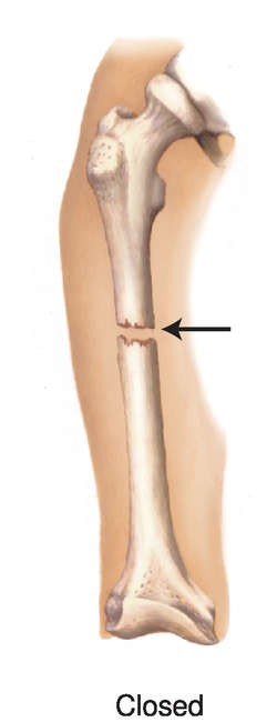
closed fracture
A fracture of the bone with no skin wound.
Colle fracture
See: Colles, Abrahamcomminuted fracture
A fracture in which the bone is broken or splintered into pieces.
complete fracture
A fracture in which the bone is completely broken (i.e., neither fragment is connected to the other).
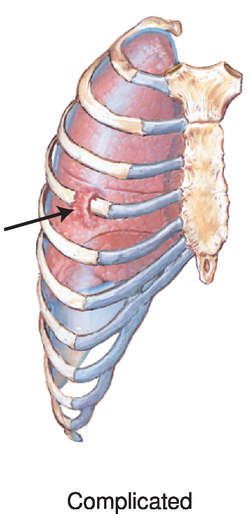
complicated fracture
A fracture in which the bone is broken and has injured some internal organ, such as a broken rib piercing a lung.
compound fracture
A fracture in which fragments of bone protrude through the skin or in which there is a break in the skin or soft tissue at the fracture site. Such a fracture exposes the wound to possible infection. Synonym: open fracture
compression fracture
A fracture of a vertebra by pressure along the long axis of the vertebral column. Such fractures, which may occur traumatically or as a result of osteoporosis, are marked by loss of bone height.
curbstone fracture
An avulsion fracture of the posterior margin of the tibia, typically due to striking the dorsal surface of the foot on an unyielding surface, such as a curb.
depressed fracture
A fracture in which a piece of bone (as of the skull or ribs) is broken and driven inward.
diastatic fracture
A fracture that follows a cranial suture and causes it to separate.
direct fracture
A fracture at a site where force was applied.
dislocation fracture
double fracture
Two fractures of the same bone.
Dupuytren fracture
See: Dupuytren, Baron GuillaumeDuverney fracture
See: Duverney, Joseph G.epiphyseal fracture
A separation of the epiphysis from the bone between the shaft of the bone and its growing end. It occurs only in skeletally immature patients. See:
Salter-Harris fracture.
fatigue fracture
Stress fracture.fissured fracture
A narrow split in the bone that does not go through to the other side of the bone.
flexion-teardrop fracture
An unstable fracture of the cervical spine in which a small fragment of the anteroinferior corner of a vertebral body avulses from the rest of the vertebra due to massive flexion applied to the cervical spine. Patients with this fracture have sustained injuries to all the spinal ligaments and usually severe spinal cord injury and quadriplegia.
fragility fracture
A fracture that occurs in a bone weakened by osteoporosis.
greenstick fracture
A fracture in which the bone is partially bent and partially broken, as when a green stick breaks. It occurs in children, esp. those with rickets. There is a compression fracture on the concave side of the bend and a tension fracture on the convex side.
hairline fracture
A minor fracture in which all the portions of the bone are in perfect alignment. The fracture is seen on a radiograph as a very thin line lying between the two segments and not extending through the bone.
hangman's fracture
A bipedicular fracture of the second cervical vertebra, often with a concomitant dislocation of the vertebra. The term originates from properly performed judicial hangings. At the moment when the dropped criminal fully extends the rope, the hangman's knot causes fracture dislocation of the upper cervical spine and transection of the spinal cord or medulla. If the knot is not made or applied properly, death is usually due to asphyxia. Contemporary hangman's fractures occur mostly in automobile accidents or athletic competitions.
hip fracture
A fracture of the proximal portion of the femur, i.e., of either the head, neck, intertrochanteric or subtrochanteric regions of the hip. Hip fracture occurs each year in approximately 225,000 Americans over 50. It is more common in women than in men due to osteoporosis and is esp. common in slender, elderly women. Mortality rates after hip fracture are influenced by the patient's age, general physical health, and the type of fracture.
Etiology
Osteoporosis predisposes an elderly person to hip fracture.
Symptoms
Pain in the knee or groin is the classic presenting sign of a hip fracture. If the femur is displaced, shortening and rotation of the leg may be present.
Treatment
Preoperatively, Buck's traction may be used in the short term to alleviate muscle spasms. An open reduction is the preferred surgical treatment. A femoral prosthesis may be used for femoral neck or head fractures. The bone takes 6 to 12 weeks to heal in an elderly patient.
Patient care
During hospitalization, general patient care concerns apply. The patient is prepared physically and emotionally for surgery according to the orthopedic surgeon's protocol, and postsurgical care and pain control (epidural or intravenous patient-controlled analgesia [PCA]) is discussed. Neurovascular status of the affected limb is assessed according to protocol and compared to the unaffected limb. The patient is referred for physical and occupational therapy and uses a walker until the bone is completely healed. Prevention and relief of pain and monitoring of postoperative complications, including infection, hip dislocation, and deep venous thrombosis or pulmonary embolism, are primary concerns. Use of an incentive spirometer is encouraged to prevent atelectasis and respiratory complications. Prophylactic antibiotics and anticoagulants are administered as prescribed, and hip precautions are implemented to prevent dislocation. These precautions include having the patient avoid hip adduction (usually by an abductor wedge), rotation, and flexion greater than 90° during transfer and ambulation activities, and by using a raised toilet seat and semi-reclining chair. The patient is typically hospitalized for 2 to 4 days and then discharged to a nursing home, subacute unit, transitional care unit, rehabilitation center, or home for rehabilitation for several weeks.

FRACTURE OF THE HUMERUS: Radiographic image of a fracture of the humerus (COURTESY of W. Robert Strauss, Jr.)
fracture of humerus
Disruption of the bony cortex of the upper arm. If the fracture is of the upper end of the humerus, the arm is abducted and splinted for about 4 weeks. Movements of the elbow and wrist are started early, and active movements of the shoulder are begun in about 3 weeks. See:
illustration;
acromiohumeral;
capitellum;
cubitus;
glenoid cavityIn a fracture of the shaft and lower end of the humerus, the limb is put in a cast in a position midway between pronation and supination with the humerus at right angles to the forearm. Movement of the shoulder, wrist, and finger is allowed.
impacted fracture
A fracture in which the bone is broken and one end is wedged into the interior of the opposing end.
incomplete fracture
A fracture in which the line of fracture does not transverse the bone.
indirect fracture
A fracture distant from the place where the force was applied.
insufficiency fracture
A stress fracture occurring in abnormal bone (e.g., osteoporotic bone) subjected to normal forces.
intracapsular fracture
A fracture occurring within the capsule of a joint.
intrauterine fracture
A broken fetal bone.
Jefferson fracture
See: Jefferson fractureJones fracture
See: Jones fracturelead pipe fracture
A fracture in which the bone is compressed and bent so that one side of the fracture bulges and the other side shows a slight fracture line.
LeFort fracture
A fracture usually involving more than one of the facial bones: maxillary, nasal, orbital, and/or zygomatic.
Synonym: mid-face fracturelover's fracture
A colloquial term for a fracture of the calcaneus, due to jumping from a height, e.g., a balcony or second-story window.
march fracture
A fracture of the metatarsals as a result of overuse.
mid-face fracture
LeFort fractureMonteggia fracture
See: Monteggia fracturenightstick fracture
A nondisplaced transverse fracture of the ulna resulting from a direct blow.
nonunion of fracture
See: nonunionoccult fracture
A fracture that is suspected based on clinical grounds (e.g., guarding, pain, and swelling) but not seen on x-rays. The fracture may be seen with bone scans or magnetic resonance imaging.
open fracture
Compound fracture.orbital blow-out fracture
A fracture of the bony floor beneath the eye. It typically results in entrapment of the inferior rectus muscle, with a consequent inability to look upward with the affected eye, which causes double vision during vertical gaze.
open-book fracture
A fracture in which the pubic symphysis is broken apart, e.g., in a high-speed, front-end automobile crash. The pelvis can separate in the front even though it remains held in place posteriorly by the spine or posterior ligaments.
overriding fracture
Overriding.pathological fracture
A fracture of a diseased or weakened bone caused by a force that would not have fractured a healthy bone. The underlying disease may be a metastasized cancer, primary cancer of the bone, or osteoporosis.
Patient care
The limbs and joints of at-risk patients are gently and carefully supported when repositioning, exercising, or mobilizing. If such patients fall or are injured, and report limb, pelvic, or back pain or inability to bear weight, the patient and the affected limb should be stabilized and - diagnostic imaging obtained.
pelvic fracture
Fracture of one of more of the bones of the pelvis, i.e. of the ilium, ischium, or pubis. Pelvic fractures occur after falls, esp. in the elderly, in whom the pelvic bones may be weakened by osteoporosis, and after high-impact trauma, e.g., motor vehicle crashes. Some pelvic fractures are accompanied by internal organ damage, esp. to the genitourinary organs. Regaining the ability to walk after pelvic fracture sometimes requires months of rehabilitation.
penile fracture
Sudden trauma to the tunica albuginea of the penis, resulting in a rupture of the corpus cavernosum and sometimes a tearing of the urethra. The injury typically occurs during sexual intercourse (or, less often, during masturbation) and may be accompanied by bleeding into the penis.
ping-pong fracture
A depressed skull fracture in a newborn or very young infant in which the bone bends inward (like a dented ping-pong ball) without breaking. It may occur during strenuous labor and delivery, or in trauma to the pliant skull in the first weeks of life.
Pott fracture
See: Pott, John Percivallpretrochanteric fracture
A fracture that passes through the greater trochanter of the femur.
Rolando fracture
See: Rolando fracturesimple fracture
A fracture without rupture of ligaments and skin.
fracture of skull
, fractured skullLoss of the integrity of one or more bones of the cranium. A fracture is classified according to whether it is in the vault or the base but, from the point of view of treatment, a more useful classification is differentiating between a simple fracture (uncommon) and a compound fracture. When a compound fracture occurs in the vault of the skull, the bone is depressed and driven inward, possibly damaging the brain. Treatment is operative. See: fracture
sleeve fracture
An avulsion fracture of the patella that typically occurs as a result of a sudden strong contraction of the quadriceps muscle group.
Smith fracture
See: Smith fracturesnowboarder's fracture
A fracture of the lateral border of the talus caused by inversion and rotation of the talus within the mortise. Signs and symptoms often mimic those of an inversion (lateral) ankle sprain.
fracture of the spine
Fracture of a vertebral body or its bony prominences. Synonym:
vertebral fracture See:
burst fracture;
compression fracture;
hangman's fracture;
Jefferson fractureTreatment
The patient is carefully assessed for evidence of neuromuscular compromise and other internal injuries. To prevent complications and promote healing, vests, casts, or halo devices may be used, depending on the location of the fracture. A program of supervised physical therapy may be needed during recovery.
Prognosis
Prognosis depends on the type of spinal fracture and associated spinal cord involvement.
spiral fracture
A fracture that follows a helical line along and around the course of a long bone.
sprain fracture
The separation of a ligament from its insertion, taking with it a piece of the bone.
See: avulsion fracturestellate fracture
A fracture with numerous fissures radiating from the central point of injury.
straddle fracture
A traumatic fracture of all four pubic rami, often associated with injury to the urethra.
stress fracture
A microfracture that appears without evidence of a single traumatic onset. This type of fracture is difficult to diagnose by standard radiography and may not become visible until 3 to 4 weeks after the onset of symptoms. Scintigraphy, CT, and/or MRI may lead to earlier identification of the fracture lines. Stress fractures occur from repetitive microtraumas (from running, aerobic dancing, or marching or other cyclical actions), from improper shoes on hard surfaces, or from inadequate healing time after stress. Stress fractures are classified as fatigue fractures or insufficiency fractures based on their etiology. Undiagnosed and untreated stress fractures may progress to frank fractures. Synonym:
fatigue fracture See:
insufficiency fractureT fracture
A fracture in which bone splits both longitudinally and transversely.
toddler's fracture
A fracture of the distal third of the tibia, sustained in a child typically aged 2 to 4. The child may limp or refuse to walk because of pain. The fracture may not be easily seen on plain radiographs.
Synonym: childhood accidental spiral tibial fracturetorus fracture
A fracture where the structure of one side of the bone is compressed, while the opposite side deflects from the growth plate, leaving the cortex intact.
Synonym: buckle fracturetranscervical fracture
A fracture through the neck of the femur.
transverse fracture
A fracture in which the fracture line is at right angles to the long axis of the bone.
trimalleolar fracture
A fracture of the lateral and medial malleoli of the ankle joint with an additional fracture of the posterior edge of the distal tibia.
tripod fracture
A fracture in which the zygoma is separated from its attachment to the maxilla and the temporal and frontal bones.
vertebral fracture
Fracture of the spine.Wagstaffe fracture
See: Wagstaffe fractureillustrationMedical Dictionary, © 2009 Farlex and Partners

)
)
)
)
)
)
)
)
)
)
)