terminal bronchiole


Also found in: Dictionary, Thesaurus, Encyclopedia, Wikipedia.
Related to terminal bronchiole: respiratory bronchiole

bronchiole

 [brong´ke-ōl]
one of the successively smaller channels into which the segmental bronchi divide within the bronchopulmonary segments. adj., adj bronchi´olar.
respiratory b's the final branches of the bronchioles, communicating directly with the alveolar ducts; they are subdivisions of terminal bronchioles, have alveolar outcroppings, and themselves divide into several alveolar ducts.
Respiratory bronchiole. From Dorland's 2000.
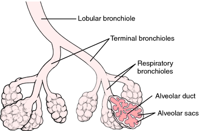
( and see color plates.)
terminal bronchiole the last portion of a bronchiole that does not contain alveoli, i.e., one whose sole function is gas conduction; it subdivides into respiratory bronchioles.
Miller-Keane Encyclopedia and Dictionary of Medicine, Nursing, and Allied Health, Seventh Edition. © 2003 by Saunders, an imprint of Elsevier, Inc. All rights reserved.

ter·mi·nal bron·chi·ole

the end of the nonrespiratory conducting airway; the lining consists of simple columnar or cuboidal epithelium without mucous goblet cells; some of the cells are ciliated; numerous Clara cells also occur.
Farlex Partner Medical Dictionary © Farlex 2012

ter·mi·nal bron·chi·ole

(tĕr'mi-năl brong'kē-ōl)
The end of the nonrespiratory conducting airway; the lining is simple columnar or cuboidal epithelium without mucous goblet cells; most of the cells are ciliated, but a few nonciliated serous secreting cells occur.
Medical Dictionary for the Health Professions and Nursing © Farlex 2012
References in periodicals archive ?
The amount of pigment in terminal bronchioles was significantly correlated with the amount of pigment in respiratory bronchioles, particularly for mineral dusts (linear regression analysis, [R.sup.2] = 0.66, p [is less than] 0.05).
Abbreviations: a, alveolus; AD, alveolar ducts; BV, blood vessel; TB, terminal bronchioles. (A) Histopathology showing lung inflammatory response to O-MWCNTs.
On microCT, lungs from patients with the centrilobular emphysematous phenotype of COPD had a reduction of 99.7% in the terminal bronchiolar cross-sectional area per lung, compared with that of the control lungs, and a reduction of 89% in the total number of terminal bronchioles per lung.
As terminal bronchioles penetrate more deeply into the lungs, they divide into microscopic respiratory bronchioles.
The first 16 subdivisions of the bronchi ending in terminal bronchioles are called the conducting airways.
Of concern is exposure to a high concentration of particulate matter size 10 and less ([PM.sub.10]), because such particles can reach the terminal bronchioles and alveoli (6).
In the lung, epithelial hyperplasia with squqmous metaplasia were noticed in the terminal bronchioles. In some areas of lung, alveolar walls were thickened due to hyperplasia of type II pneumocytes (Fig.
The virus's preference for type II pneumocytes, alveolar macrophages, and nonciliated cuboidal epithelial cells in terminal bronchioles "may contribute to the severity of the pulmonary lesion," they noted.
This case illustrates the classic findings of carbon pigment deposition around terminal bronchioles, dust macules, and mixed dust fibrosis.
The Dutch researchers noted that the virus's preference for type II pneumocytes, alveolar macrophages, and nonciliated cuboidal epithelial cells in terminal bronchioles "'may contribute to the severity of the pulmonary lesion."