Tibia Bone
TIBIA
- Tibia is the second largest bone (after femur).
PROXIMAL END (upper end)
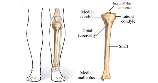
- Proximal (upper) end of tibia includes medial & lateral condyles, forming tibial plateau.
- It also includes tibial Tuberosity & intercondylar area.
- Attachments on proximal end are:
- Medial condyle: Semimembranous (posteriorly), capsule of knee joint, tibial (medial) collateral ligament (deep part), medial patellar retinaculum (anteriorly).
- Lateral condyle: illiotibial tract (anteriorly), capsule of superior tibiofibular joint.
- Tibial Tuberosity: Ligamentum patellae
- Intercondylar area (from anterior to posterior);
- Anterior horn of medial meniscus
- Anterior cruciate ligament (ACL)
- Anterior horn of lateral meniscus
- Posterior horn of lateral meniscus
- Posterior horn of medial meniscys
- Posterior cruciate ligament (PCL)
- Upper end of tibia is the growing end. The secondary center for upper end appears just before birth & fuses with shaft at around 16-18 years.
SHAFT
- Shaft of tibia is plasmoid in shape.
- It has three borders (anterior, medial & interosseous or lateral) and three surfaces (medial, lateral & posterior).
- Anterior border (also c/d shin) and medial surface are subcutaneous.
- Tibia is the commonest site of osteomyelitis from direct contamination as well as from hematogenous spread.
- Nutrient artery to tibia is the largest nutrient artery. It is a branch of posterior tibial artery.
DISTAL END
- It is slightly expanded with a projection downwards & medially i.e medial malleolus.
- Medial malleolus gives attachment to deltoid ligament (medial collateral ligament) of ankle.
- Secondary center of ossification for lower end appears during 1st year forms malleolus by 7th years & fuses with shaft by 15-17 years.
Exam Question
- Tibia is the second largest bone (after femur).
- Length of tibia is 20% of height.
- Posterior dislocation of tibia on femur is prevented by Posterior cruciate ligament
- Action of tibialis anterior is Inversion of foot.
- Upper end of tibia is the growing end. The secondary center for upper end appears just before birth & fuses with shaft at around 16-18 years.
- Tibia is the commonest site of osteomyelitis from direct contamination as well as from hematogenous spread.
Don’t Forget to Solve all the previous Year Question asked on Tibia Bone