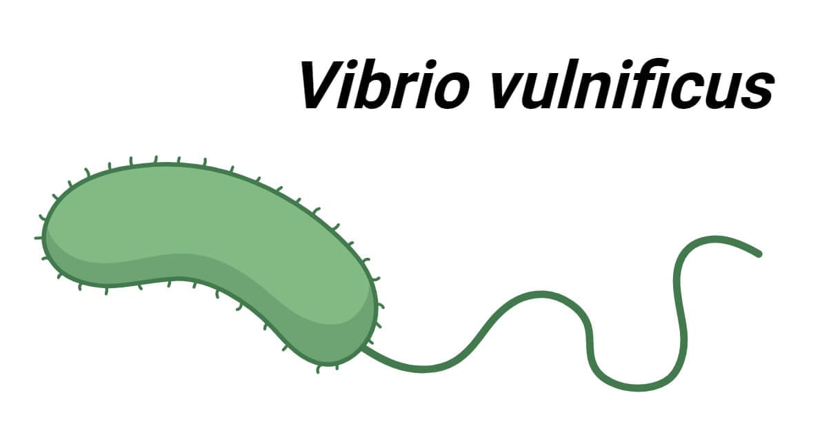
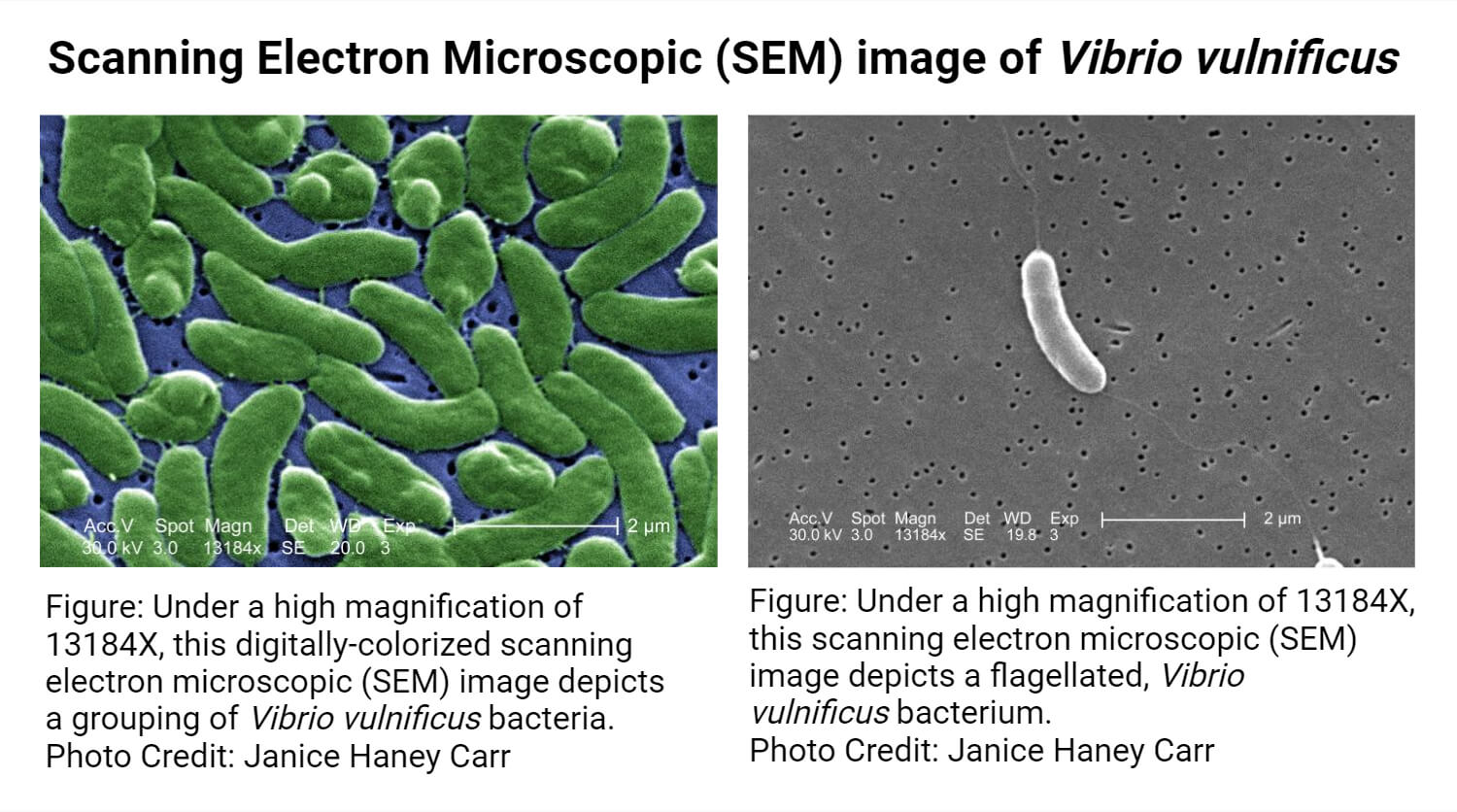
Vibrio vulnificus is a Gram-negative, facultative anaerobic, curved rod-shaped, motile, Gammaproteobacteria of the Vibrio genus in the Vibrionaceae family. It is one human pathogenic species of the Vibrio genus together with Vibrio cholerae and Vibrio parahaemolyticus.
- V. vulnificus was first described as a separate bacterial species in late 1976 when it was initially named Beneckea vulnifica and later it was named as Vibrio vulnificus in 1979. It is mainly found in warm seawater and estuarine environments. It is frequently reported to cause gastroenteritis, septicemia, and wound infections.
- Biotyping has divided V. vulnificus into three biotypes; biotype 1 is associated with human infections, biotype 2 is associated with human and fish (eel) infections, and biotype 3 is associated with wound infections in fish handlers.
Interesting Science Videos
Vibrio vulnificus Classification
| Domain | Bacteria |
| Phylum | Pseudomonadota |
| Class | Gammaproteobacteria |
| Order | Vibrionales |
| Family | Vibrionaceae |
| Genus | Vibrio |
| Species | V. vulnificus |
Habitat of Vibrio vulnificus
- V. vulnificus is a marine bacterial species. It is normally found in coastal areas, estuaries, brackish water, deltas, and other places with warm saline water globally. It is also reported from deeper ocean surfaces as well.
- Primarily, V. vulnificus is associated with marine animals like oysters (primarily), shellfish, crabs, and even higher fishes. Through such kinds of seafood, they come into contact with humans and cause disease.
Morphology of Vibrio vulnificus
- V. vulnificus shows pleomorphism. They are commonly found in two morphologically distinct forms; circular during the dormant viable non-culturable (VBNC) state and rod-shaped (curved) during the active state.
- In the VBNC state, they appear as small cocci measuring 0.3 μm in diameter. This stage is found in low-nutrition and low-temperature (5°C or below) environments.
- In a normal active state, they appear as curved bacilli measuring 3 μm long and 0.7 μm wide.
Cultural Characteristics of Vibrio vulnificus
- For the culture of Vibrio spp., the sample must be first enriched in an enrichment broth like Alkaline Peptone Water, and then it can be cultured in different mediums for selective isolation and identification.
- Diverse types of culture mediums can be used for the isolation of V. vulnificus, the most commonly used ones include TCBS (thiosulfate citrate bile salt sucrose) agar medium, CHROMagar Vibrio medium, Blood agar medium, and Tryptic Soy medium.
- Selective mediums are also available like mCPC (modified cellobiose polymixin B colistin) medium, and Vibrio vulnificus medium (VVM). This medium contains antibiotics like Polymixin B and colistin, alkaline pH, and moderate salt concentration which suppresses the growth of other organisms.
- In TCBS medium, V. vulnificus produces small, circular, green colonies.
- In Blood agar medium, V. vulnificus produces small (about 3 mm), smooth, creamy, or greyish-white colored, non-hemolytic or α- hemolytic colonies.
Biochemical Characteristics of Vibrio vulnificus
General Biochemical Tests
| General Biochemical Characteristics | Vibrio vulnificus |
| Capsule | Variable |
| Catalase | Positive (+) |
| Citrate | Negative (-) for clinical isolatesVariable for environmental isolates |
| Flagella | Positive (+) |
| Gas | Negative (-) |
| Gram Staining | Gram Negative Bacilli |
| H2S (Hydrogen Sulfide) | Negative (-) |
| Indole | Positive (+) for clinical isolatesVariable for environmental isolates |
| Motility | Motile |
| Methyl Red (MR) | Positive (+) |
| Nitrate Reduction | Positive (+) |
| Oxidase | Positive (+) |
| ONPG Test | Positive (+) (Except biotype 3 members) |
| OF (Oxidative Fermentation) | Facultative anaerobe |
| Growth on 1% to 6% NaCl | Positive (+) |
| TSI Test | A/A, no gas, no H2S |
| Urease | Negative (-) |
| Voges-Proskauer (VP) | Negative (-) |
Carbohydrate Fermentation Tests
| Carbohydrate Fermentation Tests | Vibrio vulnificus |
| L-Arabinose | Negative (-) |
| Amygdalin | Positive (+) |
| D-Adonitol | Negative (-) |
| Cellobiose | Positive (+) (Except biotype 3 members) |
| Dulcitol | Negative (-) |
| D-Galactose | Positive (+) |
| Glucose | Positive (+) |
| Inositol | Negative (-) |
| Glycerol | Negative (-) |
| Lactose | Positive (+) (Except biotype 3 members) |
| Melibiose | Negative (-) |
| Maltose | Positive (+) |
| Mannitol | Variable |
| D – Mannose | Positive (+) |
| Rhamnose | Negative (-) |
| Raffinose | Negative (-) |
| Sorbitol | Variable |
| Sucrose | Negative (-) |
| Trehalose | Positive (+) |
| Xylose | Negative (-) |
Enzymatic Hydrolysis Tests
| Enzymatic Hydrolysis Tests | Vibrio vulnificus |
| Arginine Dehydrolase | Negative (-) |
| DNase | Positive (+) |
| Esculin Hydrolysis | Variable |
| Gelatinase | Positive (+) |
| Lysine Decarboxylase | Positive (+) |
| β-Galactosidase | Positive (+) |
| Ornithine Decarboxylase | Positive (+) in clinical samplesVariable in environment isolates |
| Tryptophan Deaminase | Negative (-) |
Vibrio vulnificus Associated Diseases and Outbreaks
- Vibrio vulnificus is a deadly pathogenic Vibrio species that causes disease in humans and some marine life. In human, V. vulnificus are primarily associated with wound infections, gastroenteritis, and septicemia. It is the leading cause of death and disease associated with seafood in the USA and coastal areas and is accountable for about 90% of deaths caused by Vibrio species in those areas.
- The mortality rate in V. vulnificus infection is very high – 25% in wound infection, about 50% in septicemia, and around 33% in gastroenteritis – even with treatment. This higher mortality is mainly due to endotoxin shock and vulnerabilities in patients with liver disease, immunocompromised state, diabetes, HIV, etc.
- V. vulnificus disease outbreaks are frequently reported in the USA, usually in the summer months. The countries with the most documented cases of V. vulnificus are the United States, South Korea, Taiwan, Japan, and Mexico. Coastal areas in the Southeastern United States like Florida, Texas, Alabama, Louisiana, Mississippi, and other nearby areas have higher incidences of V. vulnificus infections. The Gulf of Mexico is another hot zone for outbreaks of V. vulnificus-associated infections. Coastal areas of Asia, the Middle East, and warm areas in Europe are also endemic zones.
Virulence Factors of Vibrio vulnificus
A variety of virulence factors have been identified in V. vulnificus which contributes to disease pathogenesis. Some of the important virulence factors are listed below.
- Polysaccharide Capsule: This helps the pathogens to escape the phagocytic effect of human immune cells.
- Endotoxin: The lipopolysaccharide of this bacterium is not potent enough to activate the immune system to produce cytotoxic TNF and cytokines; however, LPS induces inflammatory responses in wound infections. The capsular proteins are capable of triggering an immune response causing shock in the patient.
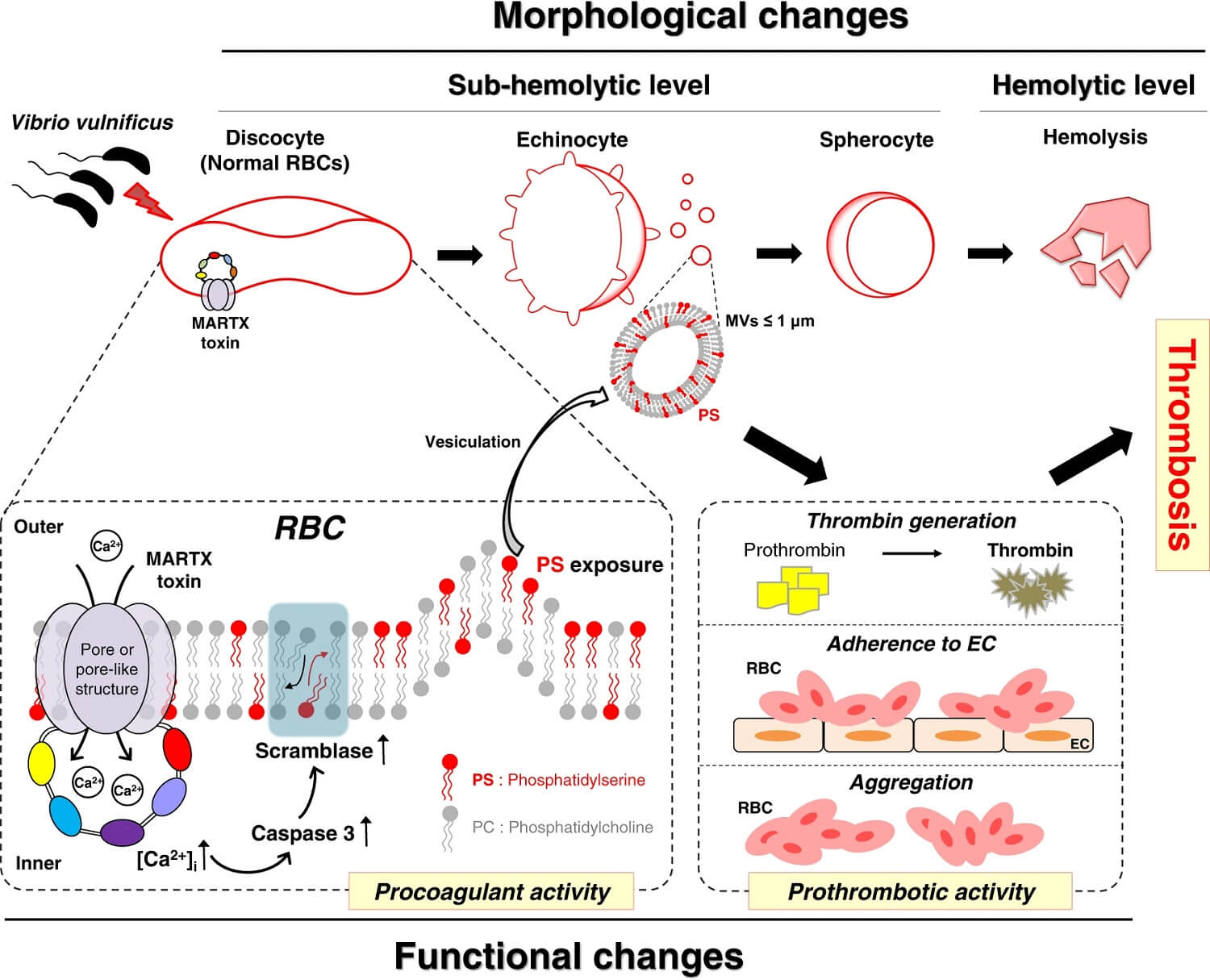
- Exotoxin: Extracellular cytolysin/hemolysin, MARTX toxin, and metalloproteases are toxic compounds secreted by this bacterium which results in host cell/tissue destruction and helps in their dissemination.
- Iron Acquisition from Transferrin: V. vulnificus can capture iron bound to transferrin and use it in their metabolism.
Laboratory Diagnosis/Identification of Vibrio vulnificus
V. vulnificus and its infections are confirmed using several techniques including culture and biochemical identification, serological analysis, and molecular analysis.
1. Microscopy and Biochemical Characterization
Clinical specimens like blood, wound swabs, stool, and tissue biopsy are cultured on a suitable medium (commonly TCBS) and the developed colonies are studied macroscopically and microscopically and characterized biochemically.
2. Immunological Detection
ELISA and agglutination tests are used to detect V. vulnificus antigens in samples and antibodies against V. vulnificus antigens in the patient’s serum.
3. Molecular Methods
Polymerase chain reaction (PCR) is the most rapid and accurate method to detect and confirm the presence of V. vulnificus in any specimen. Colony hybridization tests and DNA probe assays are also available for their rapid identification.
References
- Lydon, K. A., Kinsey, T., Le, C., Gulig, P. A., & Jones, J. L. (2021). Biochemical and Virulence Characterization of Vibrio vulnificus Isolates From Clinical and Environmental Sources. Frontiers in Cellular and Infection Microbiology, 11. https://doi.org/10.3389/fcimb.2021.637019
- Amalina, N.Z., Santha, S., Zulperi, D. et al. Prevalence, antimicrobial susceptibility and plasmid profiling of Vibrio spp. isolated from cultured groupers in Peninsular Malaysia. BMC Microbiol 19, 251 (2019). https://doi.org/10.1186/s12866-019-1624-2
- https://www.fda.gov/food/laboratory-methods-food/bam-chapter-9-vibrio
- Hsu, Y., & Tamplin, M. L. (1998). Enhanced Broth Media for Selective Growth of Vibrio vulnificus. Applied and Environmental Microbiology, 64(7), 2701-2704. https://doi.org/10.1128/aem.64.7.2701-2704.1998
- Ayrapetyan M, Williams TC, Oliver JD. Interspecific quorum sensing mediates the resuscitation of viable but nonculturable vibrios. Appl Environ Microbiol. 2014 Apr;80(8):2478-83. doi: 10.1128/AEM.00080-14. Epub 2014 Feb 7. PMID: 24509922; PMCID: PMC3993182.
- https://www.vetbact.org/species/84
- Oliver, J. D. (2002). Chapter 17 Culture media for the isolation and enumeration of pathogenic Vibrio species in foods and environmental samples. Progress in Industrial Microbiology, 37, 249-269. https://doi.org/10.1016/S0079-6352(03)80020-6
- Heng, P., Letchumanan, V., Deng, Y., Mutalib, S. A., Khan, T. M., Chuah, H., Chan, G., Goh, H., Pusparajah, P., & Lee, H. (2017). Vibrio vulnificus: An Environmental and Clinical Burden. Frontiers in Microbiology, 8. https://doi.org/10.3389/fmicb.2017.00997
- https://www.sciencedirect.com/topics/agricultural-and-biological-sciences/vibrio-vulnificus
- Linkous, D. A., & Oliver, J. D. (1999). Pathogenesis of Vibrio vulnificus. FEMS Microbiology Letters, 174(2), 207-214. https://doi.org/10.1111/j.1574-6968.1999.tb13570.x
- https://www2.mst.dk/udgiv/Publications/1999/87-7909-344-2/html/kap02_eng.htm
- https://www.tgw1916.net/Vibrio/vulnificus.html
- Strom, M. S., & Paranjpye, R. N. (2000). Epidemiology and pathogenesis of Vibrio vulnificus. Microbes and Infection, 2(2), 177-188. https://doi.org/10.1016/S1286-4579(00)00270-7
- https://www.fau.edu/hboi/research/ocean-health-human-health/microbiology/vibrio/
- Church, S. R., Lux, T., Baker-Austin, C., Buddington, S. P., & Michell, S. L. (2016). Vibrio vulnificus Type 6 Secretion System 1 Contains Anti-Bacterial Properties. PLOS ONE, 11(10), e0165500. https://doi.org/10.1371/journal.pone.0165500
- Vibrio vulnificus. (2023, September 6). In Wikipedia. https://en.wikipedia.org/wiki/Vibrio_vulnificus
- Haftel A, Sharman T. Vibrio vulnificus Infection. [Updated 2023 Jun 12]. In: StatPearls [Internet]. Treasure Island (FL): StatPearls Publishing; 2023 Jan-. Available from: https://www.ncbi.nlm.nih.gov/books/NBK554404/