History of endoplasmic reticulum:
In 1945, Porter, Claude, and Fullam used electron microscopy to observe vesicle-like structures in cell culture investigations, which marked the beginning of the history of the endoplasmic reticulum. These objects ranged in size from 100 to 150 micrometers.
Introduction of Endoplasmic reticulum:
The endomembrane system of the cell can be accounted for in large part by the overall structure. For instance, the ER can make up more than 50% of the cell’s total lipid bilayer in cells like liver hepatocytes, which are designed for protein production and detoxification. Similar to this, activated B-lymphocytes that make antibodies or pancreatic beta cells that secrete insulin both have a significant ER membrane system.
Even though their compositions might vary, the endoplasmic reticulum membranes and the outer nuclear membrane are connected. Special membrane-embedded proteins in the ER maintain the ER’s shape and curvature. This organelle interacts intimately with a variety of other organelles, which makes it an essential regulator of cell function.
Endoplasmic reticulum products are frequently packaged and further processed in the Golgi body before being secreted. This is a microscopic view of a lung tissue segment taken from a mammal. Small black circles are mitochondria that are situated close to the ER membranes.
Define endoplasmic reticulum.
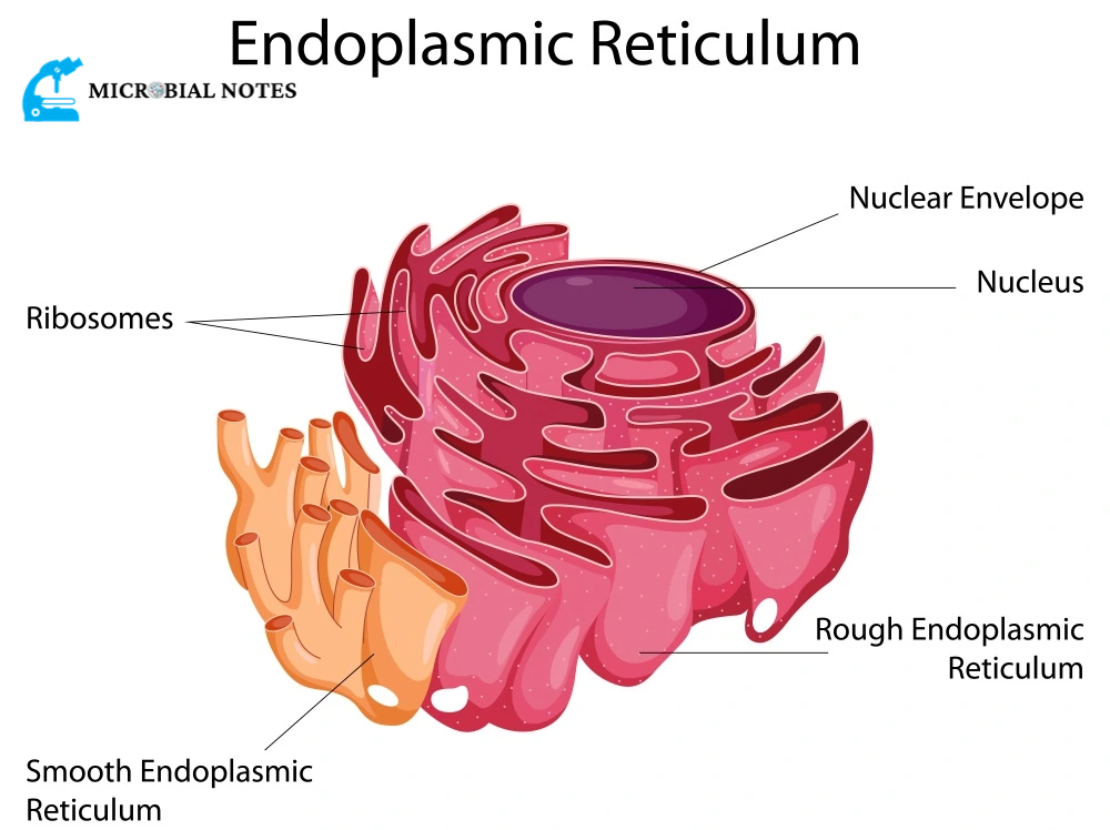
The endoplasmic reticulum (ER) is a sizable organelle composed of tubules and membrane sheets that start close to the nucleus and span the cell. Many of the products made by a cell are produced, packaged, and secreted by the endoplasmic reticulum. A section of the endoplasmic reticulum is lined with ribosomes, the building blocks of proteins.
Where endoplasmic reticulum located?
The majority of the nucleus’ instructions are processed by the endoplasmic reticulum. As a result, the endoplasmic reticulum envelops and extends from the nucleus. The endoplasmic reticulum can make up more than 50% of cells in tissues that release several substances for the rest of the body.
mRNA, or messenger RNA, is typically expressed by the nucleus and instructs the cell on how to make proteins. Numerous ribosomes, which are the main site of protein synthesis, are found in the rough endoplasmic reticulum. Protein synthesis and correct protein folding occur in this area of the organelle.
The main site of lipid synthesis is the smooth endoplasmic reticulum. As a result, it is devoid of ribosomes. Instead, it goes through a series of chemical processes to produce the phospholipid molecules required to build different membranes and organelles.
The endoplasmic reticulum in its rough form is frequently located nearer to the nucleus than the endoplasmic reticulum in its smooth form. However, a network of tiny tubules links the nucleus, and both copies to one another.
Explain the structure of the endoplasmic reticulum.
- Similar to the unit membrane of the plasma membrane, the endoplasmic reticulum membrane is fluid-mosaic and ranges in thickness from 50 to 60 Ao.
- It has been discovered that several different types of enzymes are present in the endoplasmic reticulum membranes and are necessary for a variety of significant synthetic processes. The most significant enzymes are the stearases, NADH-cytochrome C reductase, NADH diaphorase, glucose-6-phosphatase, and Mg++ activated ATPase.
- The plasma membrane, nuclear membrane, and Golgi apparatus membranes are all still connected via the endoplasmic reticulum membrane.
- The endoplasmic reticulum’s cavity is well developed and serves as a conduit for secretory products.
There are three different parts of the endoplasmic reticulum:
- Lamellar structures or cisterns
- The vesicular shape, vesicle, or
- Tubules or tubular shape.
A Cisternae:
RER typically manifests as cisternae in cells that play synthetic roles, such as those in the pancreas, notochord, and brain. The cisternae are lengthy, flattened, unbranched tubules with a diameter of 40 to 50 µm that resemble flattened sacs. They still stay bundled or staked parallel to one another.
A vesicle:
The oval, membrane-bound vacuolar structures that make up the vesicles range in diameter from 25 to 500 m. They exist in various cells but are most frequently isolated in the cytoplasm of the SER.
A tubule:
With the cisternae and vesicles, the tubules are branched structures that make up the reticular system. They typically exist practically everywhere in cells and range in diameter from 50 to 190 µm. The tubular form of ER, which is dynamic in nature and frequently observed in SER, is connected to membrane movements, fission, and fusion within the cyst cavity network.
What are the types of endoplasmic reticulum?
There are two types of endoplasmic reticulum
Smooth endoplasmic reticulum:
- The term “agranular endoplasmic reticulum” is also used to describe them.
- The smooth walls of this form of endoplasmic reticulum are a result of the ribosomes’ lack of attachment to the membranes.
- Most of those cells that are engaged in the metabolism of lipids (including steroids) and glycogen contain the smooth form of the endoplasmic reticulum. For instance, adipose cells, interstitial cells, liver cells that store glycogen, cardiac conduction fibers, spermatocytes, and leucocytes.
Rough endoplasmic reticulum:
- The ribosomes are still anchored to their membranes, which accounts for their rough walls.
- Rough ER (RER) membranes include particular transmembrane glycoproteins for ribosomes, known as ribophorins I and II, to which the ribosomes are linked while undergoing polypeptide synthesis.
- In cells that actively synthesize proteins, such as pancreatic cells, plasma cells, goblet cells, and liver cells, the rough type of endoplasmic reticulum is common.
Explain the function of the endoplasmic reticulum.
The rough endoplasmic reticulum is where protein synthesis takes place. All proteins start their translation in the cytoplasm, however, some are transported into the ER to be folded and sorted for various destinations. During translation, proteins that are translocated into the ER are frequently intended for secretion. These proteins are initially folded in the ER before being transported to the Golgi apparatus, where they can then be sent to other organelles.
For instance, in the lysosome, this method is used to produce hydrolytic enzymes. Alternatively, the cell may secrete these proteins. The digestive tract’s enzymes come from this source. The endomembrane system itself could be the third possible function for proteins that are translated in the ER. This is especially true for proteins called chaperones, which help other proteins fold. When the cell is under stress from unfolded proteins, the genes that encode these proteins are increased.
Lipid synthesis:
The smooth endoplasmic reticulum is crucial for the creation of phospholipids and cholesterol. As a result, this region of the ER is crucial for both the ER’s complex endomembrane system and the production and maintenance of the plasma membrane. The SER is rich in enzymes that are essential for the synthesis of steroid hormones as well as sterol and steroid biosynthesis pathways.
Because the adrenal gland cells secrete five separate classes of steroid hormones that affect the metabolism of the entire body, the SER is therefore highly significant in these cells. Enzymes found in the mitochondria are also involved in the manufacture of these hormones, highlighting the connection between these two organelles.
Calcium Store:
The SER is a crucial location in the cell for the storage and release of calcium. The sarcoplasmic reticulum, a modified version of the SER, creates a vast network of contractile cells like muscle fibers. Additionally, calcium ions play a role in controlling cellular metabolism and can alter cytoskeletal movements.
The broad ER network’s ability to connect with the plasma membrane and utilize Ca2+ for signal transduction and nuclear activity modulation makes it possible. The ER can also employ its calcium reserves to trigger apoptosis in response to stress in conjunction with mitochondria.
References:
Hardin, J., Bertoni, G., & Becker, W. M. (2018). Becker’s world of the cell. Pearson.
https://biologydictionary.net/endoplasmic-reticulum/
https://microbenotes.com/endoplasmic-reticulum-er-structure-types-functions/