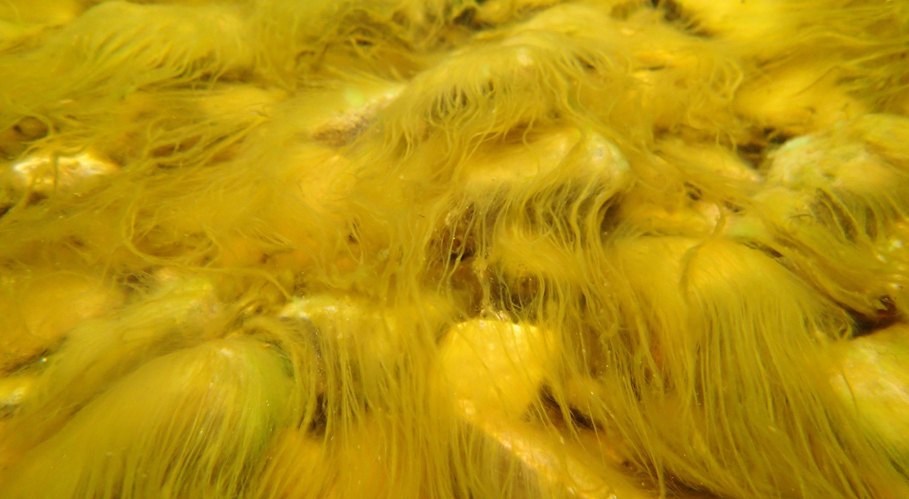
A couple of metres from the patch of diatoms I wrote about in the previous post, the mud surface had a distinct green tinge, and we collected separate samples from this patch using a piece of lens tissue (see: “Eyes wide open in Cassop’s muddy fringe …”). Under the microscope, we saw more of the elongate diatoms sedately gliding across the field of view but the most abundant organisms were green shapeshifting cells, wriggling animatedly as if searching for something that was constantly eluding them. These were cells of Euglena,probably E. ehrenberghii. Euglena belongs to the Euglenophyta, which is now placed with the Protozoa and, as such, is a relatively distant alga of other green-coloured algae (see “Origin story …”). Euglena and relatives have appeared on a few occasions in the past in this blog (see “Cassop Pond in May” and “Puzzling puddles beside the Pennine Way”) but I have never written much about the characteristics of the group as a whole.
Euglena cf. ehrenbergii from Deptford Creek throwing some shapes. Scale bar: 20 micrometres (= 1/50th of a millimetre). The picture at the top of the post shows the lens tissue in place at the edge of the creek.
The Euglenophyta were thought to have arisen when a primitive green alga was engulfed by a protozoan which, rather than digesting it straight away, decided to “farm” it inside its body so that it got a constant feed of energy rather than a one-off hit. That’s a rather cartoonish way of describing an immensely rare but important evolutionary event that required lots of internal biochemical rewiring but it gives you the gist of what happened.
I used to give my students in Nigeria an essay question: “Euglena: plant or animal. Discuss”. Obviously, there are bright green chloroplasts, legacies of its distant ancestry, but there are several “animal”-like characters too (I should add that, when I was teaching in Nigeria, the endosymbiotic theory that I discussed in “Origin Story” was not the orthodoxy that it is today). Undergraduate students, new to the world of algae, were not used to seeing “real” plants moving around and had also been taught about rigid cell walls whereas these squirming Euglena cells are clearly flexible. There is no cell wall, as such; instead, there is a protein-based “pellicle” just inside the cell membrane. This pellicle consists of overlapping strips wound helically around the cell. Movement comes from co-ordinated contraction and relaxation of these strips, accompanied by cytoplasmic streaming (which I describe in “The pros and cons of cell walls …”). Some Euglenophyta (such as the Trachelomonas we met in “Puzzling puddles beside the Pennine Way”) have a rigid outer casing called a “lorica” which serves as a de facto “cell wall” and these all retain their shape, unlike Euglena.
In the ever shifting mud of Deptford Creek, this wriggling motion means that Euglena cells can constantly adjust their position. However, it is not their only means of locomotion. Euglenophytes also have two flagella (one very short) but these are not easy to see in the Deptford Creek population. Flagella are usually considered to be aids for swimming rather than creeping, so maybe this organelle is not so useful in this habitat or perhaps it was just difficult to see with the lighting I was using.
You can also see a red eyespot in many of the cells, which helps it move towards light, and also a spherical contractile vacuole (most obvious in the top left image). This is used for regulating the concentrations of salts in a cell, a function that is essential for an organism that lives in an estuarine environment where the saltiness of the water will vary throughout a tidal cycle.
Were we to take some of these Euglena cells and grow them in a dark laboratory we would see one final “animal” characteristic that Euglena possess. If Euglena were truly a plant, trying to grow it in the dark would be catastrophic; however, Euglena can thrive, so long as there is a supply of organic carbon (such as simple sugars). You would see that the chloroplasts and eyespot would gradually wither because they were no longer needed. I describe this strategy being practised by another type of alga in “The little tarn of horrors …”
Euglena and relatives are the reliable “character actors” of the algal world, in that you see them playing small supporting roles in a great many habitats but only rarely do they get star billing. Intertidal mudflats are one such habitat: the ecological equivalent of a niche, low budget independent film that plays only at your local arthouse cinema. While the masses ogle more charismatic organisms, a few of us peer down our microscopes and watch a different type of drama unfold.
Some other highlights from this week:
Wrote this whilst listening to: The Pogues, of course.
Currently reading: Jerusalem: the Biography, by Simon Sebag Montafiore. Topical reading.
Cultural highlight: In a similar vein, we watched Tel Aviv on Fire directed by Sameh Zarabi, a black comedy set around a checkpoint at the Israeli/West Bank border. It sounds like a bleak subject for humour but it does a good job of highlighting the absurdity that underlies the unfolding tragedy in the region.
Culinary highlight: a moussaka-like dish in Caspian II, a Turkish restaurant in Heaton, delicious but unfortunately rather rushed as I had underestimated the time needed to get there on a day when Newcastle were playing at home. We made it to the match with 10 minutes to spare. 1-0 to the Toon, if you are interested.
“Fieldwork” usually involves pointing the car towards the countryside and leaving all the travails of urban living behind for a day or two. I like to stand amidst glorious scenery sucking pure air into my lungs and reflecting on how lucky I am to do the work I do. But all that changed earlier this week. My destination was not the Pennines or the Lake District but a muddy creek in Deptford surrounded by blocks of flats, old industrial premises and a viaduct that carried the Docklands Light Railway into the badlands of south London.
Just out of sight on the right of the picture at the top of the post is the low wooden building of the Creekside Discovery Centre, a charity that promotes urban wildlife. My involvement here is part of a citizen science project that will help the staff at Creekside raise awareness of the microscopic algae that inhabit Deptford Creek. It is co-ordinated by Susi Arnott, a filmmaker, whose film Moon, Mud and Microbes I took to Green Man in 2021 (see “Cover versions …”). I
The visit had to be closely-calibrated to coincide with low tide, so that we had as much time as possible wallowing on the mud looking for algae. I’m somewhat out of my comfort zone, but we soon spotted a rich chocolatey-brown patch on the mud surface that looked extremely promising. This would have been submerged at high tide, and had probably been exposed for five or six hours by the time we visited. I scraped off some of the growth to look at later, although we also tried a variant of the “lens tissue” method for sampling (see “Eyes wide open in Cassop’s muddy fringe …”). My sample turned out to be full of a large sigmoid Nitzschia, cells of which glided to and fro across the field of view as I peered down my microscope. I’m going to be circumspect about naming the species because I’m not familiar with brackish water diatoms and there are a number of possible candidates, but the genus is unmistakable.
The patch of diatoms growing on a low shelf on the embankment at the right hand side of the image at the top of the post.
I felt more at home standing in the middle of the creek itself. The bed here is rocky rather than muddy and the water, at low tide at least, is fresh. There are some distinct diatom patches on some of the stones here too, and I picked some up using a toothbrush to look at later. Again, I’m tempted to name them but, with the exception of Rhoicosphenia abbreviata (which is both distinctive and can tolerate brackish conditions), I’m reluctant to do so without having looked at cleaned frustules. There are species of Navicula, in particular, which have similar outlines and dimensions to common freshwater species but which, on close inspection are something else altogether (see: “More diatoms that like cold weather …”).
The sigmoid Nitzschia growing on mud surfaces in Deptford Creek, November 2023. Scale bar: 20 micrometres (= 1/50th of a millimetre).
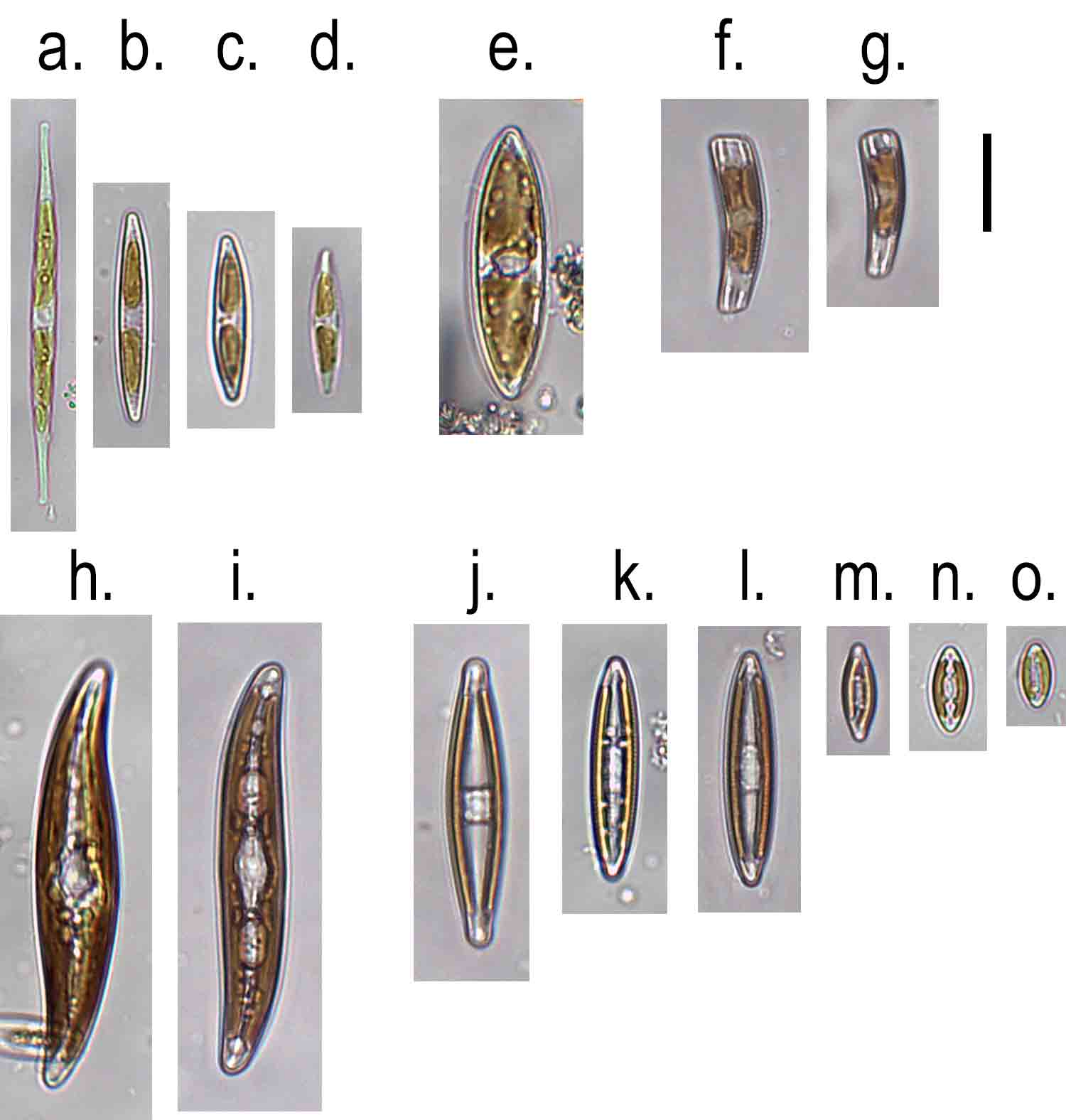
More diatoms from Deptford Creek. a. – d. Nitzschia spp.; e. Tryblionella sp.; f.g. Rhoicosphenia abbreviata; h., i. Gyrosigma; j. – o. Navicula spp.Scale bar: 20 micrometres (= 1/50th of a millimetre).
There was another diatom with a sigmoid outline in this second sample. I wish I knew what advantages this curved outline confers on an organism because this is the not the first time that I have seen two sigmoid genera, quite distantly related, living so close together. This particular organism is Gyrosigma. Whilst I need cleaned material to be sure of what species of Gyrosigma I’ve got, it is easier, ironically, to differentiate the genus from Pleurosigma when looking at live material because the plastids (chloroplasts) have different structures (see “Microscopic monsters in mud …”). Pleurosigma has two or four ribbon like plastids which have a convoluted form whereas the two plastids of Gyrosigma hug the valve outline. Despite outward appearances, these two genera belong to different families.
Both samples were dominated by diatoms that can move around, reflecting the shifting, unstable nature of the habitat within which they live. When viewed under a microscope we see this as horizontal movement but, out on the mud of Deptford Creek, this will be in three-dimensions, including a daily “commute” from the deeper parts of the mud to the surface in phase with the tides. We see them gliding serenely across a glass slide but the process is more like a climber ascending a tight “chimney” on a rock face – first one, then another of the diatom’s two raphes will be engaged, and sometimes both together. Talking of commuting, the sun was starting to descend on Deptford Creek and I had a train to catch. I emerged from the tiny oasis that the Creekside Discovery Centre nutures and joined the stream of people heading towards Deptford Station to start the journey back home.
If you are wondering why the word “floundering” appears in the title, look at the picture below. We were walking up the creek when we noticed this fellow who seemed to have got stuck in the shallow water of the creek. He didn’t seem too alarmed by our presence (or maybe he just didn’t have too many options for escape – flounder body language is not something I know much about), allowing me to get my camera into the water to take some pictures.
Some other highlights from this week:
Wrote this whilst listening to: Patti Smith’s Horses and west coast cool jazz on Chet Baker and Art Pepper’s 1956 album Playboys
Currently reading: Colm Tóbín’s Homage to Barcelona
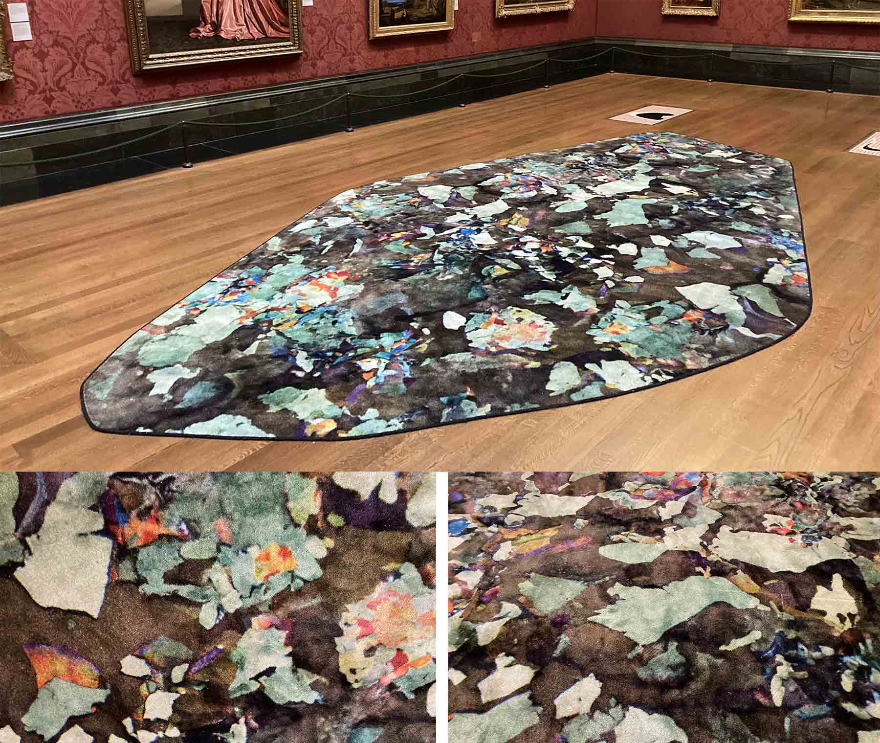
Cultural highlight: Installation by Céline Condorelli, Artist-in-Residence at the National Gallery in London, featuring a rug whose abstract patterns were based on the patterns of pigments on pictures from the gallery’s collection viewed under a microscope. The title Pentimenti (the corections) comes from the use of imaging technologies to discern the changes artists make during the process of composition.
Culinary highlight: nut roast and trimmings: Sunday lunch cooked by our daughter, followed by a rather good vegan sticky toffee pudding.
Part of Pentimenti (the Corrections) by Céline Contorelli in the National Gallery, November 2023.
The diatom genus Achnanthidium frequently appears in this blog. It is widespread in freshwaters, particularly in cleaner water, and telling the many species apart is one of the banes of my life. To cut a long story as short as is possible, members of this genus are typically between 10 and 20 micrometres (1/100th and 1/50th of a millimetre) and push the capabilities of light microscopy to the limit. Until recently, it was usually treated as part of a larger genus Achnanthes, and most specimens were treated as “Achnanthes minutissima”, a species first described in 1833 by the German botanist Friederich Kützing. Even by 1930, we find only three species included in Hustedt’s Die Süsswasser-flora Mitteleuropa. The widespread availability of scanning electron microscopes revealed a lot more variation within Achnanthes minutissima (or Achnanthidium minutissimum, as it has been known for the last 30 years or so), leading to the description of many new species. As many of their properties are difficult to resolve with a regular light microscope, identification is always challenging.
As these taxonomic studies inevitably mean that the historic “broad” Achnanthidium minutissimum is “split” to create new species, the convention when describing a new species is to refer back to the original description and the “type specimen” and explain how the new species differs from this. This means that several papers have been published recently which examine Kützing’s original publication. I’m usually focussed on the finer points of diatom morphology that these papers discuss, but last week something odd struck me about Kützing’s description of “Achnanthes minutissima”. The specimens he describes were growing “on Zygnema with Exilaria crystallina in a ditch near Aschersleben, in June.” Aschersleben is a town in Saxony in central Germany about 80 km from Nordhausen where Kützing lived for much of his life but, this detail apart, nothing else in this account of the habitat rings true. First, the drawing shows the diatom growing epiphytically on an alga that lacks the two star-shaped chloroplasts that characterise Zygnema (see photos in “A day out in Wasdale …”). Second, Zygnema and relatives typically produce copious mucilage and, as a result, have few epiphytes (the exception would be a dead or dying filament). Third: Exilaria crystallina is an old name for a diatom whose name has changed several times but which was called Ardissonea crystallina until very recently when it was transferred to Synedrosphenia. It is, however, a brackish and marine species, not one that is found in freshwaters. It is also, by diatom standards, an enormous species (up to 0.7 mm, according to the literature). It would also be very surprising to see this species growing alongside Zygnema and Achnanthidium, both of which are intolerant of brackish conditions.
This is not intended as a scathing critique. What appears in the illustration is certainly an Achnanthidium species – a small cell with a bent girdle attached to a surface by a short stalk. It is everything else in the description that set me scratching my head. Kützing was working 200 years ago with much less sophisticated equipment and, perhaps more importantly, without the network of supportive peers that modern biologists take for granted. I’m more surprised that, amidst all the attention this description has received in recent years, no-one seems to have commented on the incongruity of the habitat details. There is, it seems, an elephantine diatom (Syndrosphenia crystallina) in the corner of the room and no-one has noticed.
Reference
Lobban, C. S., Ashworth, M. P., Camacho, T., Lam, D. W., & Theriot, E. C. (2022). Revision of Ardissoneaceae (Bacillariophyta, Mediophyceae) from Micronesian populations, with descriptions of two new genera, Ardissoneopsis and Grunowago, and new species in Ardissonea, Synedrosphenia and Climacosphenia. PhytoKeys208: 103.
Some other highlights from this week:
Wrote this whilst listening to: Grace Petrie, folk/protest singer
Currently reading: The Slow Road to Tehran, an account of a journey from London to Tehran via Sudan by Rebecca Lowe.
Cultural highlight: the 1983 film Yentl, starring and directed by Barbara Streisland.
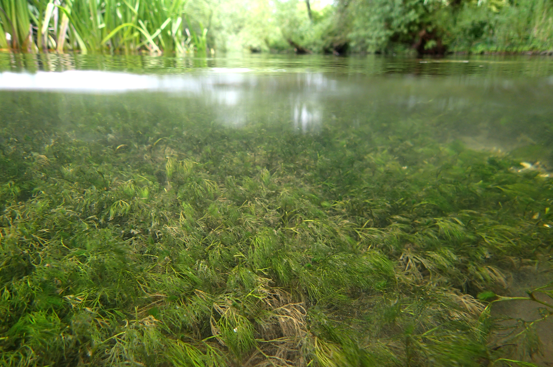
Another river, another algae-strewn boulder. This time, we’re at the River Irt, also in Cumbria, and the green algae, under the microscope, are mixed growths dominated by Mougeotia, Zygnema and Spirogyra rather than the monoculture of Oedogonium that we saw in the River Cocker. You can also see brown patches of Tolypothrix on the boulder in the foreground.
The surprise, for me at least, was the presence of conjugation tubes in the Mougeotia, bearing in mind my comments in the previous post. Did I or did I not write that “filamentous algae don’t see the need for sexual reproduction” only a few days ago, and am I or am I not now staring down my microscope at a filamentous alga doing just that? There is no obvious trigger for this: the generally-cited hypothesis is that the zygospores produced as a result of conjugation are resistant to adverse conditions; however, cool stream water in a northern English river is really a quite benign environment in which to grow. I expect to find lush growths of Mougeotia and relatives here and in other Lakeland rivers throughout the winter.
I’ve tried to capture some of this wanton behaviour in a picture that shows some of the stages that I could see. In the top left you can see some “papilla” emerging from the cells of adjacent cells then, in the centre, two papillae have met and fused to form a conjugation tube. What will happen next is that the cytoplasm of the two cells joined by the tube will migrate into the tube and fuse to form the zygote; however, I could not see this in my material. It is thought that there is some cell-to-cell signalling so that papillae grow from adjacent filaments and meet in the middle, but the nature of this signalling seems still to be a mystery.
Mougeotia from the River Irt, October 2023. A conjugation tube has formed between the two filaments at the centre of the image, and tubes have also started to grow from the filaments in the top left corner. The filaments are about 25 micrometres (1/40th of a millimetre) in diameter. The photograph at the top of the image shows submerged boulders smothered with Mougeotia and Tolypothrix on the bed of the River Irt.
In the absence of zygospores in the Mougeotia from the River Irt, I have included two images produced by Chris Carter. In both of these you can see the zygospore forming in the middle of the conjugation tube. This is in contrast to what happens to Spirogyra, a close relative of Mougeotia, where the cytoplasm from one filament (designated “male”) moves right through the conjugation tube and fuses with the “female” in her own cell.
The zygospore has a very tough cell wall and, as a result, is very resistant to adverse conditions. Having a form of sexual reproduction that is triggered by the environment seems strange but that’s partly a matter of perspective. The angiosperm lifecycle – pollen, flowers, seeds and all that caboodle – is an adaptation to a perennially unfavourable environment, when seen from an alga’s viewpoint, so the question of when it is most useful to have sex shifts from merely coping with occasional adversity to a broader need to be in phase with seasonal cycles. We are also terrestrial organisms, long conditioned to watching for seasonal prompts in the nature around us, so it is difficult for us to understand the different rhythms to which riverine algae dance.
A zygospore of Mougeotia scalaris from Yardley Chase, Northamptonshire, photographed at three focal planes by Chris Carter.
Zygospores of Mougeotia parvula from Malham Tarn, photographed by Chris Carter.
Reference
Permann, C., Herburger, K., Niedermeier, M., Felhofer, M., Gierlinger, N., & Holzinger, A. (2021). Cell wall characteristics during sexual reproduction of Mougeotia sp.(Zygnematophyceae) revealed by electron microscopy, glycan microarrays and RAMAN spectroscopy. Protoplasma258: 1261-1275.
Some other highlights from this week:
Wrote this whilst listening to: The Ghost of Tom Joad by Bruce Springsteen, celebrating our purchase of tickets to see him next May.
Currently reading: Primary Colours, novel by “Anonymous” (Joe Klein) based on Bill Clinton’s 1994 campaign.
Cultural highlight: A visit to the new Faith Museum at Bishop Auckland
Culinary highlight: meal from Sattvic Ahara, a new Indian vegetarian takeaway in the next village.
The view across Derwent Water from Keswick on the day before my October fieldwork.
I have not been to my regular haunts in the western Lake District for two months now. We usual collect samples at two month intervals but our August trip was compromised by the wet weather we had during the latter part of the summer. You can see the evidence for this in the graph below, showing how river levels fluctuated. I should point out that, for personal safety reasons, we can only go out when the river is low so even the values during the latter part of August would have made wading problematic. Samplers, it was once pointed out to me, are part of the benthos, not part of the plankton. Also, values on the river level graph are recorded at the gauging station, and will not be the same at the point where we sample. We have learned, over the years, to make a mental adjustment to get a sense of what conditions will be like when we arrive.
By early September, however, levels were low once again but, annoyingly, I had other commitments, including the course I wrote about in “Cyanobacteria inside their comfort zones …”. Anticipating river conditions based on weather forecasts and juggling with other diary commitments is all part of the job. This particular trip has been harder to organise than most. I have had to make and cancel hotel reservations on three separate occasions, and on a fourth, I drove across and stayed overnight, only to find the rivers were higher than I had hoped when I checked the hydrograph first thing in the morning.
River levels in the River Cocker (Scale Hill) from 1 August to the time of writing. Data from www.riverlevels.uk. Values are averages for each day and the dashed line is the level on the day of my visit. The photograph at the top of the post shows Oedogonium growing on submerged stones in the River Cocker at Low Lorton.
When I finally got to the River Cocker, I was greeted by the sight of lush growths of green algae across the riverbed. This turned out to be a near-monoculture of Oedogonium, which we last encountered in the River Derwent earlier in the summer (see “Borrowdale landscapes …”). It is a very common genus, albeit one that is very hard to identify to species and also one for which generalisations about ecology are difficult.
What I can say, with some confidence, is that the quantity of algae present in these streams waxes and wanes in a predictable manner, with highest values recorded in winter, but it is not so easy to say which species will proliferate on any particular occasion. That also gives us a clue about the possible reason for the patterns that we see: because the biomass fluxes happens at a number of sites across several catchments, irrespective of which species are present, it must be driven by external factors that are common to all of these sites. And because the fluxes are most extreme in rivers that are downstream of lakes (as is the case for the Cocker), we suspect that temperature plays a role. Because water has a high specific heat capacity, Crummock Water acts as a huge “heat pump”, making the water in the Cocker ever so slightly warmer during autumn and early winter than is the case in nearby rivers that do not drain out of lakes.
Filamentous algae are well placed to take advantage of this growth as they are simple and straightforward photosynthesis machines. Sunlight is trapped by their chloroplasts and converted into the building blocks of cells which divide mitotically, allowing biomass to accrue without the complications faced by more sophisticated organisms. There is a certain amount of phenological control in some filamentous algae, but this is mostly concerned with the onset of sexual reproduction (see “The intricate ecology of green slime …”). Most of the time, filamentous algae don’t see the need for sexual reproduction (see “Tales from the splash zone …”) so the Oedogonium in the River Cocker is free to take advantage of the slightly warmer water compared to nearby streams, and convert as much of the late autumn sunlight as possible. Meanwhile, the bugs that normally graze away any algae that cover submerged stones are dancing to the tunes played by their own internal clocks and lack the capacity to increase as quickly as their food supply. The result is the green riverbed that I saw in the River Cocker when I visited.
Oedogonium filaments from the River Cocker, October 2023. The lower image shows a cell with a number of cap cells. Scale bar: 20 micrometres (= 1/50th of a millimetre).
My comment about Oedogonium being able to accrue biomass without the complications faced by larger organisms needs a little qualification, because not every cell is able to divide. The photograph above shows a cell with a fine collection of “caps”, demonstrating that it has divided numerous times. There is, in other words, a first tentative step towards specialisation of cell function and, from this, we may also infer some redistribution of photosynthesis products along the filaments.
Some of these “cap cells” were conspicuously brown compared to the cells on either side. This is quite a common sight but I have found nothing in the literature that may explain what is going on. The colour is suggestive of ochre and my working hypothesis is that these cap cells are particularly metabolically active, requiring the chloroplasts in the cell to work harder than the cells on either side. This will mean that these cells evolve more oxygen and this, in turn, will mean that iron and manganese in the water are more likely to precipitate out. The lower photograph shows one of these brown cap cells colonised by diatoms (Gomphonema) and filamentous bacteria. Oedogonium often has a substantial payload of epiphytes but not usually concentrated in a few locations. Once again, what is it about the cap cells in particular that makes this a good location for other algae? The literature is silent.
More Oedogonium filaments from the River Cocker, October 2023, this time showing iron/manganese preciptitation around cap cells and associated epiphytes. Scale bar: 20 micrometres (= 1/50th of a millimetre).
I always leave my regular sampling locations curious about what I will see next time I visit. This time, however, this curiosity is leavened with a sense of trepidation as the long-term weather forecasts seem to suggest we are in for a wet autumn (driven by the El Niño in the western Pacific). As a result, I am also anticipating spending more time over the next few weeks poring over the hydrographs and weather forecasts trying to predict when river levels will be low enough to pull on my waders and get back into the river. Uncertainty will be the only certainty in my life over the next few weeks …
Some other highlights from this week:
Wrote this whilst listening to: Frankie Archer, who blends traditional folk music with electronica. I saw her on Later … with Jools Holland a couple of weeks ago and then found that she was playing at Darlington Library a few days later. Her haunting melodies have stayed with me …
Currently reading: Qian Zhonghsu’s Fortress Beseiged. Classic Chinese novel set on the eve of the Sino-Japanese war.
Cultural highlight: A Ken Loach double-bill: first, a stage adaptation of his film I, Daniel Blake at the Gala Durham, then his latest film The Old Oak at the Tyneside Cinema. The latter uses several locations in Co. Durham including Blackhall Rocks (see “County Durham’s tropical seashore”)
Culinary highlight: vegetarian tasting menu at Rebel in Heaton.
Last week saw the second presentation of the FBA’s algae identification course for 2023, with another dozen keen participants being introduced to the intriguing world of freshwater algae. The week starts with sampling in the vicinity of the FBA’s laboratory, first at Windermere itself (see “A hitchhiker’s guide to phytoplankton …”) and then, the following morning, at other sites in south Cumbria. I usually join the group heading to Cunsey Beck, which flows from Esthwaite Water to Windermere but heavy overnight rain meant that river levels were too high for safe wading. Instead, we joined Allan Pentecost on a trip to White Scar Quarry, right at the south-eastern tip of the Lake District. It is an area that Allan knows very well and I always enjoy visiting with him because I learn a lot (see, for example, “Love and sex in a tufa-forming stream …”).
Just at the edge of the quarry, a small seepage flows across a wide bedding plane and, Allan pointed out to us, an impressive range of Cyanobacteria, green algae and moss co-existed side-by-side. If you look at the photograph at the top of the post, you can see the seepage emptying onto the bedding plane just above the “d.”. At low flow conditions, there is just a gentle trickle of water running in a narrow band approximately at the centre of the picture. However, when there is more water coming down the quarry, then the water spreads out further and is also augmented by rainfall landing directly on the bedding plane. The result is a zonation, roughly akin to what would be seen on a rocky shore, albeit not for seaweeds.
Following this from the left we have:
The driest area, dominated by Nostoc commune whose ecosystem-building capabilities in otherwise adverse environments I’ve described in earlier posts (see “Landscape architects …” for the most recent of these);
An only occasionally wetted area dominated by dark brown mats of Scytonema. The colonies described in “Poking around amongst sheep’s droppings …” came from very close to here and will give you some idea of what to expect;
A slightly wetter area has reddish mats of filamentous cyanobacteria. Schizothrix, Phormidium and Homoeothrix all feature here. An example of a Phormidium growing at the air-water interface (and, therefore, presumably tolerant of desiccation) is described in “Fieldwork notes, August 2021”)
The central zone is almost permanently wet and here there is a distinct growth of the green alga; Mougeotia, a very common alga in this part of the world. Recent posts which mention this genus include “Something, somewhere, just for a moment …” and “The man who stares at algae …” (both, incidentally, describe the interplay between green algae and Cyanobacteria in Lake District streams).
From here, the sequence is reversed except, at the right-hand side of the picture frame (e.) we did not see more Nostoc commune but, instead, patches of Rivularia haematites and bright patches of the moss Philonotis fontana. I last wrote about Rivularia in “Building landscapes …”, based on another excursion that Allan led, this time in the Malham area of Yorkshire rather than in the Lake District. I wonder, in retrospect, if the moss is, itself, a result of the ecosystem-building properties of Nostoc that I mentioned above and also wrote about in “How to make an ecosystem (2)”. If I had teased apart those Philonotis clumps, would I have seen colonies of Nostoc lurking at the bottom, perhaps? We hear a lot about all the problems Cyanobacteria cause in the Lake District at the moment so it is useful, once in a while, to remind ourselves that Cyanobacteria helped to build this wonderful landscape in the first place.
Microscopic views of the zonation at White Scar Quarry, Cumbria, September 2023. a. Nostoc commune; b. Rivularia haematites; c. Phormidium sp.; d. Mougeotia sp. No scale bars, I’m afraid, as the images were grabbed ad hoc while I was teaching, but other posts referenced should all have indications of size.
Some other highlights from this week:
Wrote this whilst listening to: Africa Express Presents … The Orchestra of Syrian Musicians and Guests.
Currently reading: Maggie O’Farrell’s The Wedding Portrait, set in Renaissance Italy.
Cultural highlight: Exquisite Korean-American film Past Lives directed by Celine Song. A love story mostly set against the Manhattan skyline.
Culinary highlight: It may seem like a minor achievement, but I made the best shortbread of my life this week, following the Ayrshire Shortbread recipe in Cerys Matthew’s cookbook, Where the Wild Cooks Go …. The vegan haggis that I made from her recipe also went down well a couple of weeks ago.
Patches of Philonotis fontana on the bedding plane at White Scar Quarry, September 2023.
There is a very important distinction between the science of taxonomy and the craft of identifying organisms. Taxonomists describe, name and classify organisms, learning more about their evolutionary relationships in the process. Other biologists use the fruits of their labours but, whereas taxonomists are describing fundamental characteristics of organisms, identification is about the “art of the possible” with the equipment and material available. This mismatch is particularly acute for diatoms as state-of-the-art depends increasingly on details that cannot be discerned using light microscopy.
But there are problems even if we restrict ourselves to what can be seen with a light microscope. The face of the diatom valve is generally regarded as the most useful source of diagnostic characters for identification and so is the one most often displayed in identification guides. However, many of the diatoms that we see in our samples do not present this; rather, they lie in a manner that presents their sides (the “girdle view”). These are illustrated in taxonomic works but in far fewer numbers than for valve views. A recent paper on which I was co-author, for example, contained 193 light microscope images of Fragilaira but only nine of these (just under five per cent) were in girdle view. During the period when I was writing this post, I analysed a sample which contained over 30 per cent Fragilaria belonging to five different species, and a good proportion of these presented in girdle view so this is not a trivial problem for those of us who use diatoms to generate insights into present and past environmental conditions. Many years ago, I wrote a simple guide to identifying diatoms from girdle views and this is the basis for today’s post.
Some diatoms are commonly seen in girdle view and are much less commonly seen in valve view (e.g. Aulacoseira), some are often seen in girdle view but also occur in valve view (e.g. Eunotia, Rhoicosphenia) and some are very rarely seen in girdle view (e.g. Cocconeis). Simply knowing that you are seeing a lot more girdle than valve views is giving you some information about the three-dimensional shape of the organism you are trying to identify. Moreover, intact cells may present differently to cleaned valves, particularly if they form colonies.
Starting points
The first point to make is that the reason why most literature concentrates on the valve face is that this is much more useful for discriminating between species. Identification beyond genus is difficult if all you have is a girdle view although, sometimes, you may be able to match a girdle view to a corresponding valve view using characters that are apparent (length, striae density). You may not have an unambiguous determination, but you can establish a confident link between a particular girdle morphology and a species you have identified from valve views.
When two or more species from the same genus have similar girdle views, you can record girdles separately and then divide these amongst those species named from valve views once you have completed your analysis. I do this routinely with Achnanthidium species and a few others. In the case of Eunotia, I divide the girdle views into size categories (small, medium, large and very large) and, again, divide each amongst the size ranges in that category (some species are instantly recognizable from girdle views, others are more difficult).
Seen in valve view, the outline of a diatom can be characterised in terms of curves; seen in girdle view, it is more angular. High magnifications result in shallow depths of field which mean that you do not get an instant sense of the three-dimensional shape of a cell. For larger cells, you can use the fine focus wheel to “see” the contours of a valve outline from a girdle view which helps you match this to illustrations of valve views in identification books (see “Seeing with my fingers …“). Over time you do develop an awareness of how the different views that you get combine to give this extra dimension (“Structural engineering with diatoms …” has a sequence of photographs of Mastogloia that illustrate this).
Identification of diatoms from girdle views
First, determine the symmetry of the girdle around the longitudinal axis.
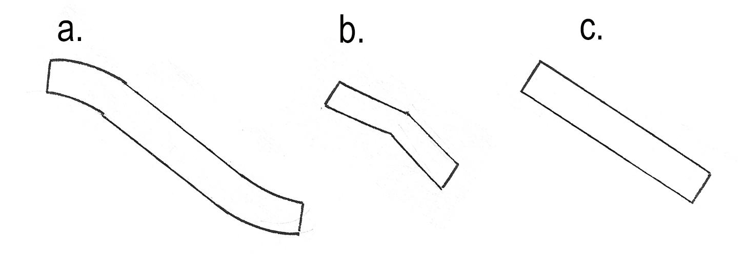
Girdle shape: a) sigmoid (rotational symmetry); b) flexed; c) straight (b & c both reflectional symmetry)
a: sigmoid: Found in a few Nitzschia spp but these rarely occur in large numbers in freshwater epilithic and epiphytic samples. All tend to be large (often > 100 µm)
b: flexed
Three groups:
b-1 Approximately parallel sided (Achnanthes, Achnanthidium, Lemnicola, Karayevia, Kolbesia, Rossithidium ,Planothidium, Psammothidium and others).
Note that a thickening is often visible on the concave (raphe) valve, indicating the position of the central raphe ends.
These are difficult to identify from girdle views. Note length, striae density (if visible) and try to match to valve views in the same sample.
Achnanthidium is often found in girdle view.
Examples of flexed girdle outlines. a. Achnanthidium (live material); b. Achnanthidium (valve and girdle view); c. Achnanthidium with central raphe thickening on concave valve indicated by arrow; d. Planothidium. Cells with flexed outlines in girdle view tend to be relatively small (< 20 micrometres) in length.
b-2: Strongly curved rapheless valve (Cocconeis)
Rarely seen in girdle view in cleaned material but sometimes seen in live material (e.g. as epiphytes on filamentous algae – see “Jammin’ in the key of algae …”)
b-3: Heteropolar: Rhoicosphenia
The concave valve has a full raphe system and the central thickening should be obvious (arrowed). The convex valve has a reduced raphe system and there is no central thickening. Note the pseudosepta protruding into the frustule.
Complete frustules of Rhoicosphenia are often encountered, even in cleaned material.
c: straight
c-1: straight, heteropolar
Gomphonema, Gomphonella, Didymosphenia: both valves have a central thickening (in contrast to Rhoicosphenia). Striae extend onto the mantle so it is possible to measure their density and compare this measurements made on valve views in order to confirm identities. Girdle views of some species have distinctive shapes.
Meridion lacks central thickenings, has distinctive costae and can form fan-shaped colonies.
Peronia lacks both central thickenings and costae and is restricted to soft-water, usually acidic habitats.
Surirella has rib-like thickenings and wavy infoldings
Some Eunotia species are also heteropolar in girdle view.
Examples of straight and flexed heteropolar girdle views: a. Rhoicosphenia abbreviata (arrow points to central thickening on concave valve); b. Gomphonema / Gomphonella (note central thickening on each valve); c. Peronia fibula; d. Meridion (note presence of costae); e. Surirella. Scale bar: 10 micrometres (= 1/100th of a millimetre).
c-2: straight, isopolar
The largest group.
c-2-i fibulae present
There are two possibilities:
Nitzschoid symmetry (many Nitzschia species) where the fibulae of the two valves are on opposite sides, so only one set are in focus at a time.
Hantzschoid symmetry (some Nitzschia species, Cymbellonitzschia, Hantzschia) where the fibulae of the two valves are on the same side so both sets are in focus at the same time.
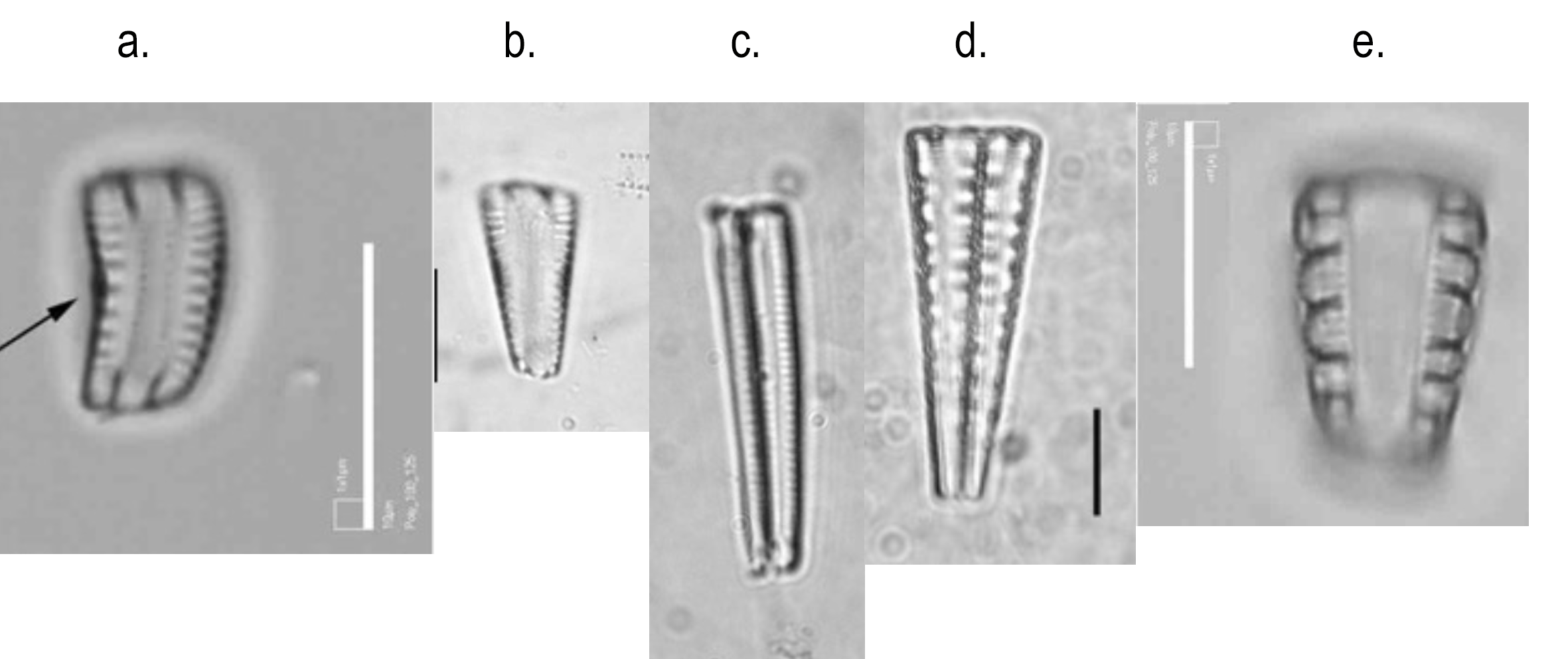
Straight, isopolar girdle views. a. Nitzschioid symmetry (Nitzschia amphibia); b. Hantzschiod symmetry (Cymbellonitzschia diluviana); c. Pinnularia subcapitata; d. Brachysira sp; e. Reimeria sinuata; f. Diadesmis confervacea; g. Odontidium hyemale; h. Fragilaria sp.; i. Staurosira construens. Scale bars: 10 micrometres (= 1/100th of a millimetre).
c-2-ii central raphe thickenings present
Several genera fall into this category. In addition to recording length and striae density you can also often see the orientation of the striae in girdle views. In the image of Pinnularia subcapitata you can see how the striae change direction from radiate to convergent as you move from the centre towards the poles. In the case of Brachysira sp, the striae may be too fine to resolve easily from a girdle view. You should also be able to see features such as pronounced central areas (as in Reimeria sinuata).
Dorsiventral genera can often be recognised by their depth (apparent through careful use of the fine focus control).
The image of Brachysira shows two recently divided cells that are yet to separate. This is quite common to see. However, a few genera of raphid diatoms (Diadesmis, Humidophila) of raphid diatoms form longer chains
c-2-iii central raphe thickenings and fibulae absent (centric and araphid diatoms plus Eunotia)
Many of these form chains, which often fail to separate on cleaning.
Diatoma and Odontidium: Measure length and costae density and compare with valve views. Note that Diatoma tends to form zig-zag chains whilst Odontidium forms straight chains (though these rarely survive cleaning).
Fragilaria and Ulnaria formlong, thin, rectangular frustules. If a swollen central area is present, you should be able to see it in girdle view. Whereas central raphe ends form a thickening on the inside of the valve surface in biraphid diatoms, the swelling leads to a outward bulging of the valve face in Fragilaira and related genera.
Straight, isopolar girdle views. a. Pseudostaurosira elliptica; b. Fragilariforma virescens; c. Tabellaria flocculosa; d. Eunotia incisa. Scale bars: 10 micrometres (= 1/100th of a millimetre).
Staurosira construens is relatively easy to recognise from girdle view because of the distinctive central swellings but several other chain-forming diatoms are difficult to assign reliably even to genus. The problem is compounded because the links between cells are strong so there are few isolated cells or valves in valve view that can provide the information needed for a reliable identification. Note that it is common for chains of cells to break along the middle of a valve. In the photo of Pseudostaurosira elliptica there are two and a half frustules (= five valves in total). Staurosirella can be distinguished by its relatively coarse striae and Tabellariaby the presence of septa.
Girdle views of Eunotia are broadly rectangular but sometimes are slightly heteropolar (see illustration of Eunotia incisa above). The raphe should just be visible towards the poles with careful focussing. The raphes (arrowed) are particularly distinctive in this example and occur closer to the ends in most species. Some Eunotias form chains but these can be distinguished from Fragilariforma (which is often found in the same samples) because chains of Eunotia end with complete frustules rather than with half-cells (Eunotia is also differentiated by its short raphes, but these can be difficult to see).
c-2-iv straight (centric diatoms)
The radial symmetry that characterises centric diatoms is not apparent when the cell is seen in girdle view, so I have included them alongside pennate diatoms with straight girdles. The chances of seeing a centric diatom in girdle view depend upon the depth of the mantle relative to the cell diameter and the propensity to form chains. Melosira and Aulacoseira both have relatively deep mantles so are often seen in girdle view. Valves of Aulacoseira tend to have distinctive areolae on the mantle, whereas those of Melosira have plain mantles. Also, Aulacoseira cells are linked by distinctive spines (see image of A. granulata below). Ellerbeckia arenaria, by contrast, has a relatively shallow mantle but cells are tightly attached, so it very often presents in girdle, rather than valve, view. Note the characteristic cross-hatched pattern on the mantles.
Girdle views of centric diatoms. a. Aulacoseira ambigua; b. Aulacoseira granulata; c. Stephanocyclus meneghinianus; d. Melosira varians; e. Ellerbeckia arenaria. Scale bar: 10 micrometres (= 1/100th of a millimetre).
Those centric diatoms that do not form chains and which have shallow mantles are relatively rarely seen in girdle view. Occasionally, you may see pairs of cells just after division that present in girdle view; one example, Stephanocyclus meneghinianus (formerly Cyclotella meneghiniana) is shown above, demonstrating the characteristic folding on the valve face.
Finally, never forget that it may not be possible to apply a species name to every single diatom that you encounter in a sample. It may only be possible to name a diatom that is naturally rare in a sample, and which does not present key differentiating characteristics to genus, and sometimes not even to that level. We strive to make unambiguous identifications of every individual in a sample but, realistically, we often have to extrapolate from those that we can identify with confidence to other cells that we assume are part of the same population but which have not presented a key feature. We can only do our best and slowly, over time, gain experience that will sharpen our judgements.
Wrote this whilst listening to: Local hero Sam Fender’s set at the Reading Festival via the BBC iPlayer.
Currently reading: Hot Protestants – a history of puritanism in England and America by Michael Winsop. Recently finished Demon Copperhead, Barbara Kingsolver’s wonderful albeit bleak retelling of David Copperfield.
Cultural highlight:Capturing the Moment – exhibition at Tate Modern looking at the interplay between painting and photography.
Culinary highlight: We have had our son and his family visiting from China over the past three weeks and have had some delicious meals cooked for us during the stay, and have also visited some good Chinese restaurants with them, including The Sichuan in London and Little Asia, a hotpot restaurant in Newcastle. Also enjoyed seeing our visitors react to traditional British cooking such as cottage pie, a full English breakfast, haggis and an Indian takeaway.
My go-to analogy for the type of freshwater ecology that I do is of being a “general practitioner” (family doctor) for lakes and rivers. I check my “patients” at regular intervals and offer them “prescriptions” to help them recover. Just occasionally, however, situations demand a different analogy, that of the hard-boiled detective, roused from semi-slumber by the shrill ring of a telephone and summoned to the scene of a crime. Sometimes there is even blood involved, or at least so it seems. I am the one who announces to the protagonists assembled in the drawing room that it is not blood at all. It is … algae.
Freshwater ecologists have a fairly good idea of what algae grow where, how they wax and wane over the course of the year and how human activities can alter these patterns. However, we also know that there is a limit to the extent that we can predict, and that there are many occasions when a single species can proliferate to the extent that members of the public can see, and be concerned by, the changes in a local water body. In most cases, these growths are benign but there are situations – cyanobacteria blooms being the best known – where there are serious implications for human and animal health.
The algae that can cause these problems can come from any one of several groups such that the initial diagnosis necessitates a good general background in algal taxonomy. We try to provide this background on the FBA course on freshwater algal identification as many of the participants work for regulators and water companies and are “first responders” to calls from concerned members of the public. On the final day, Bill Brierley gives a talk on “incidents” based on experiences from his long career in the Environment Agency.
What is striking from this talk is that representatives of all the main groups of algae [see Origin story … ] can be the culprits, along with a few groups that are not algae but which can be confused with algae. Quite a few have been covered in this blog over the years. Here’s a quick summary, along with some links.
In the Prokaryotic domain, we have the Cyanobacteria as the serial villains of the freshwater world (see: “A hitchiker’s guide to phytoplankton …”) but other groups can also cause problems. “There will be blood …” describes a bloom of Chromatium, a purple photosynthetic bacterium, which turned a pond close to where I live an alarming red colour.
However, red-coloured blooms can come from a number of Eukaryotic algal groups as well. Euglena sanguinea from the Euglenophyceae (Protozoa), in particular, can have a similar effect in standing waters and slow-moving rivers. One of the key lessons that emerged from Bill’s talk was the need for samples to be examined by a biologist with a broad understanding of algae because it would be easy to jump to the wrong conclusion if a bloom was only viewed from the shore.
A bloom of Euglena sanguinea in a pond in eastern England. Photograph: Bill Brierley. The photograph at the top of the post shows an unexplained proliferation of green algae in a river in Cumbria.
Several problem-causing organisms can be found in the Chromista (see “Unlikely bedfellows …“). The Haptophyta includes some toxin-producing groups including Prymnesium and Chrysochromulina both of which are more common in brackish and marine waters but which can extend inland, especially when salt concentrations are artificially elevated. In the summer of 2022 a bloom of Prymnesium parvum had catastrophic effects on the River Oder in Poland and Germany, killing about 360 tonnes of fish. In this case, discharges from salt mines in Poland were responsible for raising the salinity of the river and this, combined with high concentrations of nutrients and low flows, created a lethal cocktail.
Left hand images: Prymnesium parvulum (note the short, rigid “haptonema, cells about 10 micrometres long); right hand image: Chrysochromulina braunii. Photographs: Bill Brierley
Both of these genera are widespread in low-lying and brackish lakes in Norfolk and Lincolnshire and the expectation is that the favourable conditions under which they thrive will become more common as climate change continues, leading to more problems in the future.
The other group of Chromista that are often associated with problems are the diatoms. Although some marine genera do produce toxins, there is scant evidence for fish kills that are directly attributable to diatoms in freshwaters. It is more common for mass growths on the beds of rivers to attract attention from passers-by. The diatom Didymosphenia geminata is a repeat offender in this respect. Although regarded as an invasive species in some parts of the world, it is native to the UK with records extending back over 150 years (see “Journey to the headwater of the River Coquet”). Nonetheless, when conditions are right, it can produce massive growths on stream beds that can slough off and float downstream, looking uncannily like untreated sewage. Members of the public frequently call the Environment Agency about this “problem” but it is one that can be easily diagnosed.
The final major evolutionary lineage which contains algae is the Plantae and, in particular, the Chlorophyta. As for the diatoms, most of the occasions where the public call in with problems are due to mass growths that are symptoms of a general ecosystem malaise rather than being directly harmful to freshwater life. Examples include “blanket weed” (Cladophora glomerata), ”gutweed” (Ulva flexuosa – see “Loving the low flows …”) and “water net” (Hydrodictyon reticulatum” – see “Casting the net wide …”) but, in truth, many green filamentous and thalloid algae can form conspicuous growths when conditions are right.
One green alga that is responsible for fish kills is Botryococcus braunii which is quite widespread in the UK and which can form yellow-orange (sometimes red) growths in standing waters which can be confused with ferrous deposits. Reports of fish kills are mostly from warmer parts of the world but the potential for similar occurrences in northern Europe will increase as the climate warms.
Botryococcus braunii in a pond in eastern England. Photograph: Bill Brierley.
Bill’s talk on algae “incidents” is the last one on our course, and is a good place to finish. Through the week, we take the students on a journey through the world of algae. This can be a bewildering experience for a beginner, as so much has changed in recent years. We speak about insights obtained from molecular biology and the challenges we face when dealing with “cryptic” species. Each of the tutors brings some specialist knowledge and it is, therefore, important to end with a talk that emphasises the value of being a “generalist”, of knowing enough about what differentiates the major groups to be able to go into an unknown situation and make a tentative diagnosis by themselves.
The other lesson is that, rather than categorise these as “nuisance” or “problem” algae, we see that most are proliferating due to human-induced circumstances. For Prymnesium parvum, three separate stressors are involved, all generated by human actions. Paraphrasing Stephen Sondheim from West Side Story, “Society’s played him a terrible trick, And sociologically [ecologically?] he’s sick”. Algae, as ever, are misunderstood and are the symptoms, not the reasons, for ecosystem malaise.
References
Chiang, I. Z., Huang, W. Y., & Wu, J. T. (2004). Allelochemicals of Botryococcus braunii (chlorophyceae) 1. Journal of phycology 40: 474-480.
Edvardsen, B., & Paasche, E. (1998). Bloom dynamics and physiology of Prymnesium and Chrysochromulina. NATO ASI SERIES G ECOLOGICAL SCIENCES41: 193-208.
Free, G., Van de Bund, W., Gawlik, B., Van Wijk, L., Wood, M., Guagnini, E., … & Stielstra, H. (2023). An EU analysis of the ecological disaster in the Oder River of 2022. EUR 31318 EN, Publications Office of the European Union, Luxembourg.
Sobieraj, J., & Metelski, D. (2023). Insights into Toxic Prymnesium parvum Blooms as a Cause of the Ecological Disaster on the Odra River. Toxins15: 403.
Some other highlights from this week:
Wrote this whilst listening to: Gryphon, early seventies classical-folk-rock fusion.
Currently reading: The Girl who Played with Fire by Stieg Larsson. Revisiting this book a decade or so after it was written. It is still a tautly-written thriller but the tech references are now very dated.
Cultural highlight:The Barbie Movie. I have been, I promise you, a fan of Greta Gerwig since I first saw Francis Ha, so don’t judge me.
Culinary highlight: Delicious ten-course tasting menu at Rebel, new(ish) restaurant in Heaton, Newcastle
At the start of the month I wrote about J.M.W Turner’s visit to Borrowdale in the English Lake District and reflected not just on what he saw but on what he might have seen (see “Borrowdale landscapes …”). I started with a view very similar to the one he painted (my camera was pointed upstream whilst his easel was positioned so that he could see the view downstream). I then focussed on successively finer and finer detail, finishing with views photographed and drawn using a microscope.
I’ve now taken this a step further and reassembled the parts that I viewed through the microscope into a “landscape”, of sorts, a highly magnified view of the stream bed. The photograph I included in the post showed a patchwork of green and brown, the former being filamentous algae (Oedogonium) and the latter mostly diatoms. I’ve depicted the Oedogonium mostly on the left of the picture (including one cell releasing a motile zoospore) with a mixture of the diatoms Tabellaria flocculosa and Fragilaria pectinalis on the right hand side. To give a sense of scale, the Tabellaria cells are about a 50th of a millimetre long.
As I was painting this, however, I was reminded of Kenneth Clark’s statement that “… love of creation cannot really extend to the microbe” (see: “Is this the first microscopic landscape painting?”). I disagreed when I first read it, and I disagree now, quoting David Attenborough in reply: “no-one will care about what they have never experienced”. Turner did not experience the microscopic world; we are, therefore, in no position to comment on how he would have responded as an artist. He sought out visual experiences and his art is a response to this – first via field sketches and then via oil paintings worked up sometimes years later in a studio. But his art strives to do more than describe what he saw, as a scientist of his era would have wanted to do. He was much influenced by ideas of the sublime, that nature could and should provoke strong emotions, and that it was his duty, as an artist to capture both nature and those emotions on canvas.
We could argue that our response to a landscape can be divided into a component that we can understand and explain, and a component that is mysterious. Enlightenment scientists wanted to swing the balance towards the former – to understand and explain the types of rocks, the identities of the trees and plants and so on. By the end of the eighteenth century, however, artists were moving in a different direction, encapsulated by Keat’s articulation of “negative capability” – that a thinker should be “capable of being in uncertainties, mysteries, doubts, without any irritable reaching after fact and reason”. Turner would have looked through a microscope not with a scientist’s urge to explain and understand but with an artist’s capacity to accept that there is much that is still unknown. It was not, for him, about a compulsion to understand, as an Enlightenment scientist would have felt; rather it was about accepting that there was a limit to the extent that a landscape – large or small – could be broken down into explainable units. Modern ecologists share these emotions, even if their language is very different. They might prefer to say “uncertain” rather than “mysterious” but the essence is the same: we can often generalise about a situation (a Lake District stream, for example) but still not predict exactly what species we will find, or in what quantities they will occur. Negative capability seems to be something that an ecologist should cultivate – recognising the point where the benefits of greater detail diminish and we are at risk of losing the big picture.
Some other highlights from this week:
Wrote this whilst listening to: Beth Orton and Self-Esteem – in readiness for our trip to Green Man later this month. And Sean-Nós Nua, a rather wonderful album of traditional Irish folk songs by Sinéad O’Connor who died this week.
Currently reading: Black-eyed Blonde, a Philip Marlowe novel written in the style of Raymond Chandler by Benjamin Black.
Cultural highlight:Miss Saigon at the Crucible Theatre, Sheffield.
Culinary highlight: Bibimbap, at Ginseng, a Korean restaurant in Sheffield following our theatre visit.
There are not many ecosystems that are unique to the British Isles; hedgerows and hay meadows may define a certain view of the English landscape but they can be found well beyond these islands. Similarly, heather moorlands are common to other countries that share a damp north Atlantic seaboard. Chalk streams may be as close as we can get to a quintessential English landscape, as 76% of all chalk streams in the world are found in England. The most northerly of these are the headwaters of the River Hull (also known as West Beck) in the East Riding of Yorkshire.
The Hull rises on the Yorkshire Wolds, a series of low hills that lie between the Vale of York and the North Sea, underlain by the same band of Cretaceous chalk that creates the famous chalk streams of southern England. The Hull rises from springs near the town of Driffield, then flows mostly southwards to join the Humber at Kingston-upon-Hull. The lower part of the river is navigable; the upper part runs parallel to the Driffield Navigation, allowing the river itself to display the classic characteristics of a chalk stream.
Ranunculus calcareous on the bed of the River Hull at Wansford, June 2023 (shown in the photograph at the top of the post).
You can see these in the photographs above: As chalk streams are fed mostly by aquifers rather than surface runoff, the flow is very steady, meaning that there is a gravelly substratum which supports a rich macrophyte flora. This creates a lot of habitats for invertebrates which, in turn, support good trout and salmon fisheries. Two anglers who I met at the riverbank had already caught a two kilogram brown trout.
However, many chalk streams no longer fit this template of a thriving ecosystem due to a combination of over-abstraction and pollution. The River Hull, itself is classified as “moderate status” due, in part, to the fish and also to the condition of the macrophytes. Digging a little deeper, the Environment Agency’s reason for the condition of the macrophytes was “suspect data” and “method change” which, with no further explanation, does not get us much further forward. Phytobenthos, by contrast, are classified as “high status” which also seems surprising.
Diatoms from the River Hull at Wansford, June 2023: a. Ellerbeckia arenaria; b. Melosira varians; c. Fragilaria sp.; d. Diatoma vulgaris. Scale bar: 20 micrometres (= 1/50th of a millimetre).
I was curious to see what types of diatoms were thriving under these conditions and whether these justified a classification of “high status”. One of the easiest ways to collect diatoms from this type of habitat is to pull off a few shoots of submerged vegetation and put these into a plastic bag with a little stream water. Shaking the bag vigorously for 20-30 seconds is enough to dislodge the diatoms and I can then decant the brown suspension from the bag and put them into a cool box for the journey back to my study.
Not all the diatoms I saw could be named with confidence from live material but there were enough that could be recognised to make me question whether this stream was in high status for phytobenthos. There were several long chains of Melosira varians, for example, which thrive in warm, nutrient-rich conditions, and also plenty of Navicula tripunctata. The chain of Fragilaria could not be identified with confidence from live material but the Ellerbeckia arenaria was unmistakable. The latter, again, is more common in enriched than in pristine waters.
There were also plenty of Cocconeis placentula (sensu lato) which is a common epiphyte on submerged vegetation, and some large Cymbella species along with a few Nitzschia and some Amphora.
Diatoms from the River Hull at Wansford, June 2023. e. – g.: Cocconeis placentula sensu lato; h. Encyonema silesiacum; i. Nitzschia linearis; j. Cymbella lanceolata; k. – m. Navicula tripunctata; n. Gomphonema sp. Scale bar: 20 micrometres (= 1/50th of a millimetre).
Our idea of what constitutes “high status” or “moderate status” is derived from comparing the biota we find at any particular stream with that in similar streams in remote areas where the impact of humans is minimal. That is a real problem when dealing with chalk streams (see “Tales from prehistory”); however, the River Hull offers us the possibility of an alternative insight. In 1932, a pioneering biologist, Roger Butcher, visited the Hull and documented the algal flora in detail. His approach was different to mine – he left glass microscope slides in the stream to be colonised by algae, rather than collecting from macrophytes – but he did sample from locations close to Wansford. His paper gives us an insight into what algae lived in the river 90 years ago, and what might have changed.
The main difference is that he found more Achnanthidium minutissimum than I saw in my sample. This is a species complex that is associated with streams with low levels of human impact and, interestingly, one that we found to be more abundant in old samples we collected from herbaria than on modern samples from the same locations. So this points, perhaps, to a drop in condition since Butcher wrote his paper. Two important caveats: I’m not using methods identical to those that the Environment Agency use today and I’m not using the same methods as Butcher. So I don’t want to read too much into this finding, although the portents are not good. If a stream is classified as “high status” then I come to a sample with some expectations about the species I am going to see. If I see none of those, then I think we have grounds to be concerned.
References
Butcher, R. W. (1940). Studies in the ecology of rivers: IV. Observations on the growth and distribution of the sessile algae in the River Hull, Yorkshire. Journal of Ecology 28: 210-223.
Yallop, M., Hirst, H., Kelly, M., Juggins, S., Jamieson, J., & Guthrie, R. (2009). Validation of ecological status concepts in UK rivers using historic diatom samples. Aquatic Botany, 90: 289-295.
Some other highlights from this week:
Wrote this whilst listening to: More from Glastonbury 2023. Really enjoyed Weyes Blood, Ezra Collective and Cat Stevens.
Currently reading: The Unseen by Norwegian novelist Roy Jacobson
Cultural highlight: still Glastonbury
Culinary highlight: A straightforward but excellent ragu sauce at Burro, a restaurant in an old windmill on the edge of Riccall, near York. Overnight stop on the trip that took me to the River Hull