


Product details
Synonyms = 4 kDa / 89 kDa myeloperoxidase; EC 1.11.1.7; EC1.11.2.2; fj80f04; MPO; mpx; myeloid-specific peroxidase; Myeloperoxidase; Myeloperoxidase heavy chain; Myeloperoxidase light chain
Antibody type = Mouse monoclonal / IgG
Clone = MSVA-692M
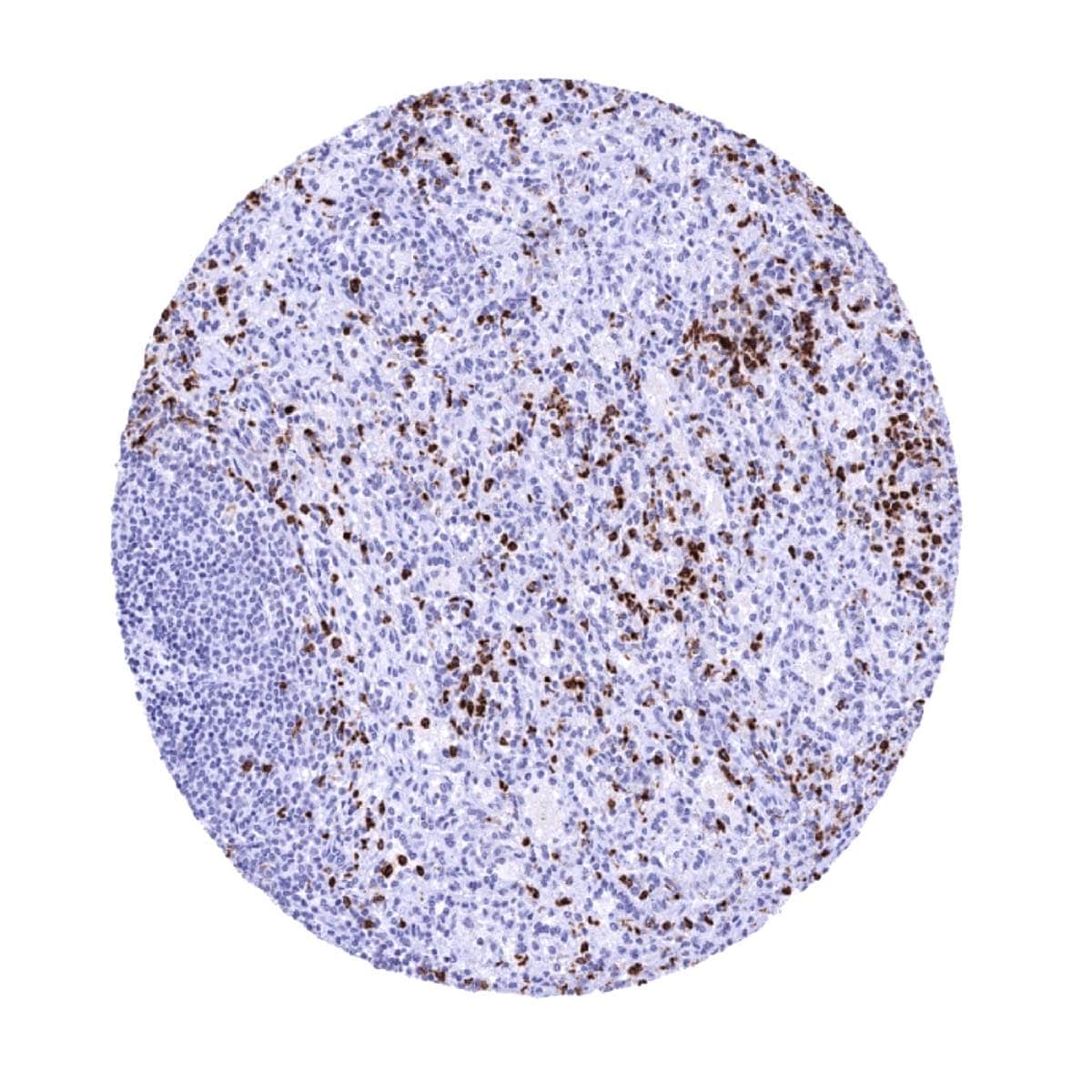
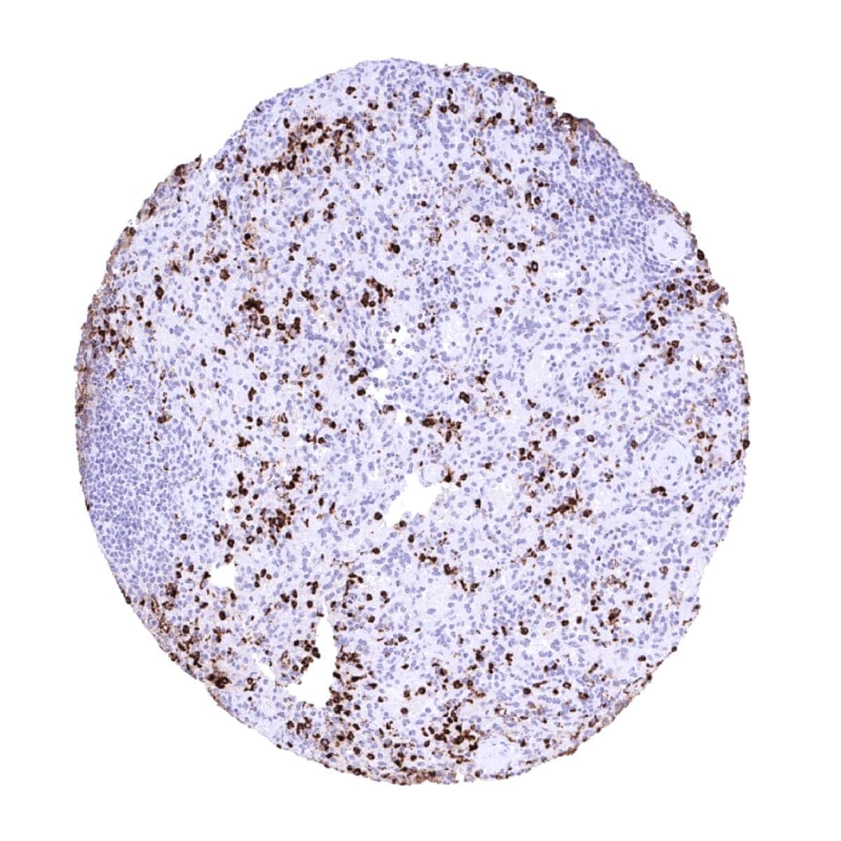
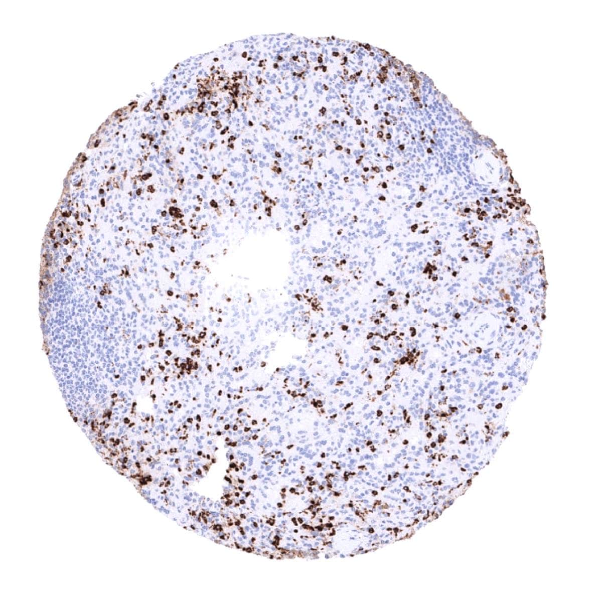
Positive control = Spleen: Numerous granulocytes with strong MPO staining should be seen within the red pulp of the spleen.
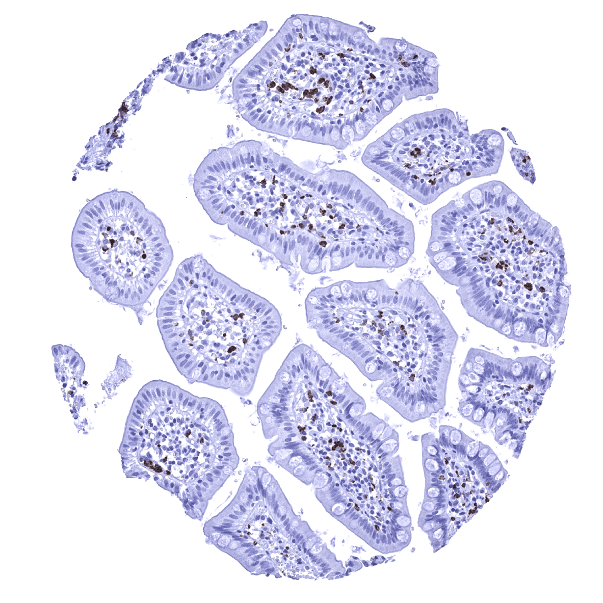
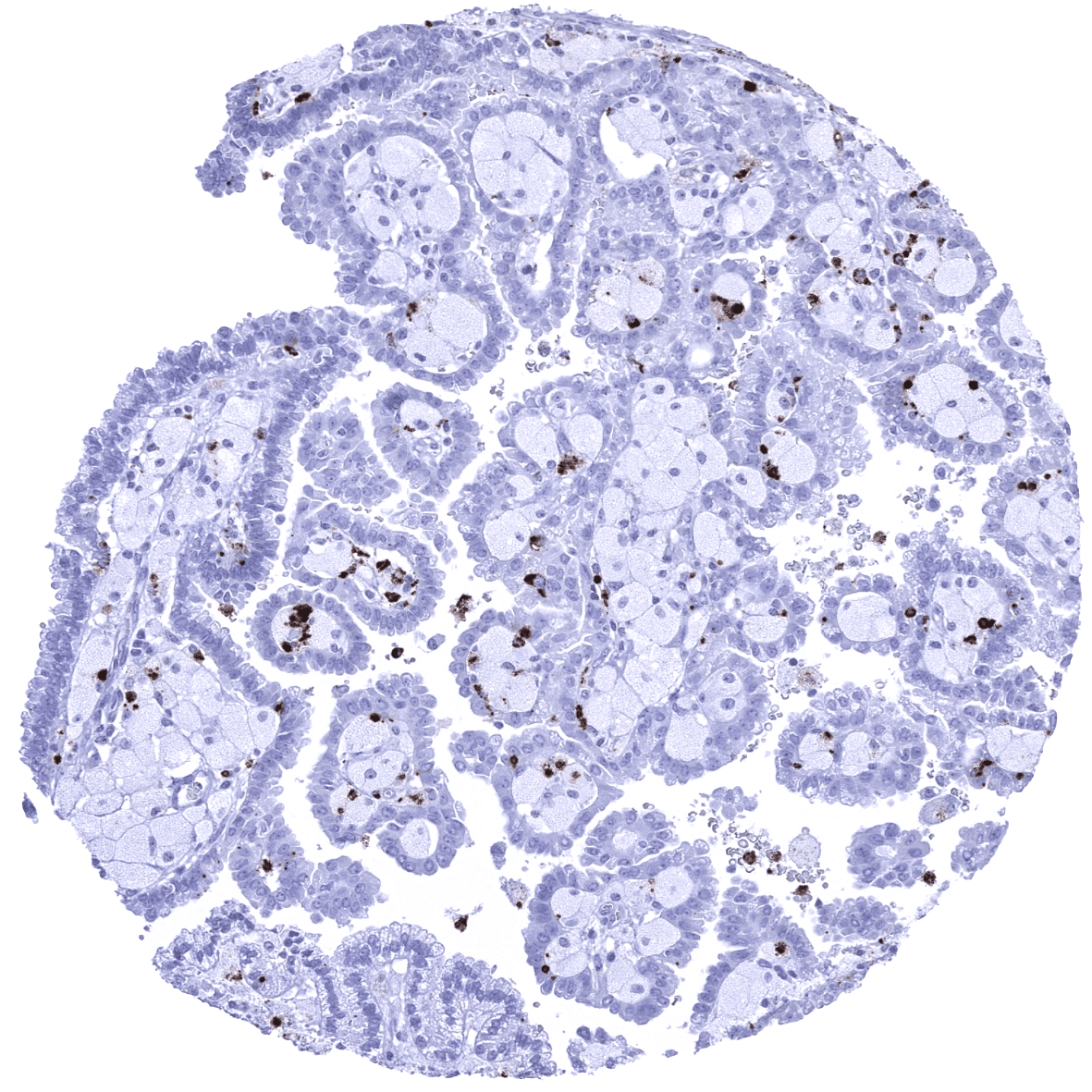
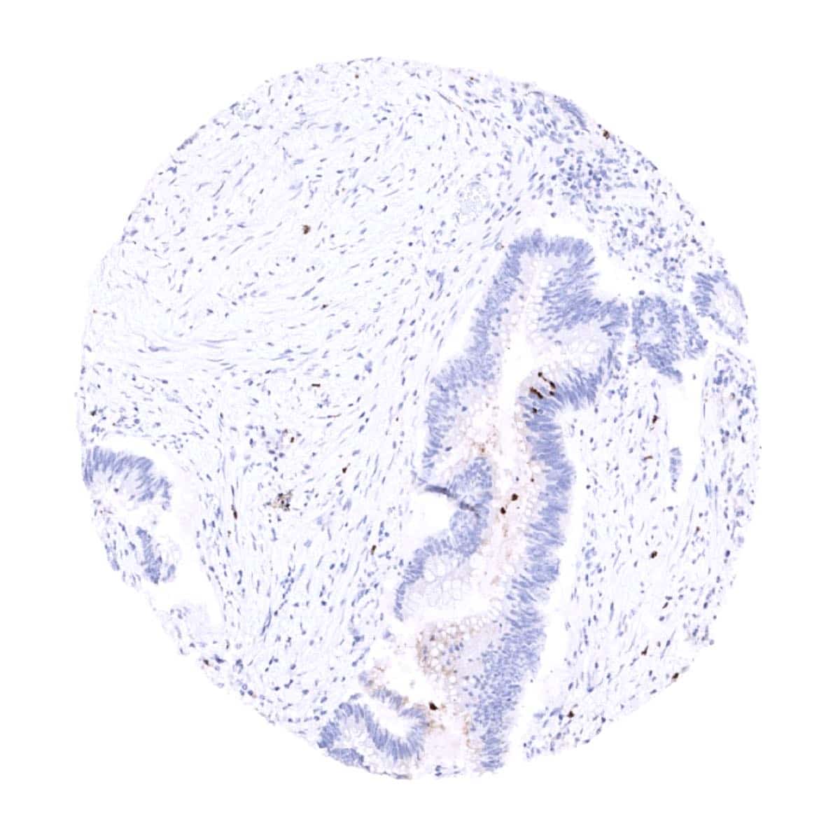
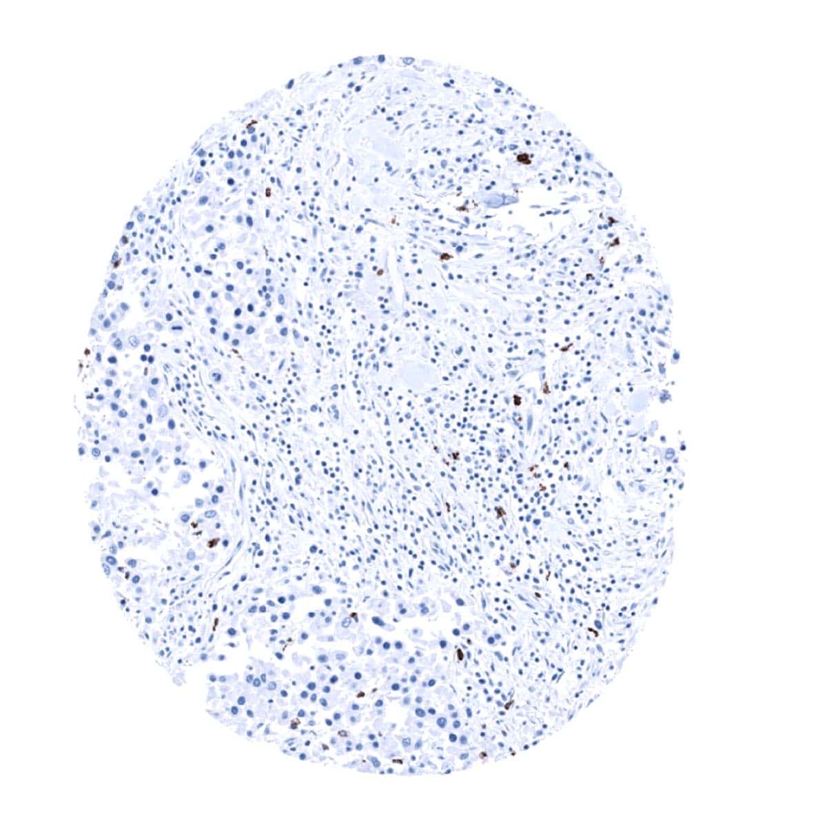
Negative control = Colon: MPO immunostaining should be absent in all epithelial cells of the colon while MPO positive granulocytes may be seen within capillaries or in the stroma of the lamina propria.
Cellular localization = Lysozome
Reactivity = Human
Application = Immunohistochemistry
Dilution = 1:100 – 1:200
Intended Use = Research Use Only
Relevance of Antibody
MPO is a expressed in granulocytes and their precursor cells.
Biology Behind
Myeloperoxidase (MPO) is a 150 kDa peroxidase enzyme coded by the MPO gene on chromosome 17q23.1. MPO is most abundantly expressed in neutrophil granulocytes and stored in azurophilic granules. It comprises about 5% of the dry mass of neutrophil granulocytes. MPO is released to provide defense against invading pathogens. Its antimicrobial activity involves the production of acid. Antibodies against MPO which are also termed anti-neutrophil cytoplasmic antibodies (ANCAs) have been implicated in various types of vasculitis. MPO is considered a possible therapeutic target for coronary heart disease, inflammatory bowel disease and other conditions.
Staining Pattern in Normal Tissues
A strong myeloperoxidase immunostaining is seen in neutrophil granulocytes while other granulocytes show a somewhat weaker and more variable staining. MPO positivity is also seen in granulocyte precursor cells in the bone marrow. MPO positive granulocytes occur in most normal tissues, either within blood vessels or also within the tissue. MPO positive material can be seen in the lumina of prostatic glands in some samples. In addition, a very weak cytoplasmic staining is seen in gastric glands which is considered a tolerable cross-reactivity.
These findings are largely consistent with the RNA and protein data described in the Human Protein Atlas (Tissue expression MPO)
Positive control: Spleen: Numerous granulocytes with strong MPO staining should be seen within the red pulp of the spleen.
Negative control: Colon: MPO immunostaining should be absent in all epithelial cells of the colon while MPO positive granulocytes may be seen within capillaries or in the stroma of the lamina propria.

Staining Pattern in Relevant Tumor Types
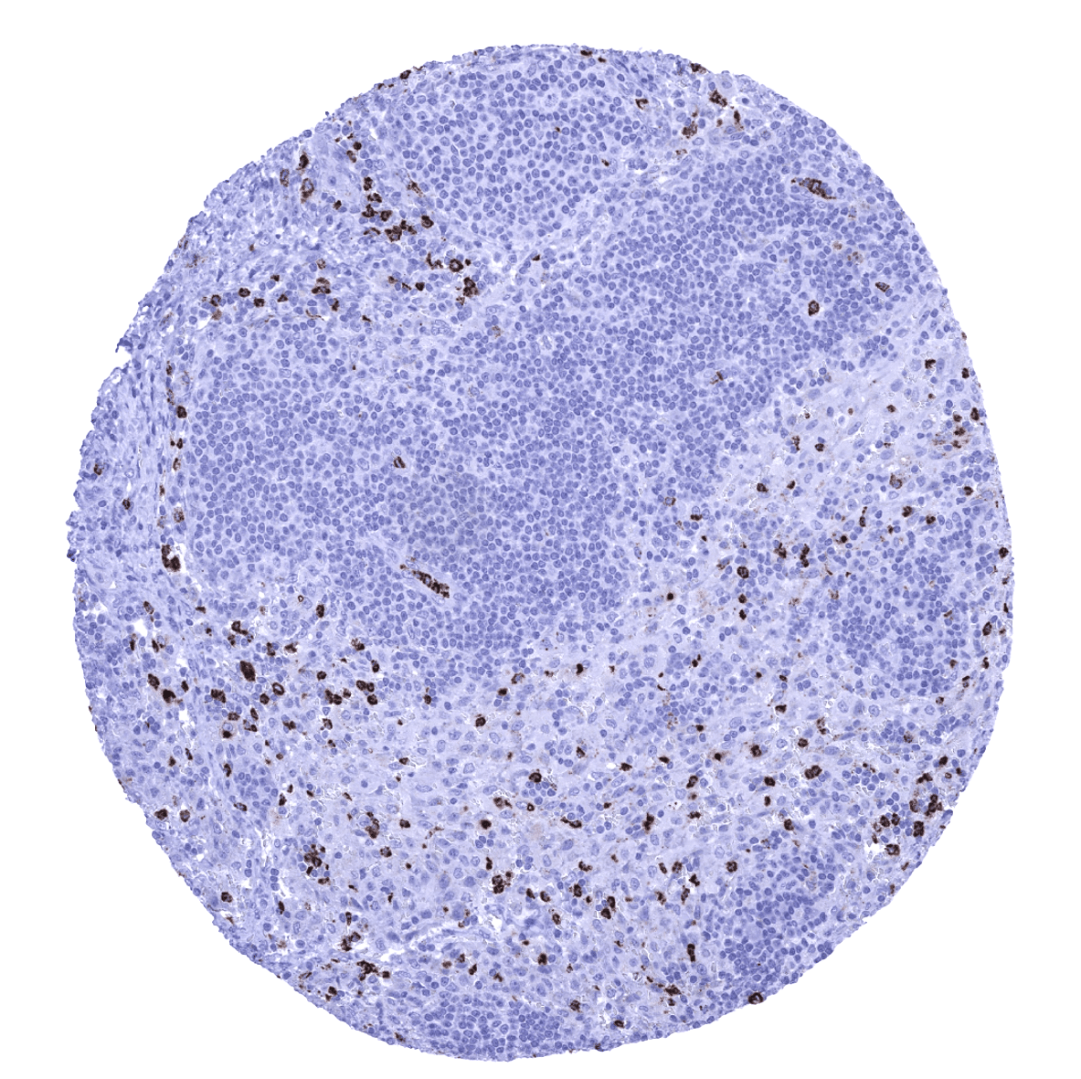
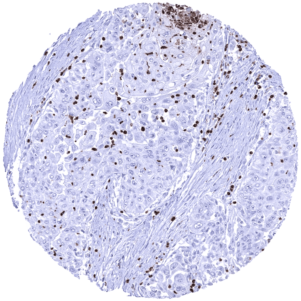
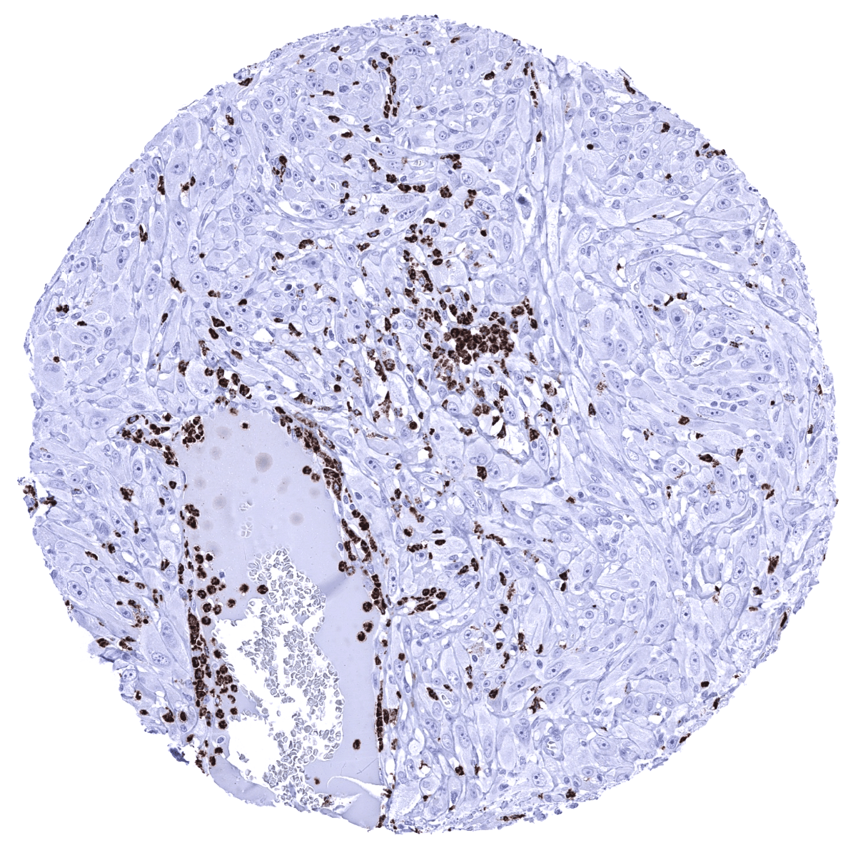
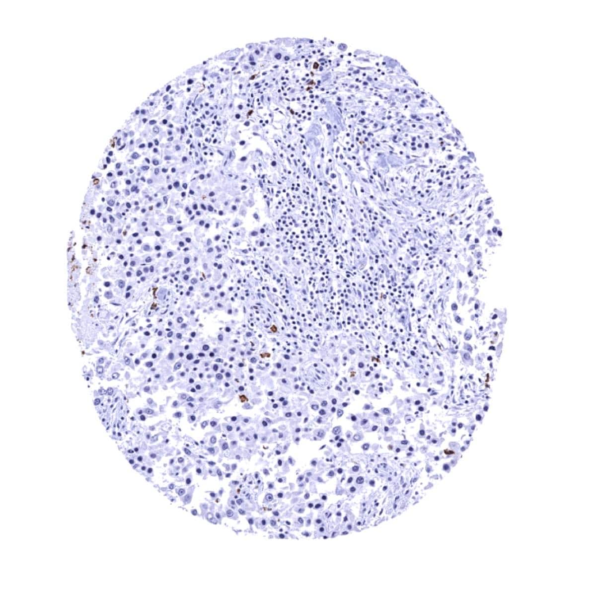
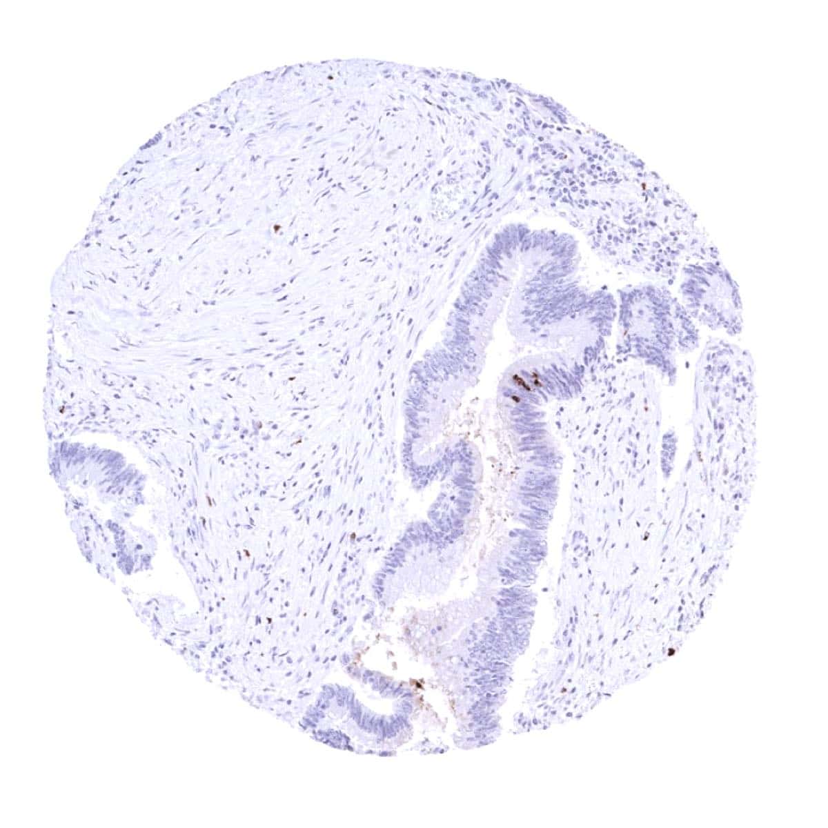
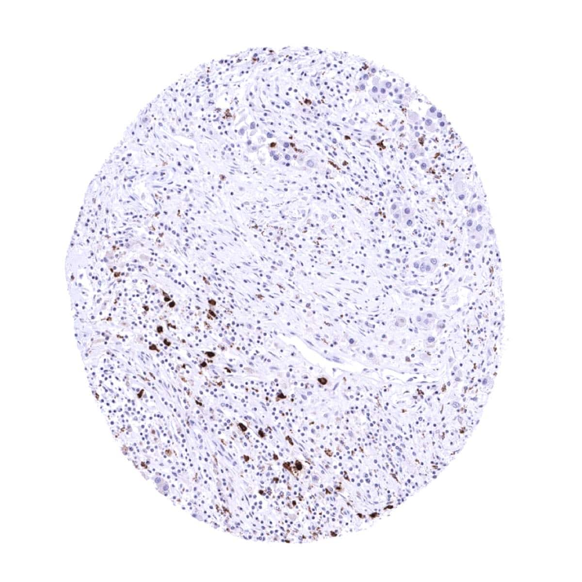
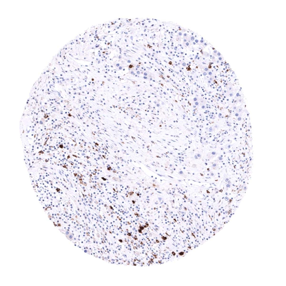
A variable content of MPO positive granulocytes is seen in cancers of various types. MPO expression by cancer cells is not seen.
The TCGA findings on MPO RNA expression in different tumor categories have been summarized in the Human Protein Atlas.
Compatibility of Antibodies
No data available at the moment
Protocol Recommendations
IHC users have different preferences on how the stains should look like. Some prefer high staining intensity of the target stain and even accept some background. Others favor absolute specificity and lighter target stains. Factors that invariably lead to more intense staining include higher concentration of the antibody and visualization tools, longer incubation time, higher temperature during incubation, higher temperature and longer duration of the heat induced epitope retrieval (slide pretreatment). The impact of the pH during slide pretreatment has variable effects and depends on the antibody and the target protein.
All images and data shown here and in our image galleries are obtained by the manual protocol described below. Other protocols resulting in equivalent staining are described as well.
Manual protocol
Freshly cut sections should be used (less than 10 days between cutting and staining). Heat-induced antigen retrieval for 5 minutes in an autoclave at 121°C in pH 7,8 Target Retrieval Solution buffer. Apply MSVA-692M at a dilution of 1:150 at 37°C for 60 minutes. Visualization of bound antibody by the EnVision Kit (Dako, Agilent) according to the manufacturer’s directions.

Agilent / Dako – Autostainer Link 48
Pretreatment in PT-Link for 30 minutes at 95°C (pH high); FLEX peroxidase blocking for 5 minutes (room temperature), MSVA-692M 1:300 for 20 minutes (room temperature), FLEX+ mouse/rabbit (LINKER) for 15 minutes (room temperature), horseradish peroxidase (HRP) for 20 minutes (room temperature), FLEX DAB+Sub-Chromo for 10 minutes (room temperature), FLEX hematoxylin for 5 minutes (room temperature).
These images reflect stainings by the protocol described above. It is of note that a comparable staining result can also be obtained by different protocols. In general, a longer pretreatment, a longer incubation time of the primary antibody, a higher antibody concentration, and a longer incubation time of FLEX+LINKER result in stronger staining, potentially at the cost of more background staining. Modifications of the protocol with a strengthening effect on staining intensity in combination with changes of other parameters that result in lower staining intensity can result in a comparable result as shown above.
Leica – BOND RX
Dewax at 72°C for 30 seconds; Pretreatment in Bond Epitope Retrieval Solution (ER2 – EDTA pH9) for 20 minutes at 100°C; Peroxidase blocking for 5 minutes (room temperature), MSVA-692M 1:450 for 15 minutes (room temperature), Post primary (rabbit anti mouse) for 8 minutes (room temperature), Polymer (goat anti rabbit) for 8 minutes (room temperature), mixed DAB refine for 10 minutes (room temperature), hematoxylin for 5 minutes (room temperature).
These images reflect stainings by the protocol described above. It is of note that a comparable staining result can also be obtained by different protocols. In general, a longer pretreatment, a longer incubation time of the primary antibody, a higher antibody concentration, a higher temperature during incubation, and a longer incubation time of Post primary and or the Polymer result in stronger staining, potentially at the cost of more background staining. Modifications of the protocol with a strengthening effect on staining intensity in combination with changes of other parameters that result in lower staining intensity can result in a comparable result as shown above.
Roche – Ventana Discovery ULTRA
Pretreatment for 64 minutes at 100°C (pH 8,4); CM peroxidase blocking for 12 minutes (room temperature), MSVA-692M 1:150 for 20 minutes at 36°C, secondary antibody (anti-mouse HQ) for 12 minutes at 36°C, anti-HQ HRP for 12 minutes at room temperature, DAB at room temperature, hematoxylin II at room temperature for 8 minutes, bluing reagent at room temperature for 4 minutes.


These images depict staining results obtained by the protocol described above. It is of note, that the Ventana machines generally require higher antibody concentrations than other commonly used autostainers because the antibodies are automatically diluted during the procedure. Various other protocols can result in an identical result as shown above. A longer pretreatment, a longer incubation time of the primary antibody, a higher antibody concentration, a higher temperature during incubation, and a longer incubation time of secondary antibody and or the anti-HQ HRP result in stronger staining, potentially at the cost of more background staining.
Potential Research Applications
- Visualization and quantification of granulocytes for assessing the clinical role of these cells.
- The distinction of granulocytes in multicolor immunofluorescence is important in immune-oncological panels for the evaluation of the microenvironment of cancers.
Evidence for Antibody Specificity in IHC
There are two ways how the specificity of antibodies can be documented for immunohistochemistry on formalin fixed tissues. These are: 1. comparison with a second independent method for target expression measurement across a large number of different tissue types (orthogonal strategy), and 2. Comparison with one or several independent antibodies for the same target and showing that all positive staining results are also seen with other antibodies for the same target (independent antibody strategy).
Orthogonal validation is not completely suited for validation of granulocytes markers because these cells can be found in every organ. Findings obtained by MSVA-692M do, however, fit with data derived from three independent RNA screening studies, including the Human Protein Atlas (HPA) RNA-seq tissue dataset, the FANTOM5 project, and the Genotype-Tissue Expression (GTEx) project, which are all summarized in the Human Protein Atlas (Tissue expression MPO). This is because the highest number of granulocytes was observed in the bone marrow and significant numbers were also seen in the spleen and the thymus. These organs were described to have significant RNA expression.
Comparison of antibodies: Specific MPO staining by MSVA-692M was suggested by a complete absence of MPO immunostaining in epithelial cells and the fact, that the distribution of positive cells identified by MSVA-692M was similar as seen by the independent antibody CAB000059 used for protein analysis in the human protein atlas. This also includes MPO positive material within prostatic glands. The only exception was the weak staining of gastric glands, which was not confirmed by CAB00059. We consider this a tolerable antibody cross-reactivity.
