
Basic Principle
There are two key muscles involved in pupillary constriction.
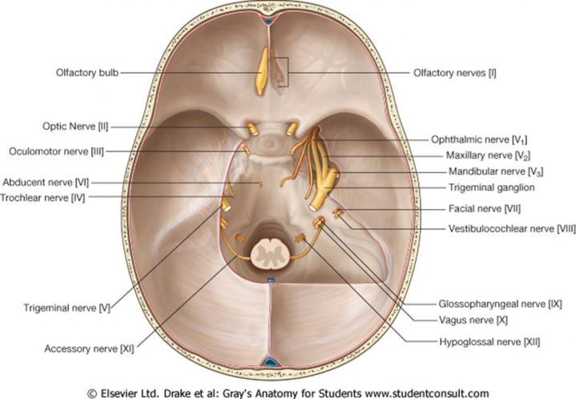
- Sphincter Pupillae- constrictor muscle that is innervated by the Parasympathetic nervous system innvervated by Oculomotor Nerve (CN3)
- Dilator Pupillae- dilator muscle that is innervated by the sympathetic nervous system
Pathway of Pupillary Light Constriction

Shining of light within the eye causes transmission via the optic nerve to synapse with neurones in the pre-tectal area rostral to the superior collicus (midbrain) forming the afferent pupillary response. Second order neurones send bilateral projections that synapse with the Edinger-Westphal Nucleus, hence forming the basis of the efferent pupillary response.
Efferent fibres leave via the oculomotor nerve and pass to the ciliary ganglion and to the short ciliary nerve to constrictor fibres.
Pathway of Pupillary Light Dilation

The Sympathetic Fibres arise initially from the ipsilateral hypothalamus through the midbrain and synapse on the C8/T1 Nerve Roots.
Second order neurones passes over the apex of the lung and synapse at the superior cervical ganglion.
They ascend with the external and internal carotid artery to supply the iris via the long ciliary nerve.
Sudomotor fibres run with the external carotid artery to supply sweat fibres to the face.
Pathway of Accomodation
The mechanism is not as clear cut, but as a finger draws closer from distant, both medial rectus contract, lens adopts a more convex shape followed by pupillary constriction.
This involves both Edinger-Westphal nucleus and relationships between the 3rd and 6th nerve.