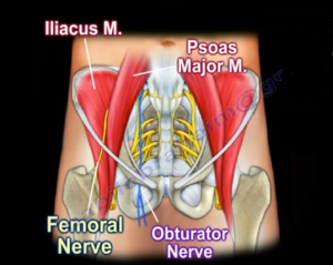
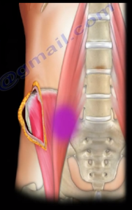
The iliacus and the psoas are the main hip flexors supplied by the femoral nerve which lies between the two muscles. The obturator nerve is medial to the psoas. The psoas arises from the transverse process of the lateral aspect of the vertebral bodies between the 12th thoracic vertebrae and the 5th lumbar vertebrae. The psoas runs downward across the pelvic brim and then passes deep to the ilioinguinal ligament 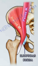 where it then forms a tendon past the hip joint capsule which inserts into the lesser trochanter of the femur. The iliacus arises from the upper two-thirds of the iliac fossa and joins the psoas to insert in the same tendon as the posas muscle. Both muscles are in the extraperitoneal space, or referred to as the iliopsoas compartment. The iliopsoas tendon is separated from the hip joint capsule by the iliopsoas bursa.
What causes an abscess of the iliopsoas muscle?
A primary abscess is caused by a hematogenous spread of the infection. The infection starts in the muscle itself. In a secondary abscess, the infection spreads from another area to the psoas muscle. For example, the infection may travel from the spine when it is infected by tuberculosis (Pott’s disease). This historically is the cause of this abscess. It can also spread from the SI joint, kidneys, or bowels. The iliopsoas abscess may initially present with sign and symptoms in the buttock, hip, or thigh. Such signs and symptoms may be obscure, nonspecific, and misleading.
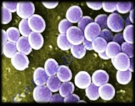 Staph aureus is the cause of iliopsoas abscesses in 88% of primary types. Polymicrobial infections are usually the cause in the secondary types. CT scans are usually the
 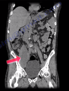 diagnostic modality of choice.
An abscess of the iliopsoas muscle is a diagnostic dilemma with a difficult diagnosis that is often delayed. The patient may be lying supine with the hip flexed and refuses to move, resisting any attempt for examination. With psoas involvement, the hip appears to be flexed, with limited and painful range of motion. This diverts attention away from the abdomen or pelvic source of the abscess. The patient may have a low grade fever and cannot straighten the leg. A high index of suspicion and performing the Psoas sign is necessary for diagnosis.
Psoas Sign
The patient is positioned on their side and the hip is extended to see if there is pain present in the iliopsoas region. The psoas sign is used in diagnosis of appendicitis but is also helpful in diagnosing a psoas abscess.
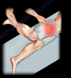
The iliopsoas abscess manifests itself as pain in the abdomen, flank, or groin area, as well as pain in the lower back. Flexion posture of the hip is also commonly noted. These abscesses are rare and present with vague clinical features.
Treatment
Percutaneous drainage is done if the abscess is simple, small, and single. Otherwise, an open drainage is the procedure of choice. When performing an open drainage of the abscess, a splitting incision is made along the iliac crest for easy access to the iliacus, psoas, and SI joint. Other incisions may be needed such as the anterior approach in the front of the hip or the posterior approach. Other sites for drainage depend on the location and extension of the abscess.
Risk factors for developing an iliopsoas abscess:
- Diabetes
- Immunosuppression
- Trauma
- Renal failure
- IV drug abuse
- Older individuals and AIDS patients
