Pediatric Genitourinary Rhabdomyosarcoma

  • Etiology: malignant neoplasm of striated muscle
  • Imaging: arise from bladder or prostate or vagina
  • MRI: intermediate to hypointense on T1WI, intermediate to hyperintense on T2WI, enhances heterogeneously, improved conspicuity on DWI
  • Complications: metastasis to lung
  • Clinical: most common childhood soft tissue sarcoma, 50% head and neck / 30% genitourinary / 20% musculoskeletal, embryonal cell type seen in infants is most common, alveolar cell type seen in older children and affects musculoskeletal system and is more aggressive, orbital has best prognosis

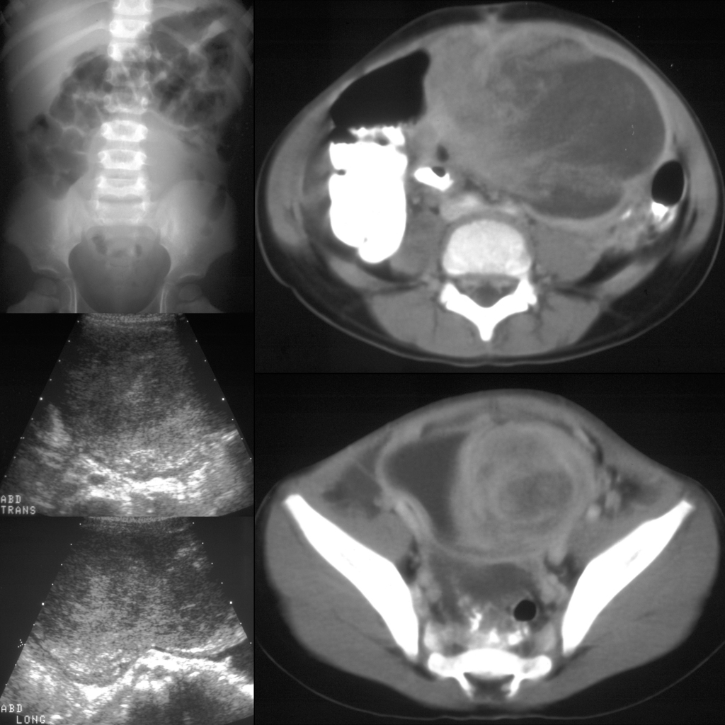
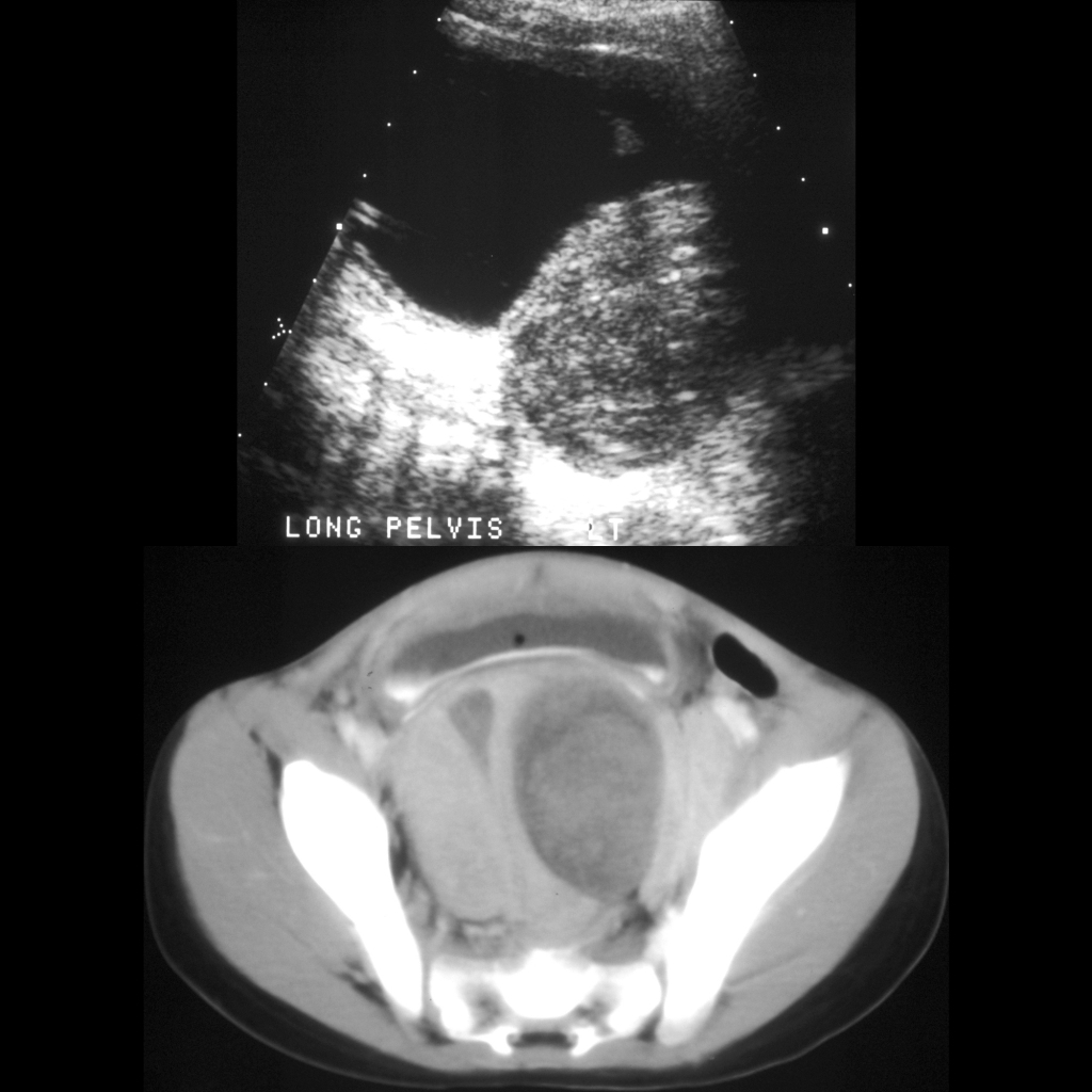
Radiology Cases of Genitourinary Rhabdomyosarcoma

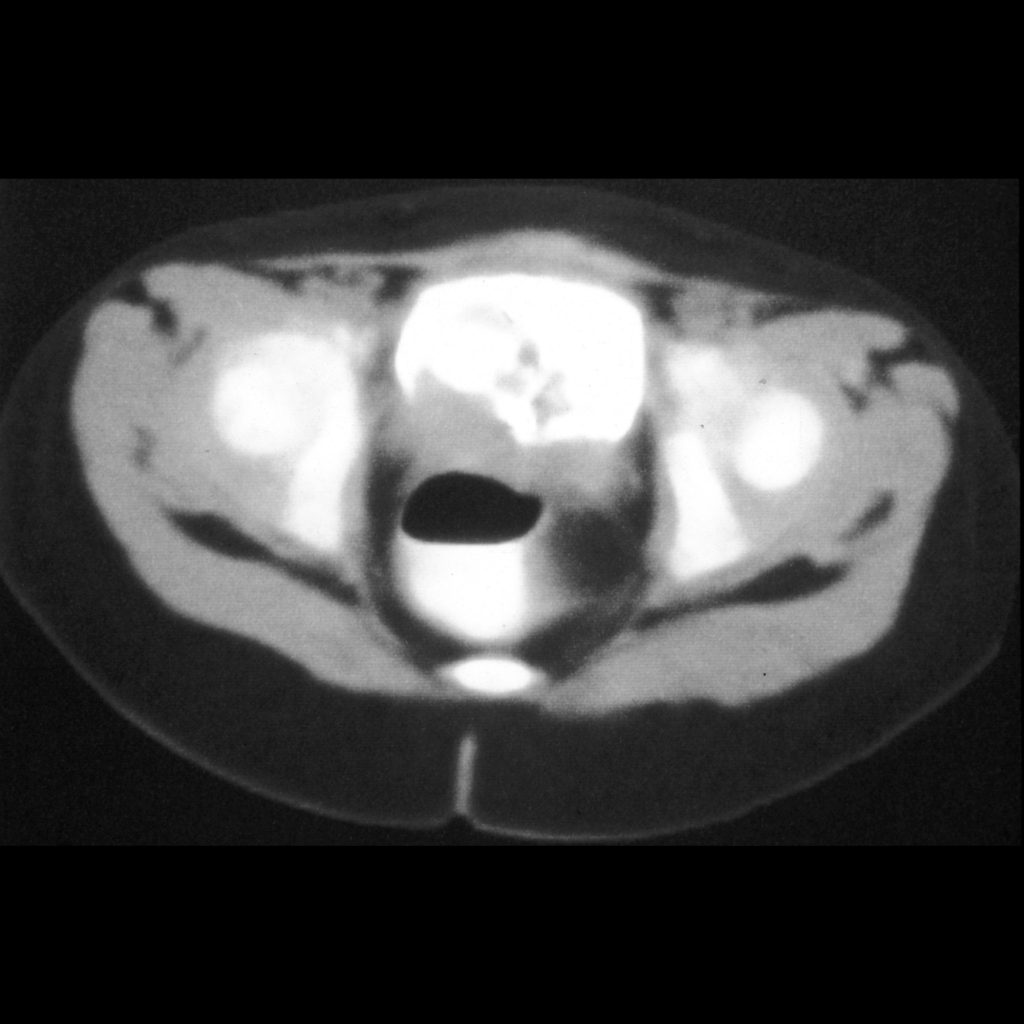
Radiology Cases of Bladder Rhabdomyosarcoma

IVP and CT of rhabdomyosarcoma of the bladder
AP image from the excretory phase of a vintage intravenous pyelogram (above) shows bilaterally normal appearing renal collecting systems and ureters. There is a multilobulated circumferential filling defect in the base of the badder. Axial CT with contrast of the abdomen shows the mass to almost complete fill the base of the bladder.
AXR and US and CT of rhabdomyosarcoma of the bladder
AXR (above left) shows a soft tissue mass in the mid abdomen displacing the bowel loops superiorly. Transverse and sagittal US of the mass (below left) show a solid, homogenous, lobulated mass. Axial CT with contrast of the abdomen (below right) shows a large heterogeneous mass in the mid to left abdomen with a low density center and swirling enhancement. There was a suggestion of direct tumor invasion into the right rectus muscle anteriorly (above right) and the bladder inferiorly.

Radiology Cases of Prostatic Rhabdomyosarcoma

US and CT of rhabdomyosarcoma of the prostate.
Sagittal US of the pelvis (above) shows an anechoic distended bladder superiorly and an echogenic mass posterior and inferior to the bladder. Axial CT with contrast of the pelvis (below) shows a round heterogenous necrotic mass in the center of the pelvis that is displacing the bladder superior and anteriorly.
CT of prostatic rhabdomyosarcoma
Axial CT with contrast of the abdomen shows a soft tissue mass arising from between the bladder and rectum that is invading into the posterior wall of the bladder, resulting in filling defects within the bladder.

Clinical Cases of Genitourinary Rhabdomyosarcoma

Clinical Cases of Sarcoma Botryoides

Clinical image of sarcoma botryoides
Clinical image shows a tan mass with frond-like excrescences protruding from the vagina.

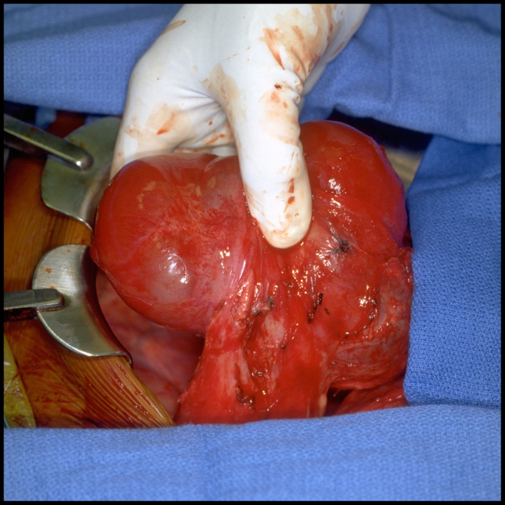
Surgery Cases of Genitourinary Rhabdomyosarcoma

Surgery Cases of Bladder Embryonal Rhabdomyosarcoma

Surgical image of rhabdomyosarcoma of the bladder
Surgical image shows the mass (between the surgeon’s fingers) to be extraperitoneal in location, originating from the left posterior aspect of the dome of the bladder. The mass did not appear to invade any normal structures except where it originated in the bladder, which is along the inferior edge of the image.

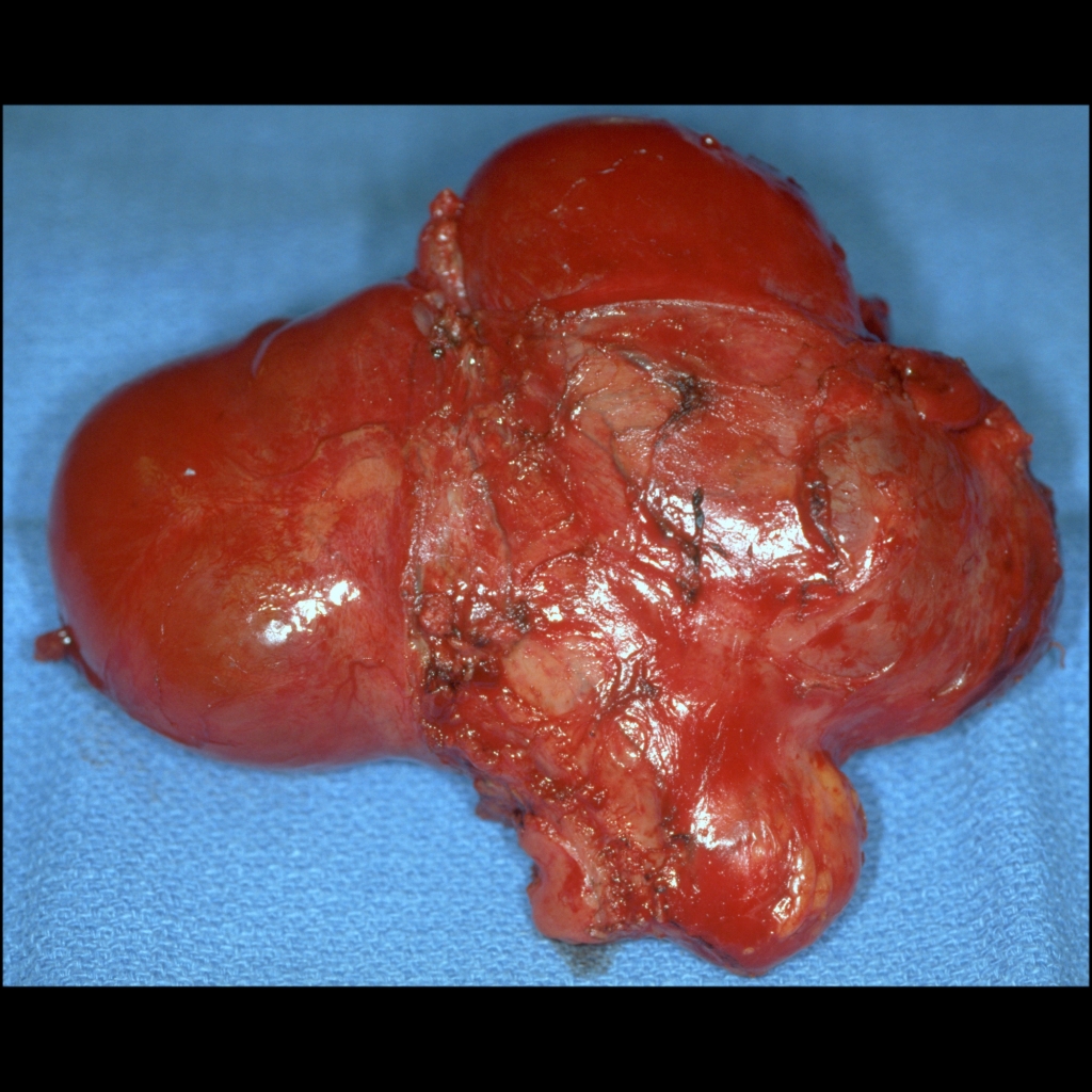
Gross Pathology Cases of Genitourinary Rhabdomyosarcoma

Gross Pathology Cases of Bladder Embryonal Rhabdomyosarcoma

Gross pathology image of embryonal rhabdomyosarcoma of the bladder
Gross pathological image shows a red / pink lobulated mass that was 14 x 11 x 7.5 cm in size with an area of the bladder dome attached to the mass inferiorly. There was widespread necrosis in the center of the mass.

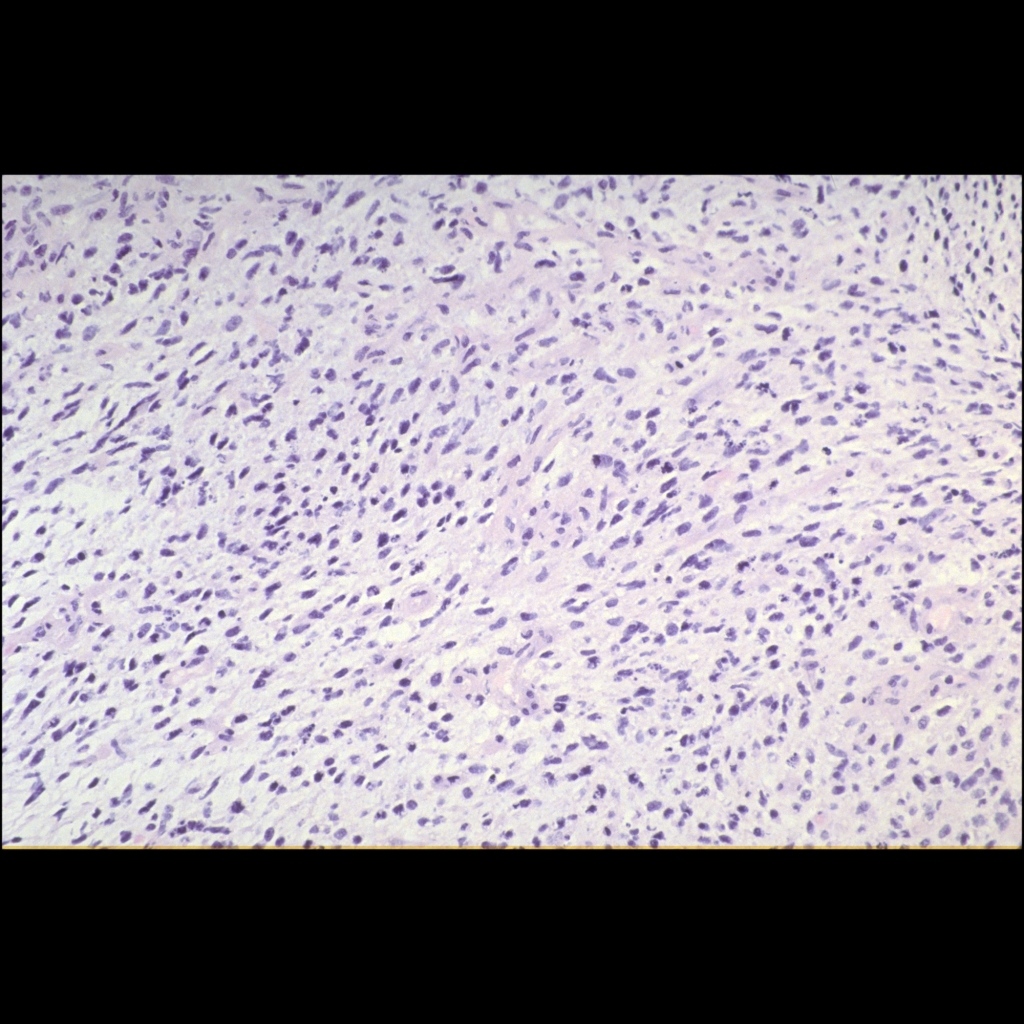
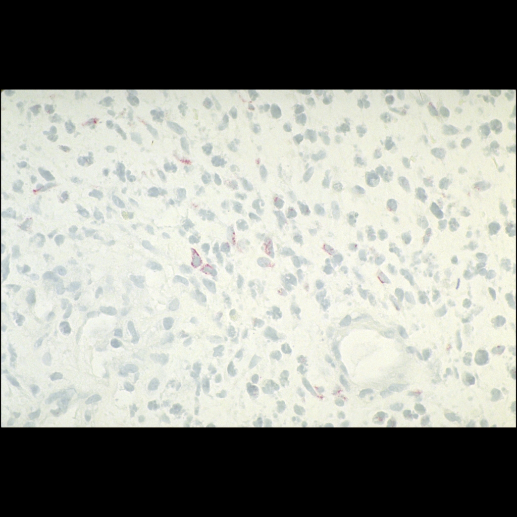
Histopathology Cases of Genitourinary Rhabdomyosarcoma

Histopathology Cases of Bladder Embryonal Rhabdomyosarcoma

Histopathology image of rhabdomyosarcoma
Histopathological image H&E stained section shows a loosely cellular neoplasm with cells characterized by small, hyperchromatic nuclei and minimal cytoplasm.
Histopathology image of rhabdomyosarcoma
Histopathological image shows in the absence of strap cells with identifiable cross-striations, a diagnosis of rhabdomyosarcoma is frequently made by the documentation of muscle specific antigens (i.e. desmin as illustrated here – red color) by immunoperoxidase staining.