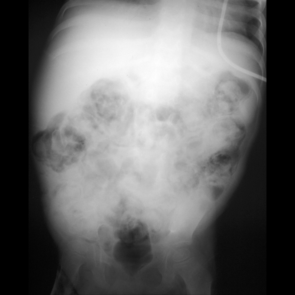
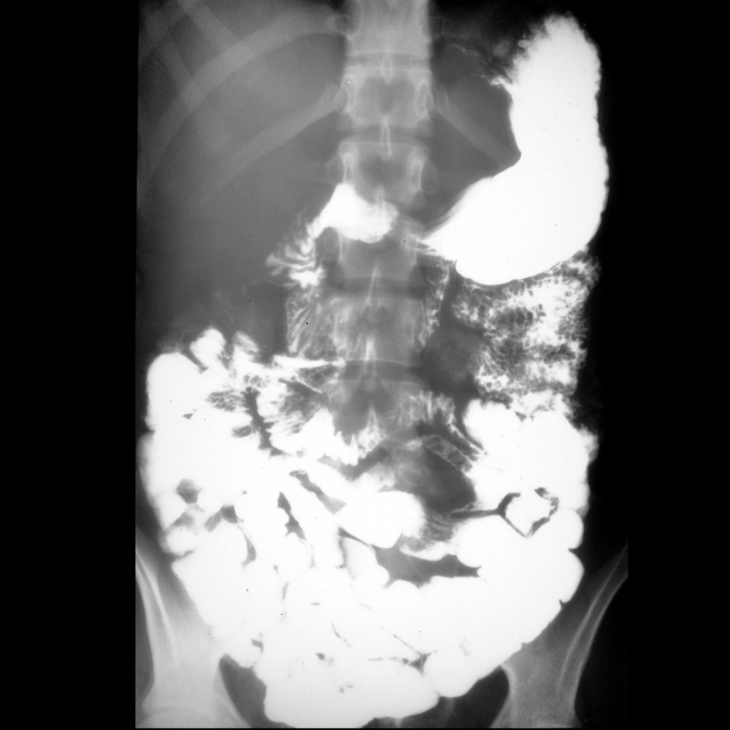
- Etiology: in immune suppressed patient who is status post bone marrow transplant, functionally competent introduced T-cells attack immunocompromized host cells in skin, liver, GI tract from esophagus to rectum with small bowel always involved
- US: small and large bowel wall thickening, mainly submucosal, especially in ileocecal region, discontinuous distribution in ~ 50% helps differentiate it from typhlitis
- CT:
— Generalized small bowel + large bowel thickening due to inflammation and sloughing of mucosal membranes with mucosal enhancement + submucosal enhancement – halo sign, submucosal edema without perienteric stranding, prominent vasa recta (comb sign), fluid-filled dilated bowel
— Ascites, hepatosplenomegaly, periportal edema, mucosal enhancement of gall bladder + bladder - Clinical: acute graft versus host disease (GVHD) occurs up to 30 days post bone marrow transplant, chronic GVHD occurs months post bone marrow transplant, symptoms include abdominal pain / nausea / vomiting / severe secretory diarrhea, also have rash / jaundice / severe mucosal inflammation
Radiology Cases of Graft Versus Host Disease