Pediatric Retropharyngeal Abscess

  • Etiology: pharyngitis or upper respiratory infection -> retropharyngeal adenitis -> supprative lymph node rupture into retropharyngeal space -> abscess, bacterial
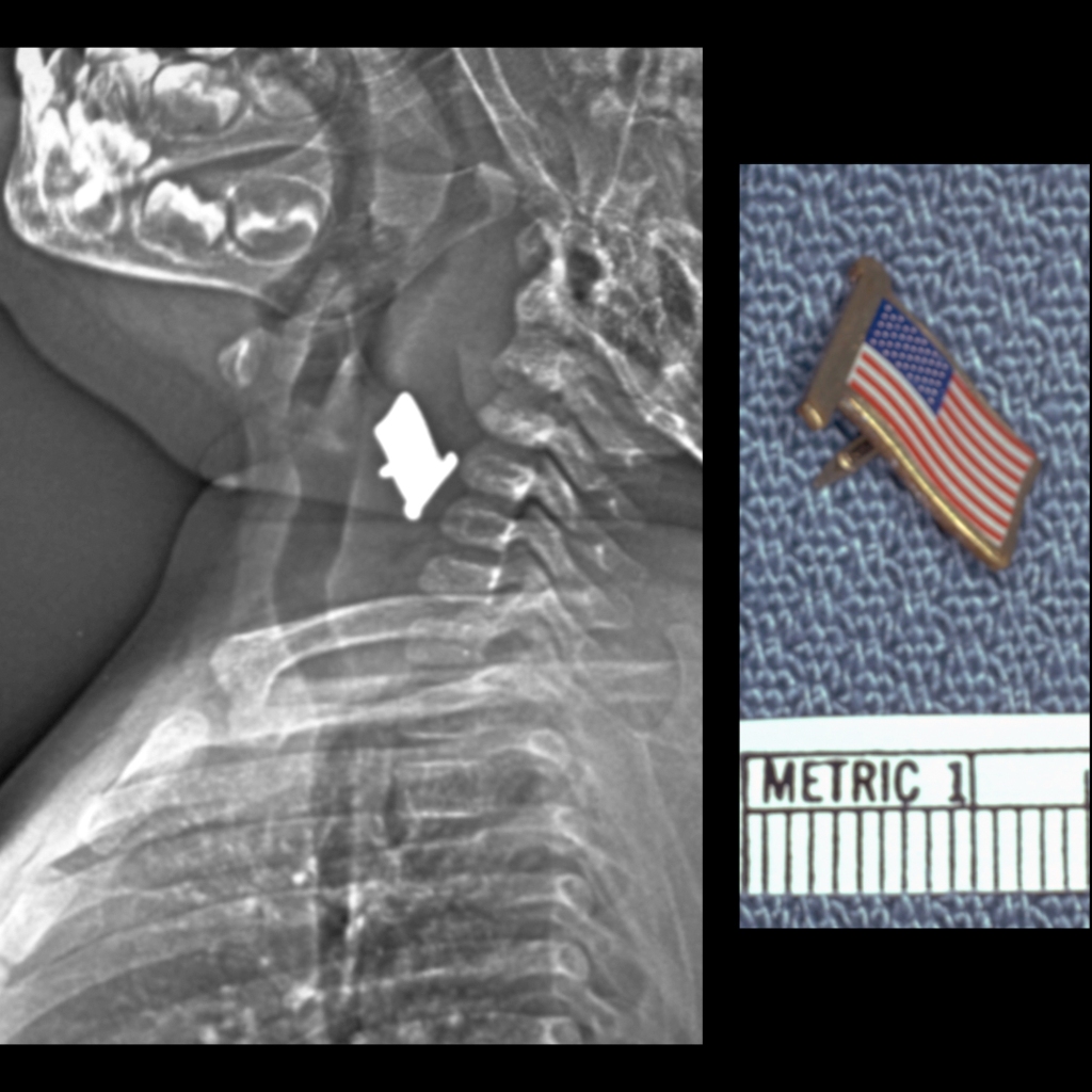
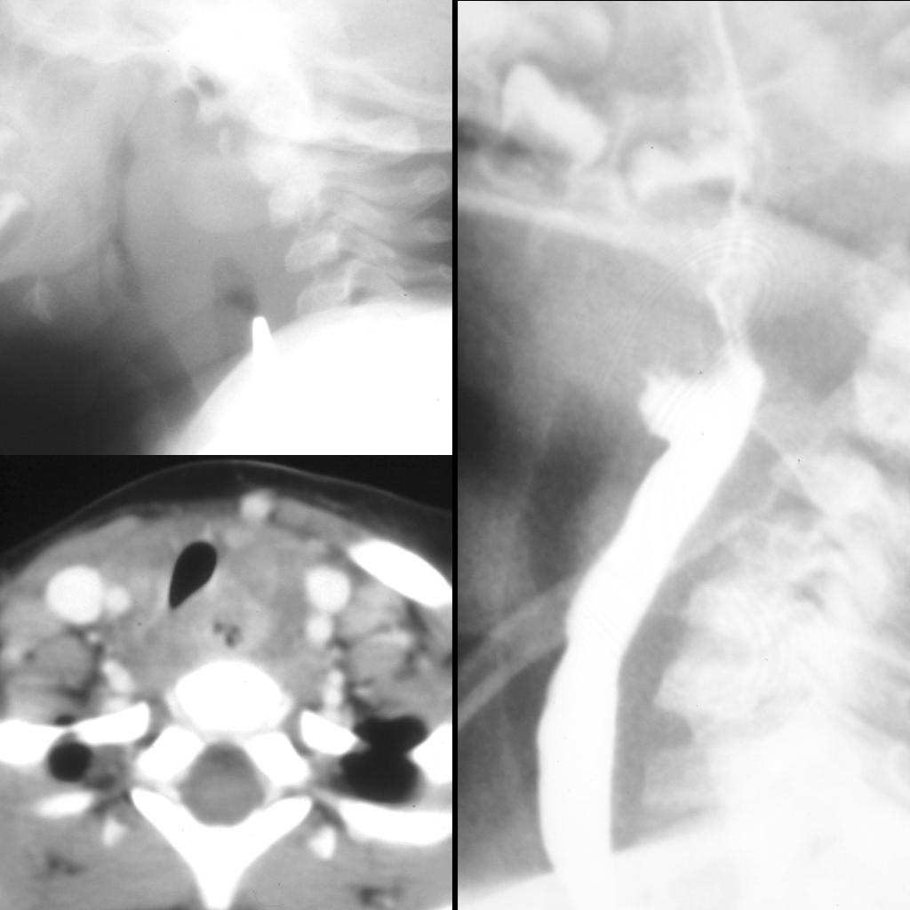
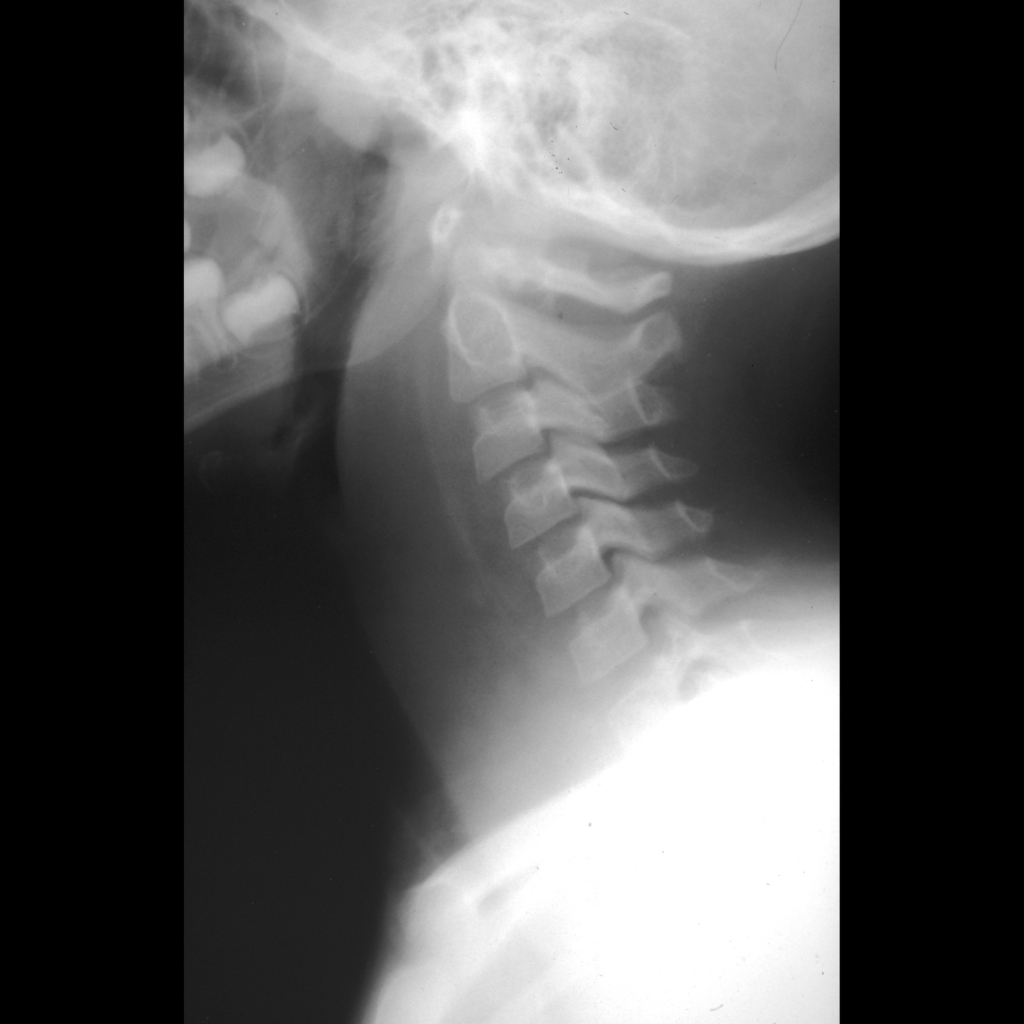
  • Imaging: normal prevertebral space is < 1/2 vertebral body at C2, soft tissue thickening of > 1 cervical vertebral body, to diagnose retropharyngeal mass need inspiratory image taken in extension, measurements less important than whether retropharynx changes with respiration, always look for retained foreign body
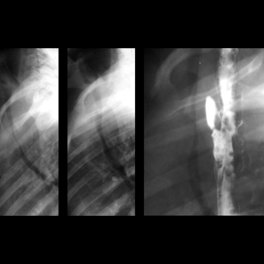
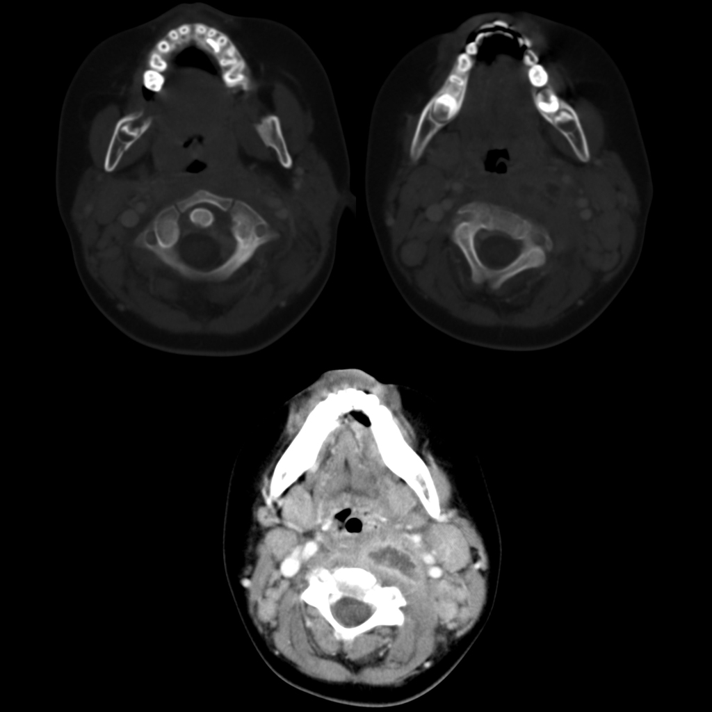
  • CT: helpful to look for complications – size of abscess, vascular complication, mediastinal extension
  • DDX: pseudothickening – physiological tracheal buckling which can be seen on lateral neck images obtained on expiration + flexion – so beware of false positives due to expiration / flexion – airway radiograph should always be taken in inspiration + extension, retropharyngeal cellulitis / phlegmon, penetrating trauma, vertebral osteomyelitis, mass, anasarca
  • Complications: airway compression, arteritis, jugular vein occlusion (Lemierre syndrome), atlanto-axial rotatory subluxation, extension into mediastinum
  • Treatment:
  • Clinical: seen in infancy and young children – 6 months to 6 years (older children + adults have peritonsillar + parapharyngeal abscess), 50% cases between 6-12 months, fever / stiff neck / dysphagia / stridor

Radiology Cases of Retropharyngeal Abscess

Radiograph of retropharyngeal abscess
Lateral radiograph of the airway taken in inspiration and extension shows massive thickening of the prevertebral soft tissues.
CT of atlanto-axial rotatory subluxation due to retropharyngeal abscess
Axial images with bone windows from a CT with contrast of the neck shows the C1 vertebral body (above left) rotated approximately 30 degrees to the right in relation to the C2 vertebral body (above right). Axial image with soft tissue windows (below) from the same CT shows a low density fluid collection with an enhancing rim to the left of midline in the retropharyngeal space.
CT of retropharyngeal abscess extending into mediastinum
Axial (upper), coronal (lower left) and sagittal (lower right) CT with contrast of the neck shows a low density fluid collection in an enlarged retropharyngeal space that extends inferiorly deep into the mediastinum, best seen on the sagittal image.

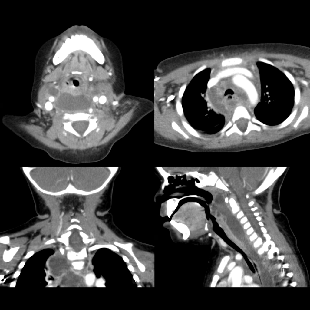
Radiology Cases of Chronic Gastrointestinal Foreign Body Causing Retropharyngeal Abscess

Pathological image of an esophageal foreign body
Lateral radiograph of the airway (left) shows a radiopaque foreign body in the cervical esophagus which is associated with a large amount of retropharyngeal soft tissue swelling. Gross pathological image (right) shows a metal flag-shaped pin that was endoscopically retrieved from the patient’s esophagus.
Radiograph and CT and UGI of chronic esophageal foreign body and retropharyngeal abscess
Lateral radiograph of the airway (above left) shows a radioopaque coin on edge in the esophagus at the level of C5. There is retropharygeal soft tissue swelling around the foreign body and air anterior to the coin in the retropharyngeal soft tissue. Axial CT with contrast of the neck after foreign body removal (below left) better shows the retropharyngeal fluid and air collection anterior to the vertebral body and causing some mass effect on the trachea. Lateral image from an upper GI exam (right) shows that the esophagus communicates with the retropharyngeal fluid and air collection.
CXR and Upper GI of esophageal foreign body
Lateral spot images from an upper GI show fixed narrowing of the trachea (left + middle) with an esophageal perforation / fistula arising from the anterior wall of the esophagus (right). Endoscopy done earlier in the day removed a set of plastic wheels from a toy car which was felt to have been present for a long time as the esophagus was ulcerated and edematous in the area of perforation.