- Etiology: abnormality of neuronal migration
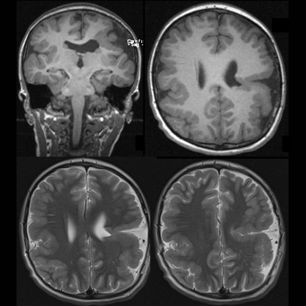
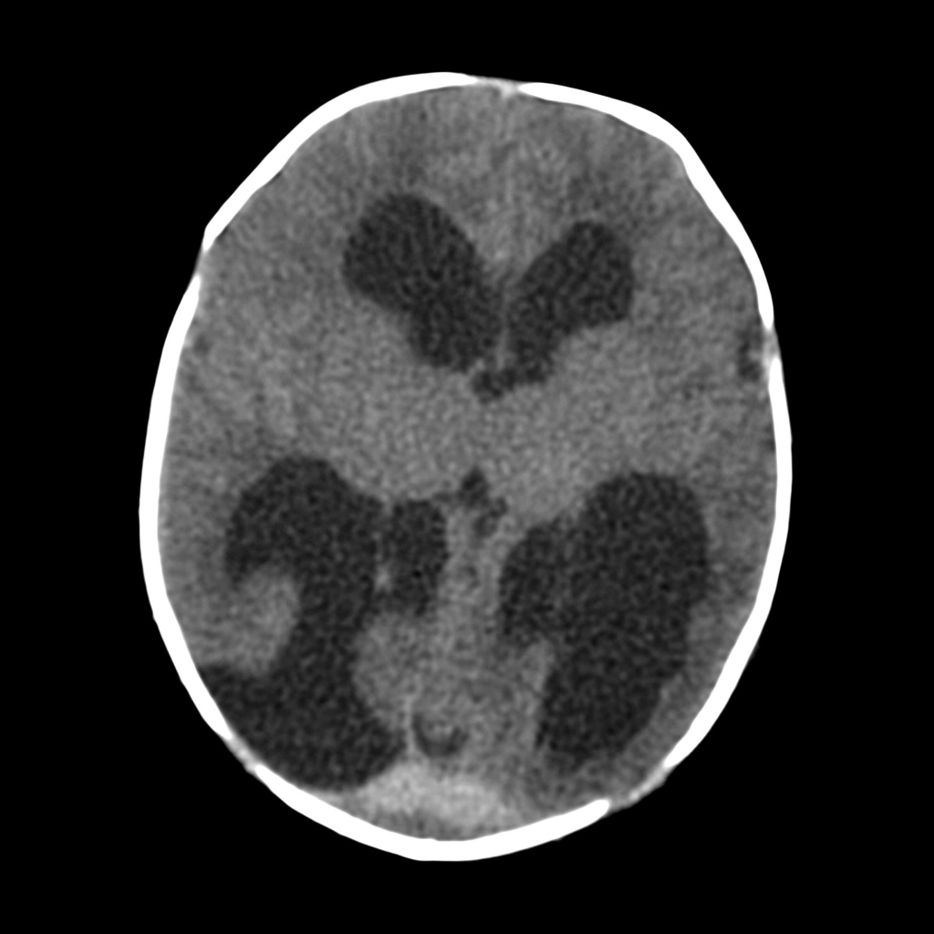
- Imaging: gray matter lined clefts from ependyma of lateral ventricle to cortical surface – a pial-ependymal seam, +/- abnormal adjacent cortex / gyri, usually dimple at ventricular surface, if cleft walls are in apposition = closed lip (Type I), if cleft walls are separated by CSF = open lip (Type II), unilateral or bilateral, often near pre or post central gyri, associated with optic nerve hypoplasia / absent septum pellucidum / septo-optic dysplasia
- DDX:
- Complications:
- Treatment:
- Clinical: often have seizures or hemiparesis or developmental delay, open lip – more common than closed lip, open lip more commonly bilateral, closed lip more commonly unilateral
Radiology Cases of Schizencephaly