Wilms Tumor

  • Etiology: renal origin, 30% of unilateral and 100% of bilateral Wilms tumor are due to nephrogenic rests
  • Imaging screening: in hemihypertrophy and Beckwith Wiedemann syndrome – baseline at 6 months, get US every 3 months until 8 years old, becoming larger or rounder suggests malignant degeneration
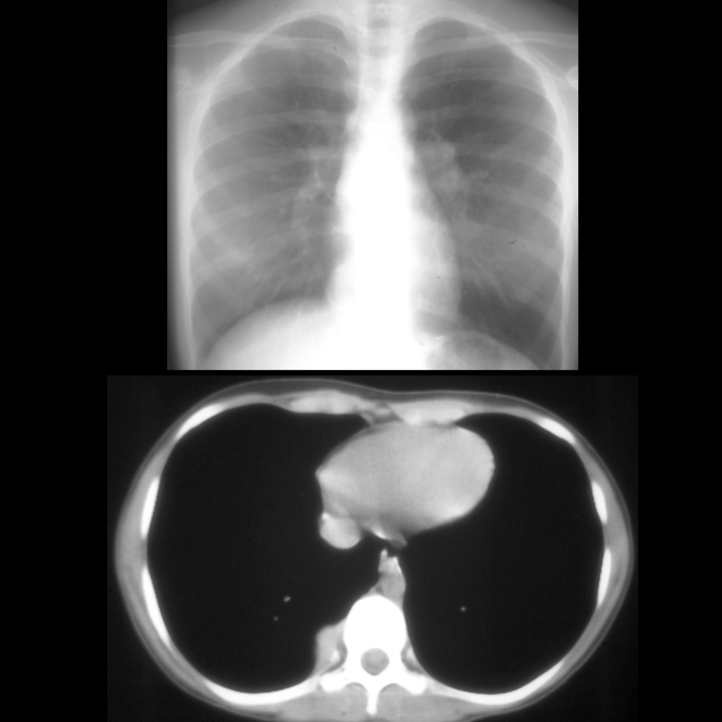
  • Imaging at presentation: establish the stage of tumor – local / locoregional / metastatic, look for tumor rupture and ipsilateral / contralateral synchronous tumor and vascular invasion in renal vein and IVC, chest CT for pulmonary metastatic disease, MRI superior for detecting bilateral renal disease
  • Imaging: well circumscribed, round, heterogenous, some cystic components, may contain small amounts of fat, fine calcifications in 9%, multicentric in 10-15%, enhance less than normal parenchyma, venous invasion, claw sign, deforms the collecting system showing it is an intrarenal mass, pushing tumor that displaces vessels
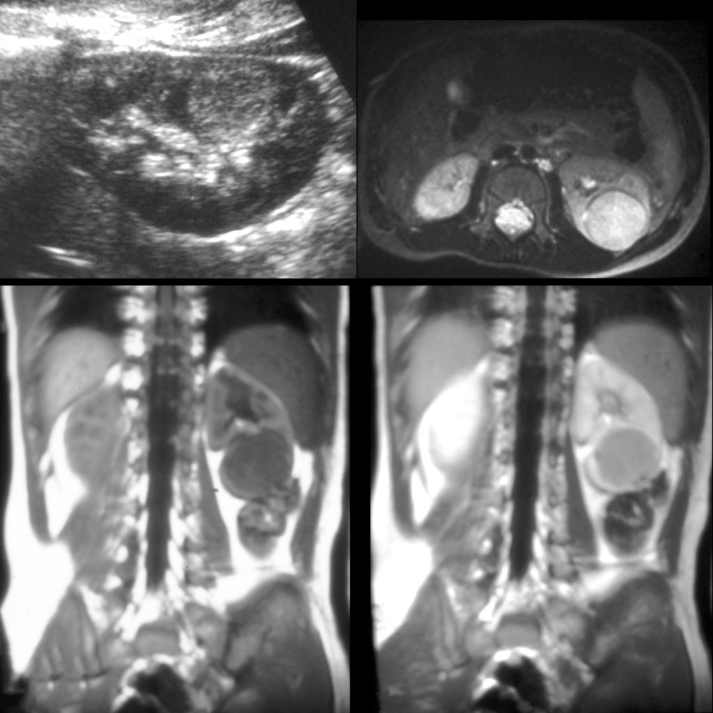
  • MR: T1 isointense and T2 hyperintense
  • Complications: regional lymph nodes, renal vein / inferior vena cava / right atrium tumor thrombus, contralateral kidney – synchronous or metachronous 10%, metastasis to lung / liver / bone in 12%
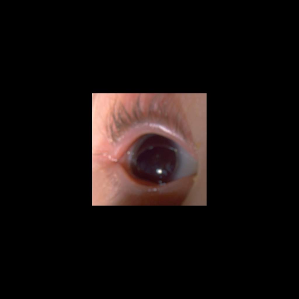
  • Clinical: most common abdominal malignancy of childhood, 87% of kidney masses in children, peak at 3 1/2 years, 80% < 5 years, rare in neonates, palpable mass in 75-95%, presentation most commonly as palpable mass and infrequently with pain / hematuria / constitutional symptoms, associated with Beckwith-Wiedemann syndrome (macroglossia, omphalocele, visceromegaly – liver / kidneys / pancreas, gigantism / hemihypertrophy / Wilms tumor in 10%), bilateral Wilms is virtually always genetic or syndrome associated

Radiology Cases of Wilms Tumor

Radiology Cases of Unilateral Wilms Tumor

MRI of Wilms tumor
Sagittal US of the left kidney (upper left) shows a round hyperechoic lesion in the lower pole of the kidney. Axial T2 MRI without contrast of the abdomen (upper right) and coronal T1 MRI without (lower left) and with (lower right) contrast of the abdomen shows a well-circumscribed, solid T1 hypointense and T2 isointense mass in the lower pole of the left kidney that enhances minimally.
CT of Wilms tumor with lung metastases, liver metastases, and IVC invasion
Axial (above right), coronal (below middle) and sagittal (below right) CT with contrast of the abdomen shows a large heterogenous non-calcified mass that fills the entire left side of the abdomen. The inferior vena cava (to the right of the aorta) was distended with tumor thrombus. Multiple liver (above left) and lung (below left) lesions are also seen.
MRI of Wilms tumor
AXR (above left) shows displacement of the bowel out of the right side of the abdomen. Sagittal US of the right kidney (above right) shows a large right renal mass that spares the upper pole of the right kidney. Coronal T1 MRI with contrast of the abdomen (below left) shows a large mass that is heterogenous in appearance that arises from the lower pole of the right kidney and that is demonstrating a claw sign superiorly. Axial T2 MRI (below right) again shows the heterogenous nature of the mass due to hemorrhage and necrosis.

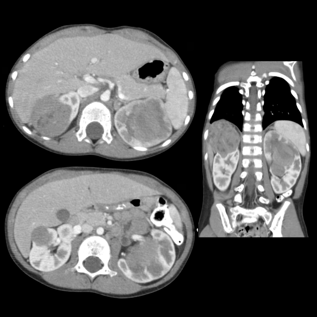
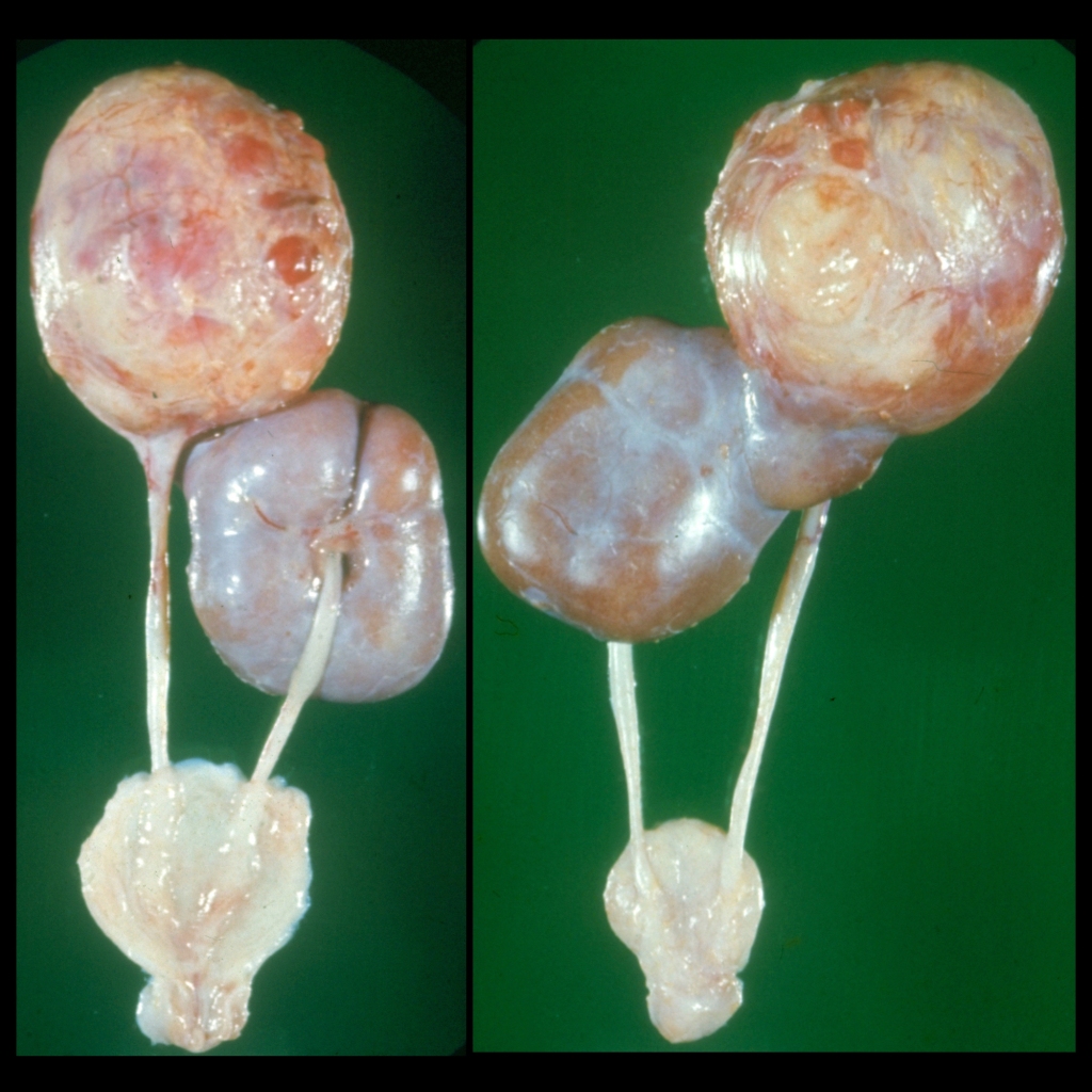
Radiology Cases of Bilateral Wilms Tumor

CT of bilateral Wilms tumor
Axial (left) and coronal (right) CT with contrast of the abdomen shows a large rounded solid heterogenous mass arising from the upper pole of the right kidney and a large rounded solid heterogenous mass arising from the center of the left kidney.

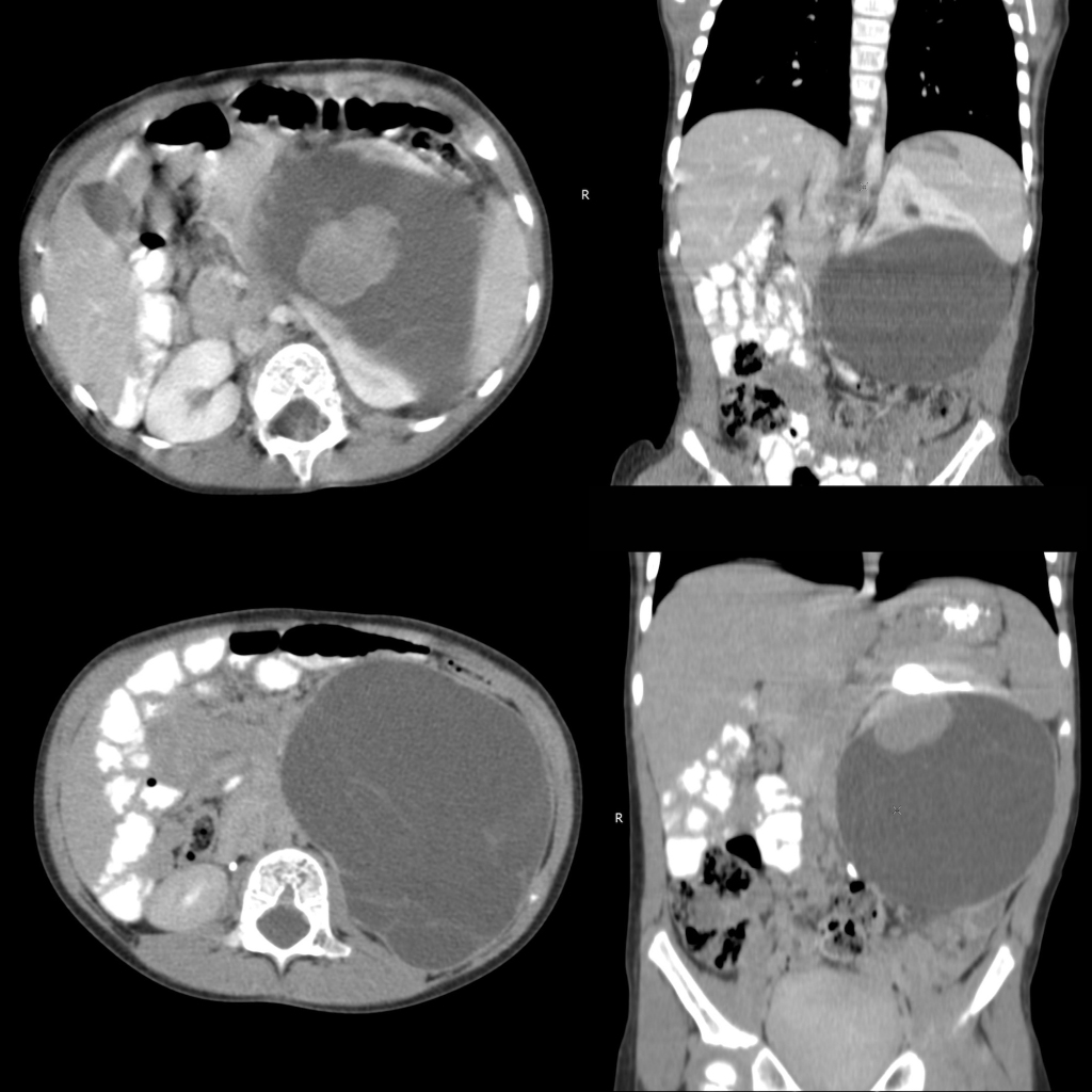
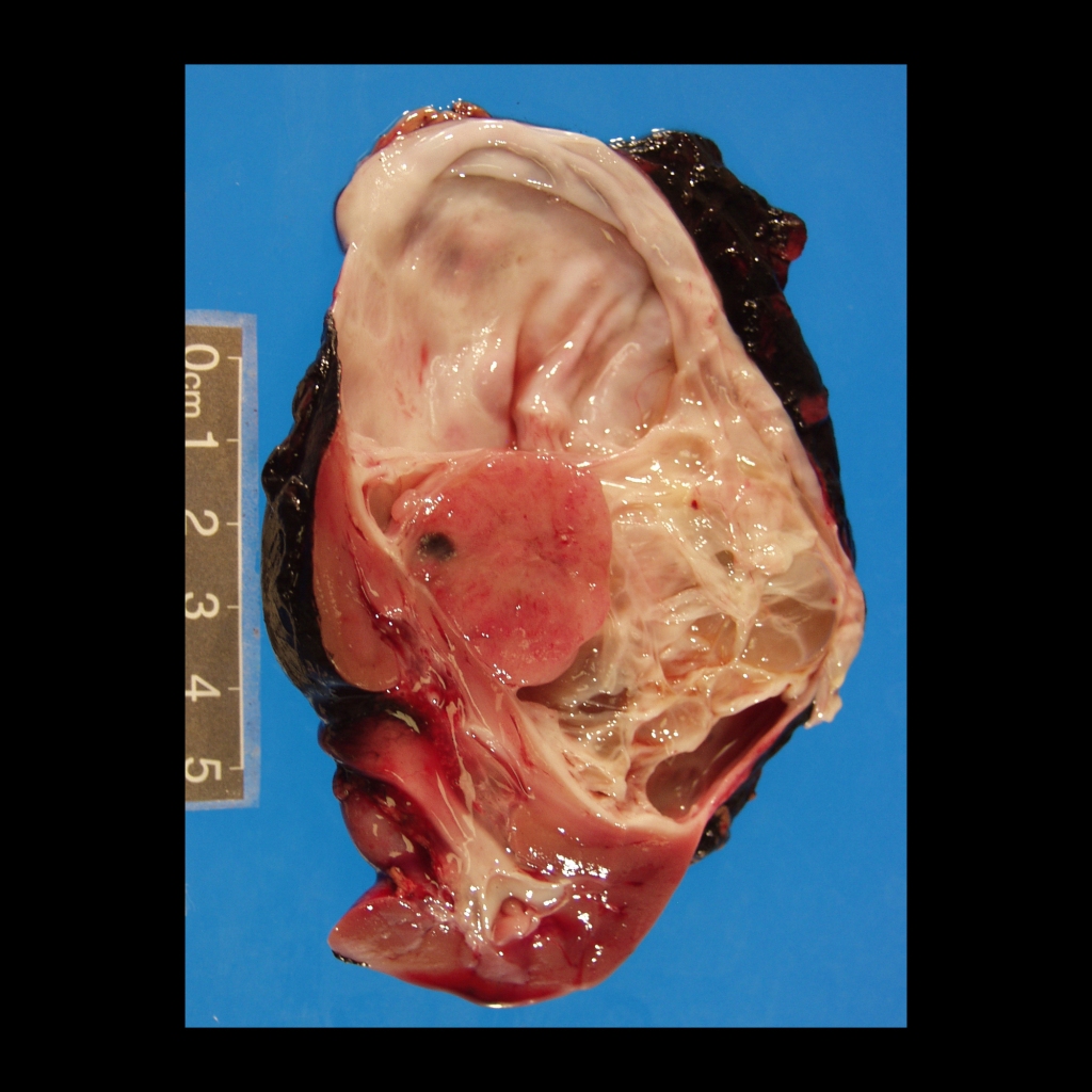
Radiology Cases of Cystic Wilms Tumor

CT of cystic Wilms tumor
Axial (left) CT with contrast of the abdomen shows a large cystic mass with septations in the left side of the abdomen. Coronal immediate image (above right) shows a claw sign proving the left kidney is the organ of origin of the mass. Coronal delayed image (below right) shows the mass has a mural nodule superiorly within it.

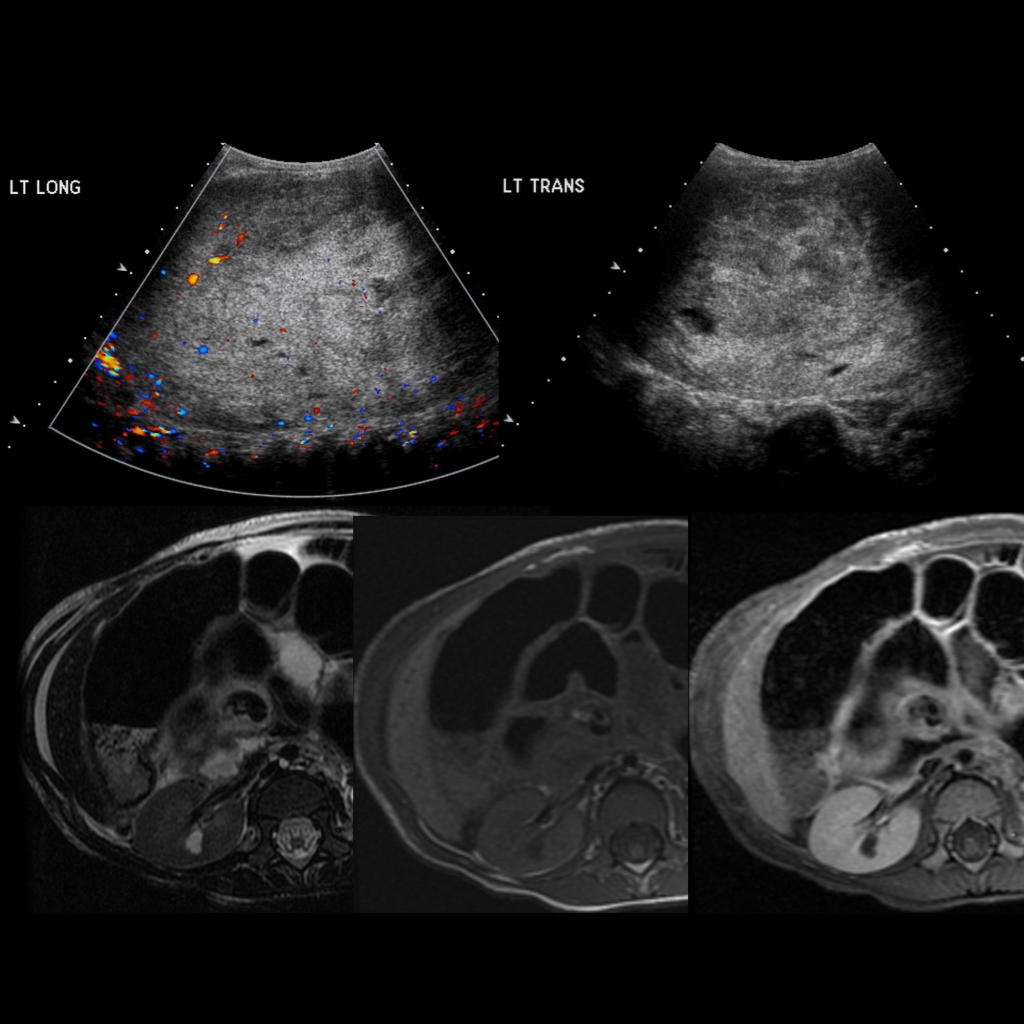
Radiology Cases of Wilms Tumor in the Left Kidney and Nephroblastomatosis in the Right Kidney

US and MRI of nephroblastomatosis
Sagittal (above left) and transverse (above right) US images of the left kidney show it to be enlarged and echogenic in appearance with little recognizable normal renal parenchyma. Axial T2 (lower left), T1 (lower middle) and T1 post contrast (lower right) MR images of the right kidney shows a small round lesion that is hyperintense on T2, isointense on T1, and that does not enhance after the administration of contrast. Two additional identical appearing lesions were seen in the right kidney.

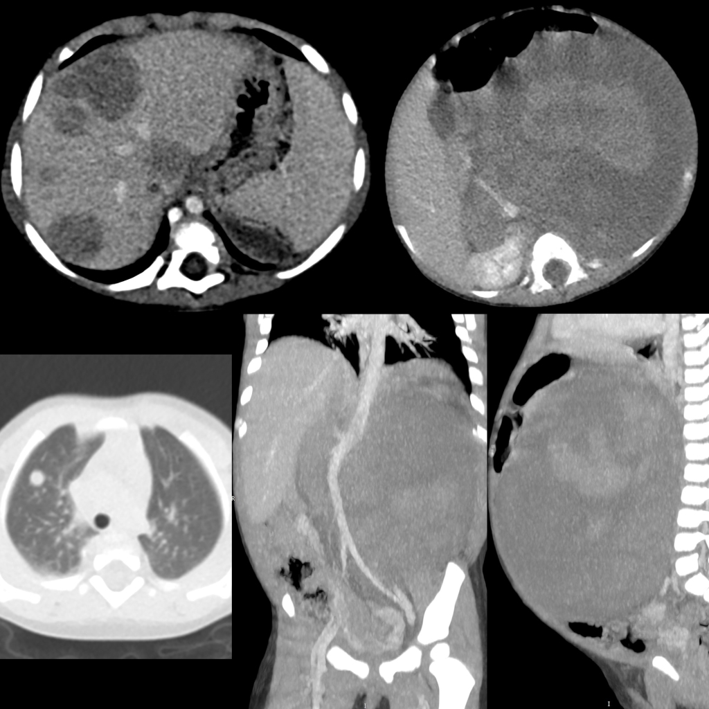
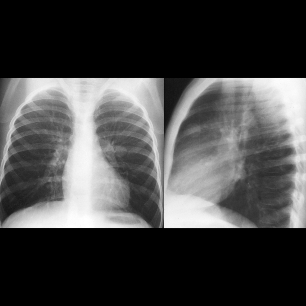
Radiology Cases of Lung Metastases in Wilms Tumor

CXR of lung metastasis in Wilms tumor
CXR PA shows a large round opacity just lateral to the left pulmonary artery which is located anteriorly on the lateral view.
CXR and CT of lung metastasis in Wilms tumor
CXR AP (above) shows a soft tissue density projecting in the right cardiophrenic angle. Axial CT with contrast of the chest (below) shows a soft tissue mass in the right posterior costophrenic sulcus.
CT of Wilms tumor with lung metastases, liver metastases, and IVC invasion
Axial (above right), coronal (below middle) and sagittal (below right) CT with contrast of the abdomen shows a large heterogenous non-calcified mass that fills the entire left side of the abdomen. The inferior vena cava (to the right of the aorta) was distended with tumor thrombus. Multiple liver (above left) and lung (below left) lesions are also seen.

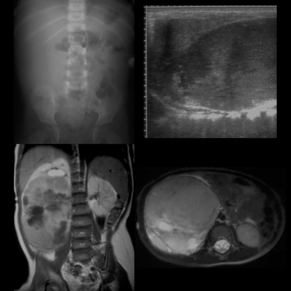
Radiology Cases of Hepatic Metastases in Wilms Tumor

CT of Wilms tumor with lung metastases, liver metastases, and IVC invasion
Axial (above right), coronal (below middle) and sagittal (below right) CT with contrast of the abdomen shows a large heterogenous non-calcified mass that fills the entire left side of the abdomen. The inferior vena cava (to the right of the aorta) was distended with tumor thrombus. Multiple liver (above left) and lung (below left) lesions are also seen.

Radiology Cases of Inferior Vena Cava Invasion in Wilms Tumor

CT of Wilms tumor with lung metastases, liver metastases, and IVC invasion
Axial (above right), coronal (below middle) and sagittal (below right) CT with contrast of the abdomen shows a large heterogenous non-calcified mass that fills the entire left side of the abdomen. The inferior vena cava (to the right of the aorta) was distended with tumor thrombus. Multiple liver (above left) and lung (below left) lesions are also seen.

Clinical Cases of Wilms Tumor

Clinical image of aniridia in a patient with Wilms tumor
Clinical image shows absence of the iris.

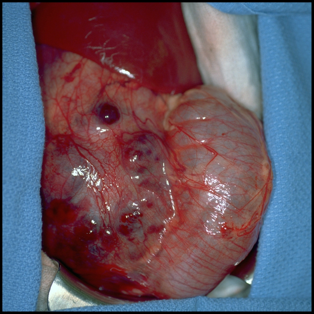
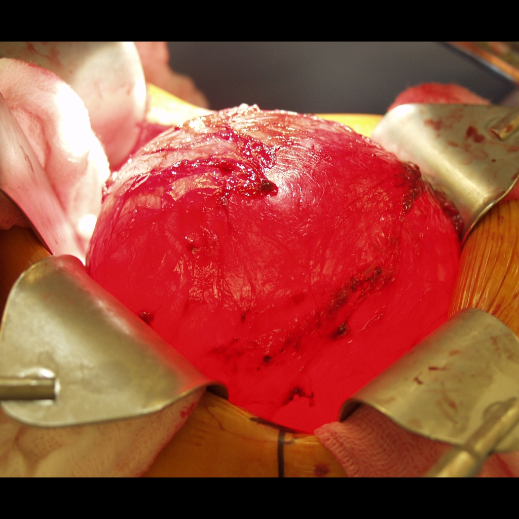
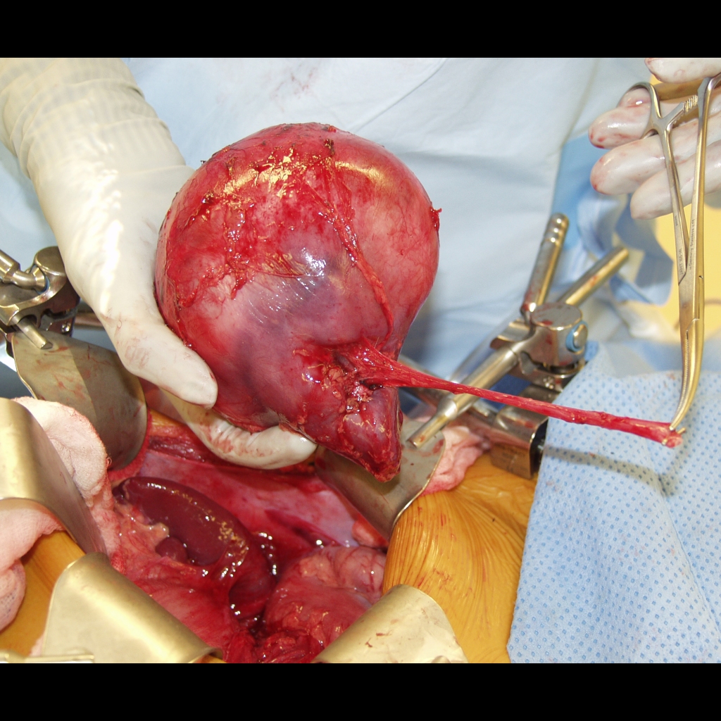
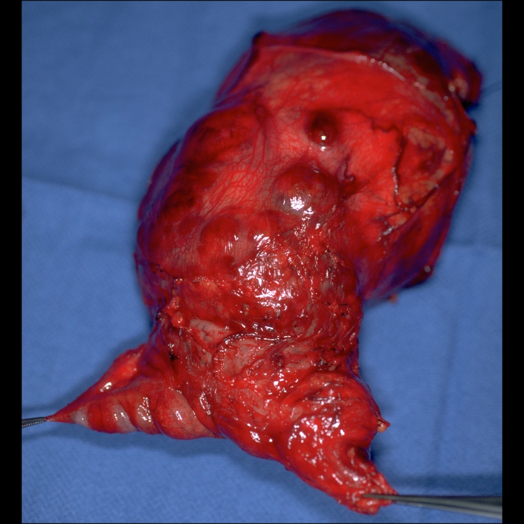
Surgical Cases of Wilms Tumor

Surgical image of Wilms tumor
Surgical image shows a large heterogenous mass in the right kidney (to the right of the surgical field) which was intimately adherent to the ileum and cecum (to the left of the surgical field).
Surgical image of cystic Wilms tumor
Surgical image shows a large mass arising from the left kidney which is being effectively transilluminated by the overhead lights, demonstrating its primarily cystic nature.
Surgical image of cystic Wilms tumor
Surgical image shows a large fluid-filled mass arising from the left kidney just after it has been resected along with the left ureter.

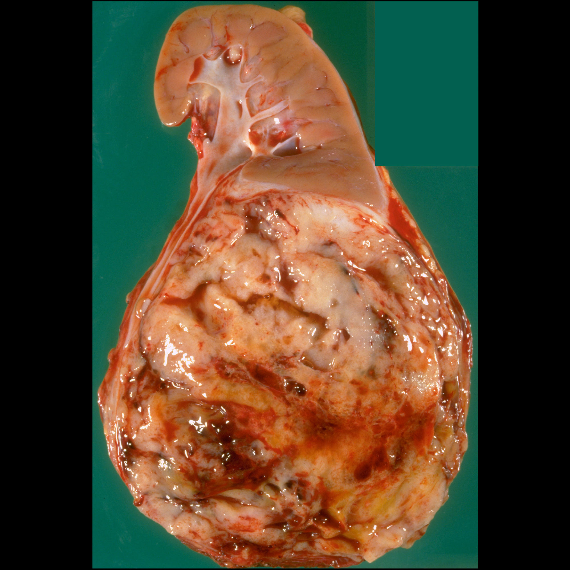
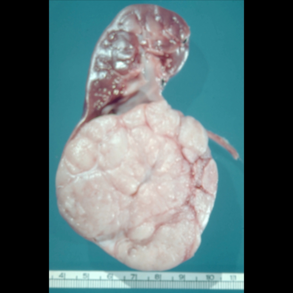
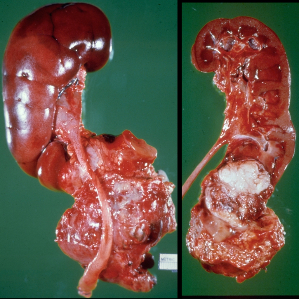
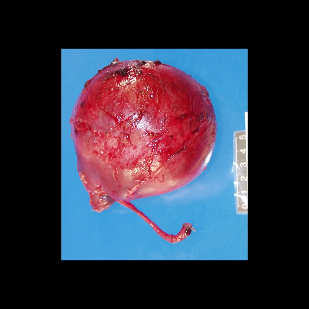
Gross Pathology Cases of Wilms Tumor

Gross pathological image of Wilms tumor
Gross pathological image shows a whitish-tan mass with areas of hemorrhage and necrosis arising from the lower pole of the kidney. Note the compression of the adjacent renal tissue which manifests itself on imaging as the renal “claw” sign.
Gross pathology image of Wilms tumor
Gross pathological image after right nephroureterectomy and en bloc ileocolectomy shows a large heterogenous mass in the right kidney which was intimately adherent to the cecum (held by the forceps on the right) and the ileum (held by the forceps on the left).
Gross pathology image of Wilms tumor
Sectioned gross pathological image shows a large, heterogenous, round mass arising from the inferior pole of the right kidney. Note the claw sign of the renal parenchyma draped along the lateral margin of the mass.
Gross pathology image of Wilms tumor
Gross pathological image (left) shows a large, heterogenous mass arising from the inferior pole of the right kidney. Note the claw sign of the renal parenchyma draped along the medial and lateral margins of the mass on the sectioned gross pathological image (right).
Gross pathology image of cystic Wilms tumor
Gross pathological image shows a large left renal mass along with the left ureter with the upper part of the mass being ballotable, demonstrating its primarily cystic nature.
Gross pathology image of cystic Wilms tumor
Sectioned gross pathological image after drainage of fluid shows the preserved lower pole of the left kidney (above), the replacement of the upper and middle poles by a cystic tumor containing multiple septations which is now decompressed, and a mural nodule (center of image).
Pathology image of Wilms tumor and renal crossed fused ectopia
Post-mortem gross pathological images obtained anteriorly (on the left) and posteriorly (on the right) show a mass in the upper pole of the right kidney. The upper pole of the left kidney is fused to the lower pole of the right kidney.

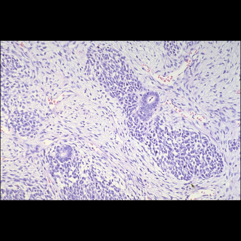
Histopathology Cases of Wilms Tumor

Histopathology image of Wilms tumor
Histopathological image H&E stained section shows the classic triphasic histologic appearance of these lesions as they recapitulate normal nephrogenesis – stroma (spindle shaped cells), blastema (islands of undifferentiated small round blue cells) and epithelium (tubular / glandular structures); mitoses and necrosis are frequent with no adverse prognostic significance.