Computed Tomography Incidentalomas

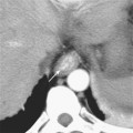
CHAPTER 7 Computed Tomography Incidentalomas


The discovery of small, incidental, asymptomatic lesions during the routine review of computed tomographic (CT) images is a common and often frustrating occurrence in routine clinical practice. Such lesions are often referred to as indeterminate, nonspecific, or “too small to characterize.” Because most busy radiologists cannot devote more than a brief moment to determining the significance and subsequent evaluation of such lesions, this section is dedicated to “incidentalomas.” We do not advocate a single approach to small incidental lesions because recommendations remain in flux. Each individual radiologist must determine the degree of uncertainty he or she is willing to tolerate, taking into consideration patient anxiety and prognosis, the personality and typical practices of the referring physician, and the number of malpractice attorneys preying on the local medical community. Regardless, one should always use common sense. If a lesion has a small chance of being malignant, recommending further costly or potentially morbid tests is of no immediate benefit to a patient with significant comorbidities of an acutely life-limiting nature. Likewise, incidental lesions should not be viewed as an opportunity for revenue building through the recommendation of frequent follow-up imaging studies. In contrast, with the increasing availability of minimally invasive therapies for malignant tumors, early detection and diagnosis of incidental malignant neoplasms has taken on new imperative.


LIVER


When faced with a small, nonspecific liver lesion, it is helpful to determine whether the patient has risk factors for a malignant hepatic tumor (primary or secondary). Most patients in this category can be identified by asking if they have a history of chronic liver disease (e.g., viral hepatitis, cirrhosis, hemochromatosis, primary sclerosing cholangitis) or known extrahepatic primary malignancy. The definition of a liver lesion that is “too small to characterize” varies in the literature but typically includes lesions smaller than 2 cm in greatest dimension. With the widespread use of thin-section multidetector CT, the size criteria for “too small to characterize” has diminished. Common causes of “too small to characterize” liver lesions include hepatic cysts, hemangiomas, and bile duct hamartomas.


Patients with No Known Cancer or Chronic Liver Disease


In the absence of risk factors such as cancer or chronic liver disease, the likelihood that a small, incidental, indeterminate liver lesion is malignant is extremely low, regardless of the age of the patient or number of lesions. Therefore, we do not automatically recommend additional evaluation of incidental nonspecific lesions smaller than 1 cm in diameter in patients without risk factors for malignant disease, provided no other compelling indications exist. Examples of lesions that might require additional evaluation include multiple lesions in an immunocompromised patient or a lesion that shows evidence of growth.


Large lesions detected incidentally in asymptomatic healthy individuals have a greater chance for being malignant than small lesions. In a study of 107 asymptomatic patients with hepatic lesions incidentally discovered with a variety of imaging techniques, Liu and colleagues found that 58% of lesions were malignant. On multivariate analysis, male sex, age older than 50 years, and tumor size greater than 4 cm were predictive of malignant disease.


Ultrasound may be a reasonable initial step to evaluate a low-attenuation liver lesion in an accessible location in an average-size patient. Lesions less than 5 mm are unlikely to be detected with ultrasound. However, up to two thirds of lesions larger than 5 mm detected with CT can be located by ultrasound, provided the CT scan is used to direct the search. Targeted ultrasound is much more likely to succeed for a lesion in a favorable location (e.g., not the dome of the liver) and in a person of average body habitus. If ultrasound reveals a simple cyst, no further evaluation is necessary. Targeted ultrasound of an indeterminate hepatic lesion may also help to determine whether sonographically guided biopsy is an option.


Magnetic resonance imaging (MRI) has the highest sensitivity and specificity of available hepatic imaging techniques but is typically reserved for the evaluation of patients with risk factors for malignant tumor or infection. MRI is capable of distinguishing between benign and malignant lesions in the majority of cases referred for a small indeterminate lesion discovered with CT. However, MRI is costly and should be reserved for cases when further characterization of a lesion is likely to affect patient management. Such situations include oncology patients for whom hepatic resection or an alternate chemotherapy regimen is contemplated.


Nuclear medicine techniques are rarely helpful in the evaluation of small, incidentally discovered hepatic lesions because of limited spatial resolution.


Patients with Known Extrahepatic Cancer


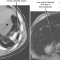
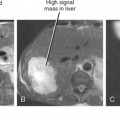
Even in patients with known primary malignancy, a small incidental lesion discovered with CT is more likely benign than malignant (Fig. 7-1). In patients with known primary cancer, a lesion considered “too small to characterize” by CT has a greater than 80% chance of being benign according to most studies. The actual number can be expected to vary with the population studied, the criteria used to define small or “too small to characterize,” the imaging technique, and the interpreting individual. Patients with breast cancer with one or more small (≤15 mm) hypoattenuating lesions on a baseline contrast-enhanced CT scan without other evidence of hepatic metastases are no more likely to experience development of subsequent hepatic metastases than patients with no such lesions. In another study of patients with breast cancer, too small to characterize lesions represented benign findings in more than 90% of women. Patterson and researchers studied the MRI evaluation of “too small to characterize” liver lesions discovered with CT in patients with breast cancer and found that only 5% of such lesions were shown to represent metastases. In a study of hepatic lesions 15 mm or smaller in patients with gastric and colorectal cancer, almost 80% were benign. In that study, if patients with larger coexistent liver metastases were excluded, small hypoattenuating lesions were metastases in only approximately 2% of patients.


In some respects, it is not surprising that small incidental lesions discovered with CT are likely benign. After all, benign hepatic lesions are extremely common, and tiny, low-attenuation, simple cysts are likely to be more conspicuous against a background of enhancing liver than equally sized enhancing metastatic lesions.


A brief analysis of lesion features can sometimes help identify worrisome lesions, although no imaging features are entirely specific. Benign lesions tend to be lower in attenuation and have more discrete margins than malignant lesions, whereas target enhancement (low-attenuation border surrounding a lower attenuation center) is suggestive of metastatic disease. For patients with extrahepatic tumors, one may also take into account the stage and imaging characteristics of the primary tumor when analyzing a small focal hepatic lesion. For example, patients with known extrahepatic metastases are more likely to have liver metastases than patients with carcinoma in situ.


Patients with Chronic Liver Disease


Patients with chronic viral hepatitis or cirrhosis are at increased risk for development of hepatocellular carcinoma. Unfortunately, patients with chronic liver disease are also likely to be plagued by small, incidental, arterially enhancing lesions on dynamic, multiphase, contrast-enhanced imaging. The cause of such lesions varies and may include arterial-portal shunt, regenerative nodule, or benign neoplasm (e.g., hemangioma or focal nodular hyperplasia–like lesions). Fortunately, small (<2 cm), arterially enhancing nodules not visible on other phases of enhancement are more likely to be benign than malignant in the setting of chronic liver disease, although it may be difficult to distinguish benign nodules from hepatocellular carcinoma based on imaging criteria alone. Therefore, patients with chronic liver disease who have small arterially enhancing nodules on dynamic, multiphase, contrast-enhanced CT (or MRI) are usually followed with CT or MRI at approximately 6-month intervals. Small, arterially enhancing lesions that demonstrate intralesional washout of contrast material during the portal venous phase, a nodule-within-nodule appearance, rim enhancement, or evidence of a pseudocapsule on portal phase images should be considered malignant until proved otherwise.


PANCREAS


Pancreatic lesions are less likely to be discovered incidentally than liver lesions, although this gap is narrowing with the widespread availability and frequent utilization of multidetector CT and MRI. Incidentally discovered solid lesions of the pancreas usually require further evaluation and intervention because many such lesions will subsequently be shown to represent adenocarcinoma or neuroendocrine tumor. Given that little controversy surrounds the management of incidentally discovered solid pancreatic masses, this is not discussed here in further detail.


Incidental asymptomatic cystic lesions are a more common occurrence. Zhang and colleagues found a prevalence rate of pancreatic cystic lesions of 19.6% in a population of 1444 patients studied with single-shot fast spin-echo MR. In general, cystic lesions are less likely to be malignant than solid lesions. Asymptomatic pancreatic cysts had a 14% chance of being malignant in one study by Goh and researchers, which included cysts of any size. Distinguishing between different types of cystic pancreatic lesions is critical to patient management. For example, a simple pancreatic cyst can be managed nonsurgically, whereas mucinous cystic neoplasm is resected when possible. Lesions of serous (microcystic) cystadenoma are benign and left alone when asymptomatic.


When faced with an incidentally discovered pancreatic cystic lesion, many factors should be considered when determining subsequent management. Patient age and surgical risk, lesion size and location, and imaging characteristics of the lesion all contribute to decisions about subsequent patient management. In general, large size, thick septations, large locules, enhancing nodules, or biliary or pancreatic duct dilatation are concerning features. If a lesion is readily accessible to interrogation with endoscopic ultrasound (EUS), aspiration and cyst fluid analysis can be helpful in management decisions. The presence or level of mucin, amylase, carcinoembryonic antigen, CA-19-9, prostaglandin E2, and malignant cytology have all been investigated as potential discriminators between surgical and nonsurgical cystic lesions of the pancreas with variable results.

Stay updated, free articles. Join our Telegram channel

Mar 6, 2016 | Posted by in GENERAL RADIOLOGY | Comments Off on Computed Tomography Incidentalomas

Full access? Get Clinical Tree

Get Clinical Tree app for offline access