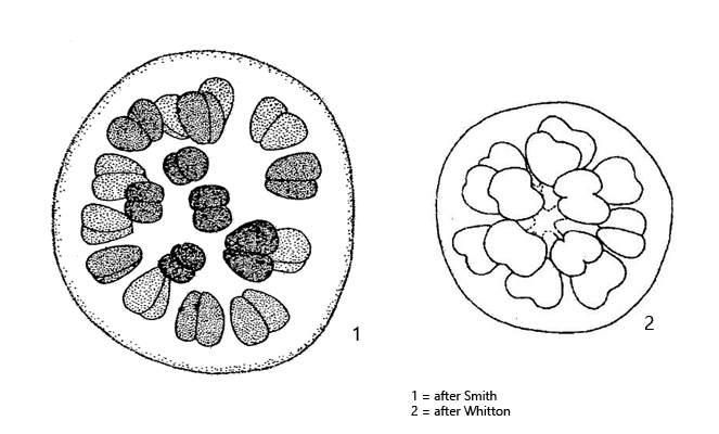
cells joined together after cell division in a cordiform shape
cells in peripherical layer at distal ends of branched, mucilaginous stalks
talks originating in center of colony
cells are separated from each other
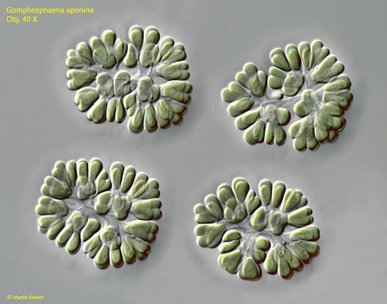
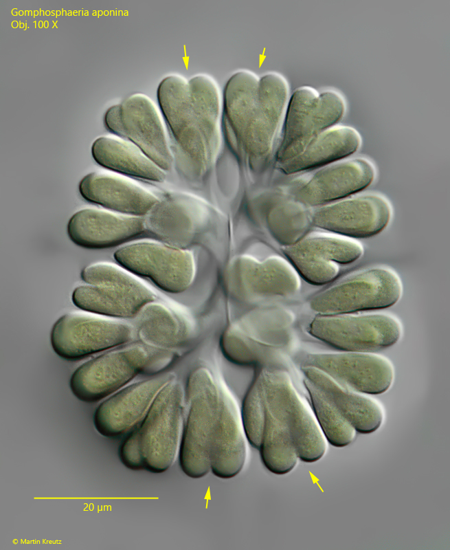
Gomphosphaeria aponina
I found Gomphosphaeria aponina among floating plant masses in the Simmelried. The center of the colony is formed by mucilaginous, branched stalks, at the ends of which are the ovoid or wedge-shaped cells. The cells remain connected after the cell division have a characteristic heart-shaped form (s. fig. 2). This peculiarity makes Gomphosphaeria aponina easy to identify. The similar genus Snowella (e.g. Snowella litoralis) also has branched stalks at the ends of which the cells of the colony are located. However, in this genus the cells do not remain connected after division.
Fig. 1:Gomphospharia aponina. D = 70–76 µm (of colonies). Overview of four colonies. Obj. 40 X.
Fig. 2:Gomphospharia aponina. D = 84 µm (of colony). A slightly squashed colony. Note the cordiform shaped cells during cell division (arrows) and the branched mucilaginous stalks in the center of the colony. The cells are 10–12 µm long. Obj. 100 X.